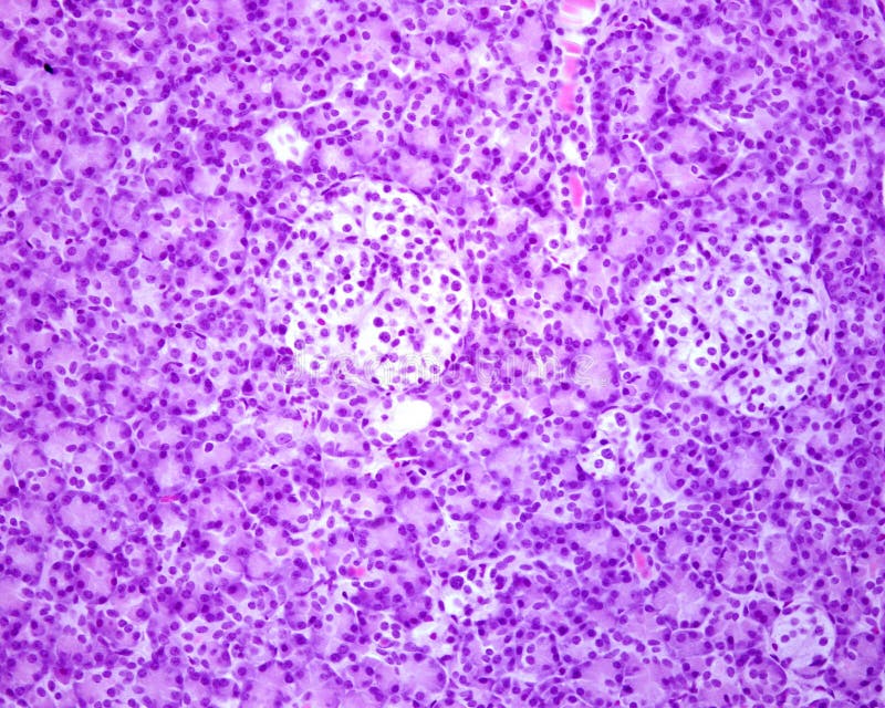
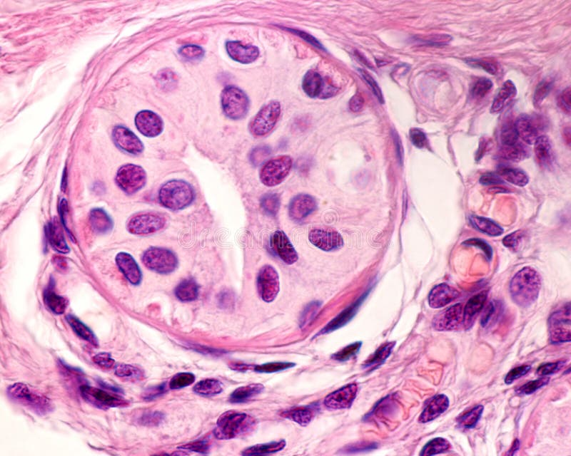
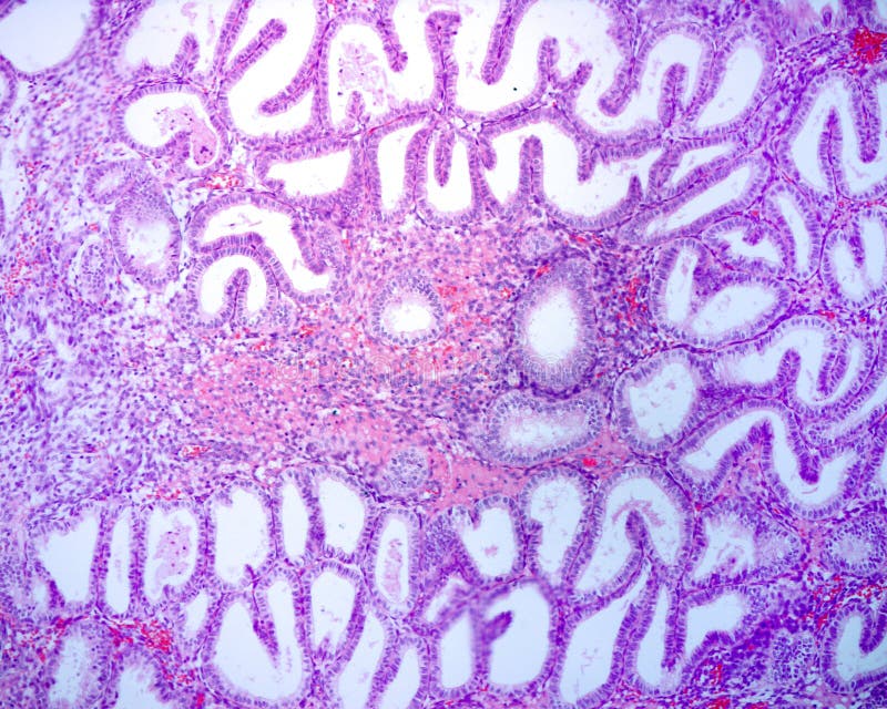
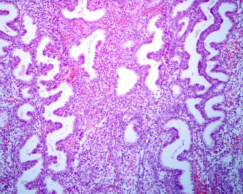
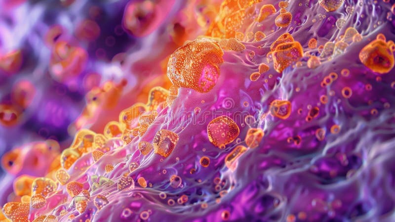
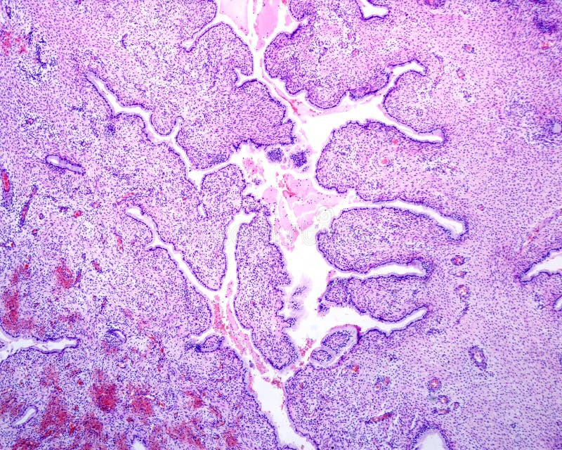
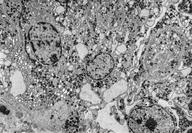
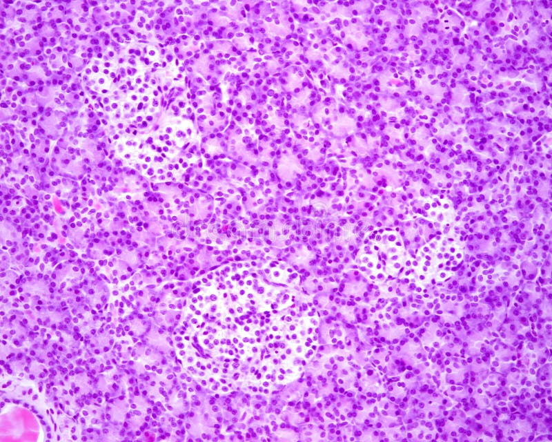
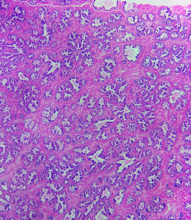
Search among 284 authentic secretory stock photos, high-definition images, and pictures, or look at other secretory unit or secretory portion stock images to enhance your presentation with the perfect visual.

Traditionally used as flavouring agent and herbal remedy. Leaves are steeped in brandy, vinegar, or water, and are said to relieve gastrointestinal and urinary tract ailments. Secretory stock images, royalty-free photos and pictures
Ovulation is when a mature egg is released from the ovary, pushed down the fallopian tube, and is available to be fertilized. Approximately every month an egg will mature within one of your ovaries. Ovulation is the release of egg from the ovaries. In humans, this event occurs when the de Graaf's follicles rupture and release the secondary oocyte ovarian cells. After ovulation, during the luteal phase, the egg will be available to be fertilized by sperm. In addition, the uterine lining (endometrium) is thickened to be able to receive a fertilized egg. If no conception occurs, the uterine lining as well as blood will be shed during menstruation. Secretory stock images, royalty-free photos and pictures
Secretory portion of sweat glands lined by two concentric layers of columnar or cuboidal epithelial cells that look rather messy and consists of a mix of light and dark staining epithelial cells. Secretory stock images, royalty-free photos and pictures
The secretory units of apocrine sweat glands are show a large lumen alveolus lined by a simple columnar epithelium with pigment granules. The apical portion of the cells are budding into the lumen. Secretory stock images, royalty-free photos and pictures
Secretory unit of an eccrine sweat gland surrounded by a pink layer of myoepithelial cells. The secretory portion is coiled appearing sectioned many times. The two profiles on top are excretory ducts. Secretory stock images, royalty-free photos and pictures
Ovulation is when a mature egg is released from the ovary, pushed down the fallopian tube, and is available to be fertilized. Approximately every month an egg will mature within one of your ovaries. Ovulation is the release of egg from the ovaries. In humans, this event occurs when the de Graaf's follicles rupture and release the secondary oocyte ovarian cells. After ovulation, during the luteal phase, the egg will be available to be fertilized by sperm. In addition, the uterine lining (endometrium) is thickened to be able to receive a fertilized egg. If no conception occurs, the uterine lining as well as blood will be shed during menstruation. Secretory stock images, royalty-free photos and pictures
False colour transmission electron microscope TEM micrograph showing chylomicrons red in enterocytes of small intestine. They appear in the Golgi apparatus pink and are secreted to extracellular spaces through secretory vacuoles. Secretory stock images, royalty-free photos and pictures
Human pituitary. Anterior lobe. Two main types of glandular cells described as acidophilic and basophilic cells. Among the cell cords, there are sinusoid capillaries of wide and irregular lumen. Secretory stock images, royalty-free photos and pictures
The squamous metaplasia of the cervix is a benign non-cancerous change consisting of a replacement of the surface columnar epithelium by a nonkeratinized stratified squamous epithelium similar to exocervix lining. Under the stratified squamous epithelium, the endocervical mucous glands still can be seen. Secretory stock images, royalty-free photos and pictures
Light microscope micrograph of a pancreas showing two islets of Langerhans. Unlike the exocrine acini that occupy most of the surface of the image, the islet of Langerhans appears as a light zone with cells arranged in cords. Secretory stock images, royalty-free photos and pictures
Secretory portion of sweat glands lined by two concentric layers of columnar or cuboidal epithelial cells that look rather messy and consists of a mix of light and dark staining epithelial cells. Secretory stock images, royalty-free photos and pictures
Neuroendocrine tumors are neoplasms that arise from cells of the endocrine and nervous systems. These neoplasms share common features, including a similar histological appearance, having special secretory granules, and often producing biogenic amines and polypeptide hormones. Bone marrow metastasis. Wright x1000. Secretory stock images, royalty-free photos and pictures
Neuroendocrine tumors are neoplasms that arise from cells of the endocrine and nervous systems. These neoplasms share common features, including a similar histological appearance, having special secretory granules, and often producing biogenic amines and polypeptide hormones. Bone marrow metastasis. HE x100. Secretory stock images, royalty-free photos and pictures
A microscopic cross-section of the human eccrine sweat gland typically displays two major segments: the deeply coiled secretory portion located in the dermis hypodermis, and the narrower duct which ascends toward the epidermis. The secretory coils are characterized by a wide lumen and a lining of larger epithelial cells, including clear cells (producing water and electrolytes) and dark cells (mucous glycoprotein secretion), all enveloped by contractile myoepithelial cells resting on the basement membrane. The duct, in contrast, features a much smaller lumen and a distinct two-layered (stratified cuboidal) epithelium specialized for the reabsorption of ions (like $ text{Na}^+$ and $ text{Cl}^-$) to produce hypotonic sweat for thermoregulation. The surrounding dermis includes abundant collagen fibers and blood vessels. Secretory stock images, royalty-free photos and pictures
Neuroendocrine tumors are neoplasms that arise from cells of the endocrine and nervous systems. These neoplasms share common features, including a similar histological appearance, having special secretory granules, and often producing biogenic amines and polypeptide hormones. Bone marrow metastasis. Wright x1000. Secretory stock images, royalty-free photos and pictures
Neuroendocrine tumors are neoplasms that arise from cells of the endocrine and nervous systems. These neoplasms share common features, including a similar histological appearance, having special secretory granules, and often producing biogenic amines and polypeptide hormones. Bone marrow metastasis. Wright x400. Secretory stock images, royalty-free photos and pictures
Human uterine cervix. Light micrograph showing, on bottom, the exocervix lined by a non-keratinised stratified squamous epithelium. On the center of the image there is the transition to endocervix, lined by a single layer of columnar cells and containing branched tubular mucous glands lined by a simple columnar epithelium. On the limit, there is a small zone of epithelial proliferation (. Secretory stock images, royalty-free photos and pictures
Light micrograph of secretory simple columnar epithelium of the rat epididymis. The cytoplasm above the nuclei shows clear mucous granules and pigment granules. The connective tissue under the epithelium shows small blood vessels. 0. 5 micrometre thick section of plastic embedded material stained with toluidine blue. Secretory stock images, royalty-free photos and pictures
Light microscope micrograph showing the Golgi apparatus in pancreatic acini stained with Cajal's formol-uranium silver method. The Golgi apparatus appears as a dark brown network located in the center of acini. Secretory stock images, royalty-free photos and pictures
Light micrograph of secretory simple columnar epithelium of the rat epididymis. The cytoplasm above the nuclei shows clear mucous granules and pigment granules. The connective tissue under the epithelium shows small blood vessels. 0. 5 micrometre thick section of plastic embedded material stained with toluidine blue. Secretory stock images, royalty-free photos and pictures
The steroid-secreting cells of the adrenal cortex have a special type of mitochondria with tubular and vesicular cristae that appear as rounded profiles instead of the elongated profiles typical of lamellar mitochondria cristae common on almost all cells. The mitochondria are surrounded by smooth endoplasmic reticulum. Lipid droplets also are frequent in that cells. Secretory stock images, royalty-free photos and pictures
Low magnification light micrograph of a pancreas showing several islets of Langerhans. Unlike the exocrine acini that occupy most of the surface of the image, the islet of Langerhans appears as a light zone with cells arranged in cords. Secretory stock images, royalty-free photos and pictures
Light microscope micrograph showing the Golgi apparatus in pancreatic acini stained with Cajal's formol-uranium silver method. The Golgi apparatus appears as a dark brown network located in the center of acini. Secretory stock images, royalty-free photos and pictures