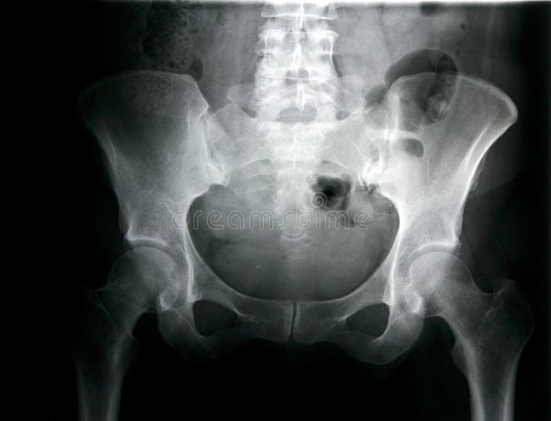
Free with trial X-ray of the pelvis with the backbone, you can see the socket of the hip joint, often used for Hip Joint Endoprosthesis, image can also be used for rheumatic diseases and Rheumatoid Arthritis, which are widespread. Pelvis stock images, royalty-free photos and pictures Backbone, pelvis, socket of hip joint, thigh bone x-ray, osteoporosis. X-ray of the pelvis with the backbone, you can see the socket of the hip joint, often used. X-ray of the pelvis with the backbone, you can see the socket of the hip joint, often used for Hip Joint Endoprosthesis, image can also be used for rheumatic diseases and Rheumatoid Arthritis, which are widespread
Free with trial Contractions. Childbrith - woman working through contractions. Pelvis stock images, royalty-free photos and pictures Contractions. Childbrith - woman working through contractions
Free with trial Female pelvis. Reproduction system anatomy isolated on white background. Pelvis stock images, royalty-free photos and pictures Female pelvis . Reproduction system anatomy. Female pelvis. Reproduction system anatomy isolated on white background. Female pelvis. Reproduction system anatomy isolated on white background.
Free with trial X-RAY true colour film of female lower thorax and pelvis. Pelvis stock images, royalty-free photos and pictures X-RAY of female lower thorax and pelvis. X-RAY true colour film of female lower thorax and pelvis. X-RAY true colour film of female lower thorax and pelvis.
Free with trial Film x-ray human's pelvis and arthritis at both hip joint (Gout , Rheumatoid). Pelvis stock images, royalty-free photos and pictures X-ray human's pelvis and arthritis at both hip joint (Gout , Rheumatoid). Film x-ray human's pelvis and arthritis at both hip joint (Gout , Rheumatoid. Film x-ray human's pelvis and arthritis at both hip joint (Gout , Rheumatoid)
Free with trial Film x-ray pelvis of osteoporosis patient and arthritis both hip. Pelvis stock images, royalty-free photos and pictures Film x-ray pelvis of osteoporosis patient and arthritis both hip
Free with trial Prostate cancer metastasis. Nuclear medicine scanning of patient with prostate cancer metastasis in areas of pelvis and spine in different colors. Pelvis stock images, royalty-free photos and pictures Prostate cancer metastasis. Nuclear medicine scanning of patient with prostate cancer metastasis in areas of pelvis and spine in different colors
Free with trial Xray of pelvic area, male. Can be seen the thigh bone, pelvis, and the ball joint between them. Also, I think, rectum, bladder, prostrate and spine (tailbone). Pelvis stock images, royalty-free photos and pictures Xray. Of pelvic area, male. Can be seen the thigh bone, pelvis, and the ball joint between them. Also, I think, rectum, bladder, prostrate and spine (tailbone
Free with trial A closeup of a physiotherapist with a stethoscope operating with a spine model with pelvis element with at dark background with digital spine image. A concept of spine diseases treatment. Pelvis stock images, royalty-free photos and pictures A doctor holding an artificial spine model. A closeup of a physiotherapist with a stethoscope operating with a spine model with pelvis element with at dark. A closeup of a physiotherapist with a stethoscope operating with a spine model with pelvis element with at dark background with digital spine image. A concept of spine diseases treatment
Free with trial Spinal column. Learning model of the human spinal column. isolated on white background. Pelvis stock images, royalty-free photos and pictures Spinal column. Learning model of the human spinal column. isolated on white background
Free with trial Doctor shows a human spine. On blue background. Pelvis stock images, royalty-free photos and pictures Doctor shows a human spine. On blue background
Free with trial X-ray of the hip and pelvis of an 83 year old woman with a history of degenerative joint disease. The x-ray shows severe DJD of the hip as well as a fracture of the neck of the femur. Pelvis stock images, royalty-free photos and pictures Hip x-ray, fracture and DJD (degenerative joint d). X-ray of the hip and pelvis of an 83 year old woman with a history of degenerative joint disease. The x-ray. X-ray of the hip and pelvis of an 83 year old woman with a history of degenerative joint disease. The x-ray shows severe DJD of the hip as well as a fracture of the neck of the femur.
Free with trial There are most popular radiological examinations ,which for diagnostic purpose during processing can be used violet color to confirm or reject pathological process in heart, bones thyroid, brain ,kidneys and other organs. Pelvis stock images, royalty-free photos and pictures Radiological diagnostic violet color elements. There are most popular radiological examinations ,which for diagnostic purpose during processing can be used. There are most popular radiological examinations ,which for diagnostic purpose during processing can be used violet color to confirm or reject pathological process in heart, bones thyroid, brain ,kidneys and other organs .
Free with trial Close up. Doctor in white gown is showing vertebrae on skeleton. Pelvis stock images, royalty-free photos and pictures Close up. Doctor is showing vertebrae on skeleton. Close up. Doctor in white gown is showing vertebrae on skeleton. Close up. Doctor in white gown is showing vertebrae on skeleton.
Free with trial Human spine in x-ray, on gray background. The lumbar spine is highlighted by red colour. Pelvis stock images, royalty-free photos and pictures The lumbar spine is highlighted by red colour. Human spine in x-ray, on gray background. The lumbar spine is highlighted by red colour. Human spine in x-ray, on gray background. The lumbar spine is highlighted by red colour.
Free with trial Human spine in x-ray, on gray background. The lumbar spine is highlighted by red colour. Pelvis stock images, royalty-free photos and pictures Spine in x-ray. Lumbar spine is highlighted. Human spine in x-ray, on gray background. The lumbar spine is highlighted by red colour. Human spine in x-ray, on gray background. The lumbar spine is highlighted by red colour.
Free with trial A gynecologist doctor probes the lower abdomen of a girl who has pain and inflammation of the reproductive system. Ovarian cyst,. A gynecologist doctor probes. Pelvis stock images, royalty-free photos and pictures A gynecologist doctor probes the lower abdomen of a girl who has pain and inflammation of the reproductive system. Ovarian cyst,. A gynecologist doctor probes
Free with trial Pelvis xray. X-ray of female pelvis. Pelvis stock images, royalty-free photos and pictures Pelvis xray. X-ray of female pelvis
Free with trial Pelvis X Ray. X ray Image Of Pelvis, Frontal view. Pelvis stock images, royalty-free photos and pictures Pelvis X Ray. X ray Image Of Pelvis, Frontal view
Free with trial Pelvis. Rontgen picture of male pelvis. Pelvis stock images, royalty-free photos and pictures Pelvis. Rontgen picture of male pelvis
Free with trial Pelvis X-ray. Xray of pelvic bones, pelvis x-ray. Pelvis stock images, royalty-free photos and pictures Pelvis X-ray. Xray of pelvic bones, pelvis x-ray
Free with trial Female Pelvis xray. Pelvis stock images, royalty-free photos and pictures Female Pelvis. Xray. Female Pelvis xray
Free with trial X-ray of the pelvis. And spinal column of a woman. Pelvis stock images, royalty-free photos and pictures X-ray of the pelvis. And spinal column of a woman
Free with trial Film X-ray of Pelvis : normal human pelvis. Pelvis stock images, royalty-free photos and pictures Film X-ray of Pelvis. Normal human pelvis
Free with trial Women doing exercises for pelvis floor in postnatal course. Pelvis stock images, royalty-free photos and pictures Women doing exercises for pelvis floor in postnatal course. Women doing exercises for pelvis floor in postnatal regression course
Free with trial Pelvis X-ray view from radiology imagery. Pelvis stock images, royalty-free photos and pictures Pelvis X-ray. View from radiology imagery
Free with trial Human's pelvis and hip joints. Film x-ray human's pelvis and hip joints. Pelvis stock images, royalty-free photos and pictures Human's pelvis and hip joints. Film x-ray human's pelvis and hip joints
Free with trial X ray image shows the outline of the flesh with pelvic bones, womb and spine beneath. Pelvis of woman in early 30s. Pelvis stock images, royalty-free photos and pictures Female Pelvis X-Ray. X ray image shows the outline of the flesh with pelvic bones, womb and spine beneath. Pelvis of woman in early 30s. X ray image shows the outline of the flesh with pelvic bones, womb and spine beneath. Pelvis of woman in early 30s.
Free with trial Regular hip and pelvis of young man. Regular hip and pelvis of a young man, isolated on black background. Pelvis stock images, royalty-free photos and pictures Regular hip and pelvis of young man. Regular hip and pelvis of a young man, isolated on black background
Free with trial Scan of a Broken Pelvis. CT Scan. Pelvis stock images, royalty-free photos and pictures Scan of a Broken Pelvis. CT Scan
Free with trial X-ray/ rtg of human pelvis - hip joint replacemen. T (hip endoprosthesis) (medical background. Pelvis stock images, royalty-free photos and pictures X-ray/ rtg of human pelvis - hip joint replacemen. T (hip endoprosthesis) (medical background
Free with trial X-ray of pelvis and spinal column. Pelvis stock images, royalty-free photos and pictures X-ray of pelvis and spinal column. X-ray of the pelvis and spinal column
Free with trial Film x-ray pelvis of osteoporosis patient and arthritis both hip. Pelvis stock images, royalty-free photos and pictures Film x-ray pelvis of osteoporosis patient and arthritis both hip.
Free with trial Female pelvis model with a cross section of the inner organ with red and blue arteries and adrenal gland as a health care and medical of the anatomy system. Pelvis stock images, royalty-free photos and pictures Female pelvis. Model with a cross section of the inner organ with red and blue arteries and adrenal gland as a health care and medical of the anatomy system
Free with trial Women doing exercises for pelvis floor in postnatal regression c. Ourse. Pelvis stock images, royalty-free photos and pictures Women doing exercises for pelvis floor in postnatal regression c. Ourse
Free with trial Film x-ray pelvis of osteoporosis patient and arthritis both hip. Pelvis stock images, royalty-free photos and pictures Film x-ray pelvis of osteoporosis patient and arthritis both hip.
Free with trial Pelvis, Human skeleton, Female Pelvis Bone anatomy, hip. The pelvis is either the lower part of the trunk of the human body, pelvic, Digital illustration. Pelvis stock images, royalty-free photos and pictures Pelvis, Human skeleton, Female Pelvis Bone anatomy, hip. The pelvis is either the lower part of the trunk of the human body, pelvic, Digital illustration
Free with trial Doctor gynecologist showing layout of female pelvis with muscles closeup. Strengthening pelvic floor muscles concept. Pelvis stock images, royalty-free photos and pictures Doctor gynecologist showing layout of female pelvis with muscles closeup. Strengthening pelvic floor muscles concept
Free with trial Computed Tomography of body, chest, abdomen, and pelvis (CT). Pelvis stock images, royalty-free photos and pictures Computed Tomography of Body. Chest, abdomen, and pelvis (CT
Free with trial X-ray of a pathologic pelvis and femurs of a dog (canine), we can see panosteitis on the femurs. Computed radiography ( CR ). Pelvis stock images, royalty-free photos and pictures DOG HIP X-RAY , PANOSTEITIS. X-ray of a pathologic pelvis and femurs of a dog (canine), we can see panosteitis on the femurs. Computed radiography ( CR. X-ray of a pathologic pelvis and femurs of a dog (canine), we can see panosteitis on the femurs. Computed radiography ( CR )
Free with trial Cross bones of leg and arm with pelvis in isolation. Pelvis stock images, royalty-free photos and pictures Cross-Bones. Cross bones of leg and arm with pelvis in isolation. Cross bones of leg and arm with pelvis in isolation.
Free with trial Xray picture. Human female pelvis hip bone xray picture. Pelvis stock images, royalty-free photos and pictures Xray picture. Human female pelvis hip bone xray picture
Free with trial Osteoarthritis of a hip joint, front view. Shown is a narrowing of the joint space and a deformation of the femoral head caused by the destruction of articular cartilage. The pelvis shows osteophyte growths (bone spurs) close to the joint. Pelvis stock images, royalty-free photos and pictures Hip - Osteoarthritis. Osteoarthritis of a hip joint, front view. Shown is a narrowing of the joint space and a deformation of the femoral head caused by the. Osteoarthritis of a hip joint, front view. Shown is a narrowing of the joint space and a deformation of the femoral head caused by the destruction of articular cartilage. The pelvis shows osteophyte growths (bone spurs) close to the joint.
Free with trial Patient refused of any operation and chemotherapy as a result of this after two years were severe spreading of metastasis to spine vertebrae pelvis and ribs. Pelvis stock images, royalty-free photos and pictures Cancer metastasis. Patient refused of any operation and chemotherapy as a result of this after two years were severe spreading of metastasis to spine vertebrae. Patient refused of any operation and chemotherapy as a result of this after two years were severe spreading of metastasis to spine vertebrae pelvis and ribs
Free with trial Hip xray. Photography of human hip and pelvis xray. Pelvis stock images, royalty-free photos and pictures Hip xray. Photography of human hip and pelvis xray
Free with trial A plain radiograph (x-ray) of the pelvic bones showing both hip joints and the lower back. Pelvis stock images, royalty-free photos and pictures Plain x-ray of the pelvis. A plain radiograph (x-ray) of the pelvic bones showing both hip joints and the lower back. A plain radiograph (x-ray) of the pelvic bones showing both hip joints and the lower back.
Free with trial Pelvis x-ray of a 65 year old female showing the repair of a hip fracture with 2 screws through the neck of the femur. On the other side there is a fracture of the lesser trochanter. Pelvis stock images, royalty-free photos and pictures Pelvis x-ray, repair of hip fracture. Pelvis x-ray of a 65 year old female showing the repair of a hip fracture with 2 screws through the neck of the femur. On. Pelvis x-ray of a 65 year old female showing the repair of a hip fracture with 2 screws through the neck of the femur. On the other side there is a fracture of the lesser trochanter
Free with trial Anatomy of human hip joint and pelvis. Human hip anatomy isolated on black background. Pelvis stock images, royalty-free photos and pictures Anatomy of human hip joint and pelvis. Human hip anatomy isolated on black background
Free with trial Colorectal cancer (also known as colon cancer, rectal cancer, or bowel cancer) is one of the most aggressive and dangerous cancer due to its spread in different parts of body. Pet/ct is very important tool for early diagnostic. Red with yellow colors in the area of pelvis with pathological tissue. Pelvis stock images, royalty-free photos and pictures Colorectal carcinoma pet /ct diagnostic. Colorectal cancer (also known as colon cancer, rectal cancer, or bowel cancer) is one of the most aggressive and. Colorectal cancer (also known as colon cancer, rectal cancer, or bowel cancer) is one of the most aggressive and dangerous cancer due to its spread in different parts of body. Pet/ct is very important tool for early diagnostic .Red with yellow colors in the area of pelvis with pathological tissue .
Free with trial Prostate cancer metastases. Impressive examination of nuclear medicine with proof of metastases in the area of spine and pelvis. Pelvis stock images, royalty-free photos and pictures Prostate cancer metastases. Impressive examination of nuclear medicine with proof of metastases in the area of spine and pelvis
Free with trial There is colorful 3D CT Urography scan with anatomical threedimensional tomography of kidneys , calyces ,ureters ,bladder, lumbar spine and pelvis , which in combination with usual X-RAY urography can help urologist in final conclusion ,concerning pathology of urological system. Pelvis stock images, royalty-free photos and pictures Ct scan 3d ct urography kidneys bladder colorful. There is colorful 3D CT Urography scan with anatomical threedimensional tomography of kidneys , calyces. There is colorful 3D CT Urography scan with anatomical threedimensional tomography of kidneys , calyces ,ureters ,bladder, lumbar spine and pelvis , which in combination with usual X-RAY urography can help urologist in final conclusion ,concerning pathology of urological system.
Free with trial A closeup of doctor operating with artificial spine model with pelvis unit of human skeleton digital human figure background. The concept of spine diseases treatment. Pelvis stock images, royalty-free photos and pictures A doctor operating with spin model. A closeup of doctor operating with artificial spine model with pelvis unit of human skeleton digital human figure background. A closeup of doctor operating with artificial spine model with pelvis unit of human skeleton digital human figure background. The concept of spine diseases treatment
Free with trial Model hip joint. Model of hip joint and pelvis isolated on white. Pelvis stock images, royalty-free photos and pictures Model hip joint. Model of hip joint and pelvis isolated on white
Free with trial Xray of the kidneys. Intravenous Urography, using contrast material showing the kidneys, Urethers, and bladder. Pelvis stock images, royalty-free photos and pictures Xray of the kidneys. Intravenous Urography, using contrast material showing the kidneys, Urethers, and bladder
Free with trial Hip xray, classic radiography. X-ray of hip, close up of coxarthrosis - plain film. Pelvis stock images, royalty-free photos and pictures Hip xray, classic radiography. X-ray of hip, close up of coxarthrosis - plain film
Free with trial Pelvic MRI. Sagital MR images of pelvis, urine bladder level. Pelvis stock images, royalty-free photos and pictures Pelvic MRI. Sagital MR images of pelvis, urine bladder level
Free with trial Adult skeleton. Details of an adult skeleton in white. Pelvis stock images, royalty-free photos and pictures Adult skeleton. Details of an adult skeleton in white
Free with trial Hip Xray - front view. Woman Hips Xrays - front view. Pelvis stock images, royalty-free photos and pictures Hip Xray - front view. Woman Hips Xrays - front view
Free with trial Spine. A model of a spinal column isolated on white. Pelvis stock images, royalty-free photos and pictures Spine. A model of a spinal column isolated on white
Free with trial The femur, or thigh bone, is the longest, heaviest, and strongest bone in the entire human body. All of the body’s weight is supported by the femurs during many activities, such as running, jumping, walking, and standing. Extreme forces also act upon the femur thanks to the strength of the muscles of the hip and thigh that act on the femur to move the leg. The femur is classified structurally as a long bone and is a major component of the appendicular skeleton. Pelvis stock images, royalty-free photos and pictures Femur bone. The femur, or thigh bone, is the longest, heaviest, and strongest bone in the entire human body. All of the body’s weight is supported by the. The femur, or thigh bone, is the longest, heaviest, and strongest bone in the entire human body. All of the body’s weight is supported by the femurs during many activities, such as running, jumping, walking, and standing. Extreme forces also act upon the femur thanks to the strength of the muscles of the hip and thigh that act on the femur to move the leg. The femur is classified structurally as a long bone and is a major component of the appendicular skeleton.
Free with trial Skin. Landscape style skin with goosebumps. Pelvis stock images, royalty-free photos and pictures Skin. Landscape style skin with goosebumps
Free with trial X-ray. Body x-ray of a dog from waist down to back legs. Pelvis stock images, royalty-free photos and pictures X-ray. Body x-ray of a dog from waist down to back legs
Free with trial Physiotherapy. Adductor problems treatment at physiotherapy. Pelvis stock images, royalty-free photos and pictures Physiotherapy. Adductor problems treatment at physiotherapy
Free with trial Scoliosis X-ray. Radiograph of a whole spine with metallic rods and hooks. Pelvis stock images, royalty-free photos and pictures Scoliosis X-ray. Radiograph of a whole spine with metallic rods and hooks
Free with trial Dog x-ray. Body x-ray of a dog, mid section with organs. Pelvis stock images, royalty-free photos and pictures Dog x-ray. Body x-ray of a dog, mid section with organs
Free with trial Dog x-ray. Body x-ray of a dog, mid section with organs. Pelvis stock images, royalty-free photos and pictures Dog x-ray. Body x-ray of a dog, mid section with organs
Free with trial Lumbar spine x-ray. With metal implant. Pelvis stock images, royalty-free photos and pictures Lumbar spine x-ray. With metal implant
Free with trial Pierced belly ring. Pierced belly, jeans and hands isolated on white. Pelvis stock images, royalty-free photos and pictures Pierced belly ring. Pierced belly, jeans and hands isolated on white
Free with trial Abdominal and pelvic angiography, vessels. Vessels of abdomen and pelvis, classical radiology, angiography. Pelvis stock images, royalty-free photos and pictures Abdominal and pelvic angiography, vessels. Vessels of abdomen and pelvis, classical radiology, angiography
Free with trial Hip bone (femur). Microscopic view of healthy normal head of femur (hip bone) showing the cartilage (gray band) and underlying bone (red) and marrow. Pelvis stock images, royalty-free photos and pictures Hip bone (femur). Microscopic view of healthy normal head of femur (hip bone) showing the cartilage (gray band) and underlying bone (red) and marrow
Free with trial There are two bean shaped kidneys in the human body. A normal kidney is about 2. 5 cm thick, 10 cm long and 5 cm wide. They are reddish-brown in color and each weighs approximately 130 gm in adults. Both kidneys receive blood from the renal artery; in a resting adult approximately 1. 2 to 1. 4 Litres of blood flows through both kidneys in one minute which means every 4 to 5 minutes all the circulating blood in the body passes through the kidneys. Kidneys help remove excess salt, water and waste products from the bloodstream. Like other endocrine organs, kidneys also secrete important hormones such as rennin and erythropoietin into the circulation. These hormones play significant roles in the production of blood cell and in the regulation of blood pressure. Pelvis stock images, royalty-free photos and pictures Kidneys. There are two bean shaped kidneys in the human body. A normal kidney is about 2.5 cm thick, 10 cm long and 5 cm wide. They are reddish-brown in color. There are two bean shaped kidneys in the human body. A normal kidney is about 2.5 cm thick, 10 cm long and 5 cm wide. They are reddish-brown in color and each weighs approximately 130 gm in adults. Both kidneys receive blood from the renal artery; in a resting adult approximately 1.2 to 1.4 Litres of blood flows through both kidneys in one minute which means every 4 to 5 minutes all the circulating blood in the body passes through the kidneys. Kidneys help remove excess salt, water and waste products from the bloodstream. Like other endocrine organs, kidneys also secrete important hormones such as rennin and erythropoietin into the circulation. These hormones play significant roles in the production of blood cell and in the regulation of blood pressure.
Free with trial Mobilisation of the spine. Physical therapist doing mobilisation of the spine. Pelvis stock images, royalty-free photos and pictures Mobilisation of the spine. Physical therapist doing mobilisation of the spine
Free with trial The uterus or uterine or womb is a major female hormone-responsive reproductive sex organ of most mammals, including humans. One end, the cervix, opens into the vagina, while the other is connected to one or both fallopian tubes, uterine tubes depending on the species. It is within the uterus that the fetus develops during gestation, usually developing completely in placental mammals such as humans and partially in marsupials such as kangaroos and opossums. Two uteri usually form initially in a female and usually male fetus, and in placental mammals they may partially or completely fuse into a single uterus depending on the species. In many species with two uteri, only one is functional. Humans and other higher primates such as chimpanzees, usually have a single completely fused uterus, although in some individuals the uteri may not have completely fused. Pelvis stock images, royalty-free photos and pictures Uterus TS_Perspective View. The uterus or uterine or womb is a major female hormone-responsive reproductive sex organ of most mammals, including humans. One end. The uterus or uterine or womb is a major female hormone-responsive reproductive sex organ of most mammals, including humans. One end, the cervix, opens into the vagina, while the other is connected to one or both fallopian tubes, uterine tubes depending on the species. It is within the uterus that the fetus develops during gestation, usually developing completely in placental mammals such as humans and partially in marsupials such as kangaroos and opossums. Two uteri usually form initially in a female and usually male fetus, and in placental mammals they may partially or completely fuse into a single uterus depending on the species. In many species with two uteri, only one is functional. Humans and other higher primates such as chimpanzees, usually have a single completely fused uterus, although in some individuals the uteri may not have completely fused.
Free with trial X-ray of a bowel obstruction of a dog (canine),we can see the pebble in the small intestine. Computed radiography ( CR ). Pelvis stock images, royalty-free photos and pictures DOG BOWEL OBSTRUCTION X-RAY. X-ray of a bowel obstruction of a dog (canine),we can see the pebble in the small intestine. Computed radiography ( CR. X-ray of a bowel obstruction of a dog (canine),we can see the pebble in the small intestine. Computed radiography ( CR )
Free with trial Water. Blue water background, light effect. Pelvis stock images, royalty-free photos and pictures Water. Blue water background, light effect
Free with trial Water. Blue water background, light effect. Pelvis stock images, royalty-free photos and pictures Water. Blue water background, light effect
Free with trial Scoliosis. Radiograph of a whole spine with metallic implants. Pelvis stock images, royalty-free photos and pictures Scoliosis. Radiograph of a whole spine with metallic implants
Free with trial A model of a human right hip joint, isolated on a white background. Pelvis stock images, royalty-free photos and pictures Hip joint. A model of a human right hip joint, isolated on a white background. A model of a human right hip joint, isolated on a white background.
Free with trial Pelvic x-ray. X-ray of pelvic and abdominal area. Pelvis stock images, royalty-free photos and pictures Pelvic x-ray. X-ray of pelvic and abdominal area
Free with trial Man holds his back in pain as he recovers from an accident. Pelvis stock images, royalty-free photos and pictures Man Pain Back. Man holds his back in pain as he recovers from an accident. Man holds his back in pain as he recovers from an accident.
 Sign in