Free with trial Anatomy of the nose and throat. Median section of human head. Anatomical model vectors Nose anatomy. Anatomy of the nose and throat. Median section of human head
Free with trial Hand holding hip with visible spine and center of back pain. Anatomical model illustrations Back pain with spine. Hand holding hip with visible spine and center of back pain
Free with trial Dermatomes vector illustration. Labeled educational anatomical skin parts scheme. Epidermis area supplied by afferent spinal nerve fibers. Cervical, thoracic, lumbar and sacral nerves division diagram. Anatomical model vectors Dermatomes vector illustration. Labeled educational anatomical skin parts. Dermatomes vector illustration. Labeled educational anatomical skin parts scheme. Epidermis area supplied by afferent spinal nerve fibers. Cervical, thoracic, lumbar and sacral nerves division diagram
Free with trial Anatomical training poster. Growth and structure of human hair. Skin and hair anatomy. Cross section of the skin layers. Detailed medical vector illustration. Anatomical model vectors Anatomical training poster. Growth and structure of human hair. Skin and hair anatomy. Cross section of the skin layers
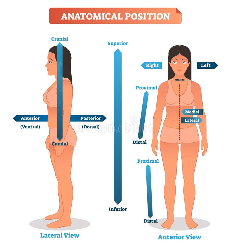
Free with trial Vector illustration of anatomical positions. Scheme of superior, inferior and proximal, distal locations, as well as medial, lateral and anterior, posterior sides. Diagram of human cranial and caudal. Anatomical model vectors Anatomical positions vector illustration. Scheme of superior, inferior and proximal, distal locations. Anterior, posterior sides. Vector illustration of anatomical positions. Scheme of superior, inferior and proximal, distal locations, as well as medial, lateral and anterior, posterior sides. Diagram of human cranial and caudal.
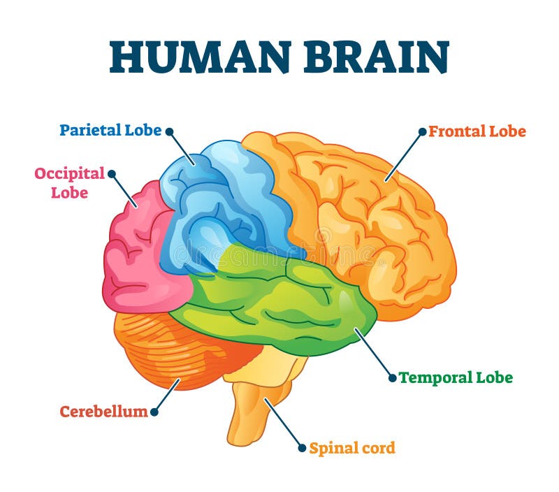
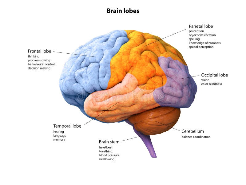
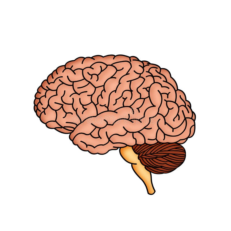
Free with trial Human brain vector illustration. Labeled anatomical educational head organ parts scheme separated by colors. Diagram with parietal, frontal, occipital and temporal lobe, spinal cord and cerebellum. Anatomical model vectors Human brain vector illustration. Labeled anatomical educational parts scheme. Human brain vector illustration. Labeled anatomical educational head organ parts scheme separated by colors. Diagram with parietal, frontal, occipital and temporal lobe, spinal cord and cerebellum.
Free with trial Chromation biological diagram vector illustration. Close-up with nucleosome, histone and DNA double helix. Science educational information. Chromosome structure elements graphic example model. Anatomical model vectors Chromation biological diagram vector illustration
Free with trial The figure represents an isolated tooth inside show where you see the blood flow and nerves, the image may be used in the field of dentistry. Anatomical model illustrations Tooth
Free with trial Spine cord vector icon isolated on white background. Anatomical model vectors Spine cord vector icon
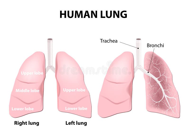
Free with trial Easy to edit illustration of Human Lungs diagram. Anatomical model illustrations Human Lungs
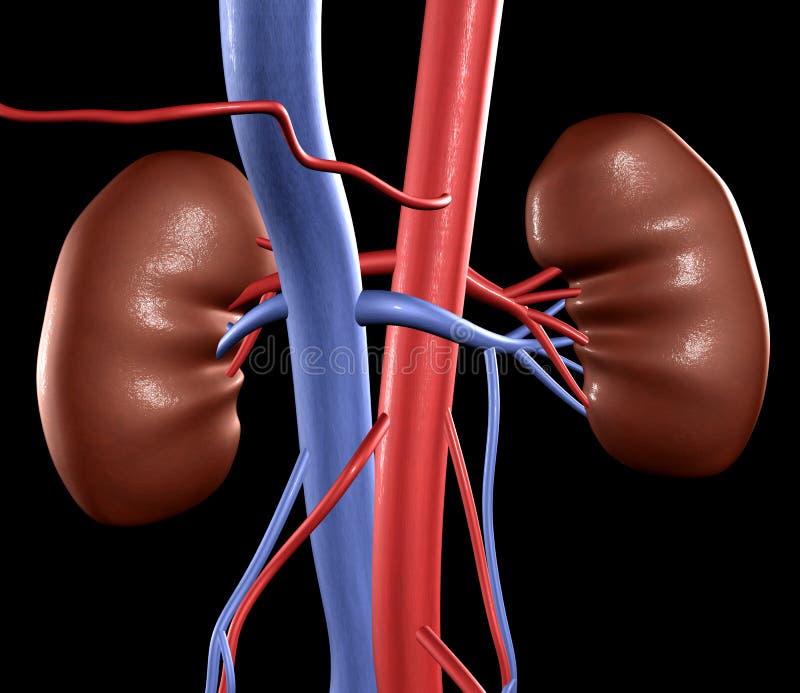
Free with trial Close-up of human kidneys with arteries and veins. Anatomical model illustrations Human Kidneys
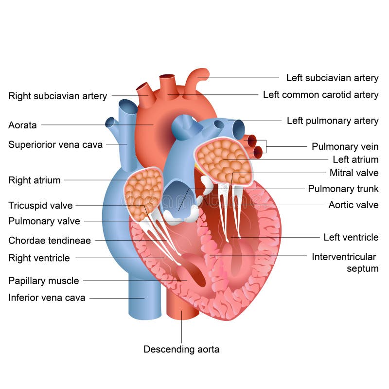
Free with trial Vector illustration of drawing of heart anatomy. Anatomical model vectors Heart Anatomy
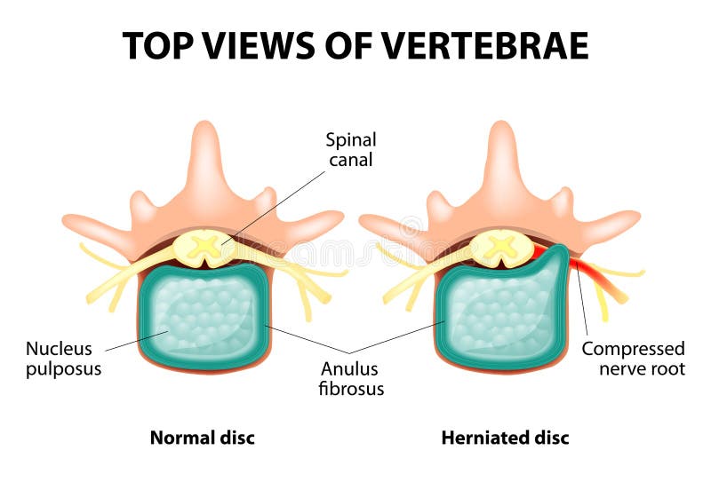
Free with trial Herniated Disc. Prolapse of intervertebral disc closeup. Anatomical model vectors Herniated Disc
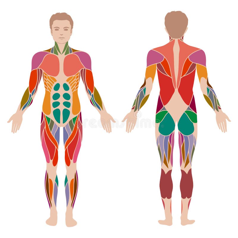
Free with trial Vector muscular human body, muscle man anatomy,. Anatomical model vectors Muscle man anatomy
Free with trial Leonardo da vinci vitruvian man vector. Illustration of vitruvian body man, classic proportion vitruvian man. Anatomical model vectors Leonardo da vinci vitruvian man vector illustration. Leonardo da vinci vitruvian man vector. Illustration of vitruvian body man, classic proportion vitruvian man
Free with trial Typical bone fractures. Vector scheme. Anatomical model vectors Typical bone fractures
Free with trial Female Reproductive System. Vector illustration flat design. Anatomical model vectors Female Reproductive System
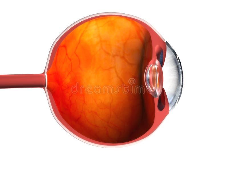
Free with trial Anatomical 3d model of a human eye. Anatomical model illustrations 3d eye. Anatomical 3d model of a human eye
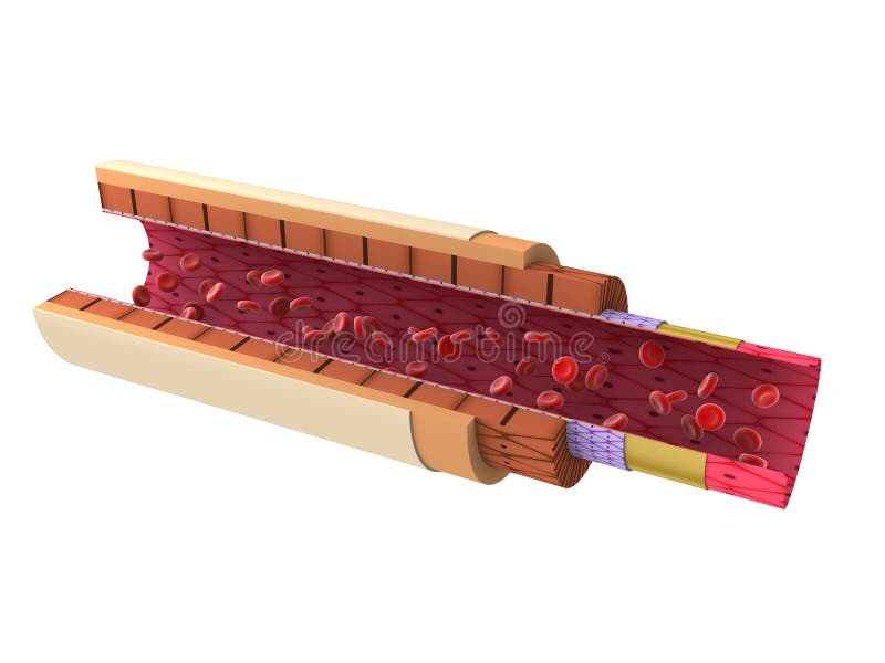
Free with trial Anatomical 3d model of a human artery. Anatomical model illustrations 3d artery. Anatomical 3d model of a human artery
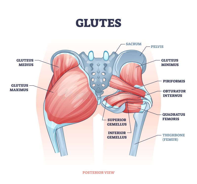
Free with trial Glutes as gluteal body muscles for human buttocks strength outline concept. Labeled educational anatomical scheme with physical skeletal and gluteus medius, maximus and minimus vector illustration. Anatomical model vectors Glutes as gluteal body muscles for human buttocks strength outline concept
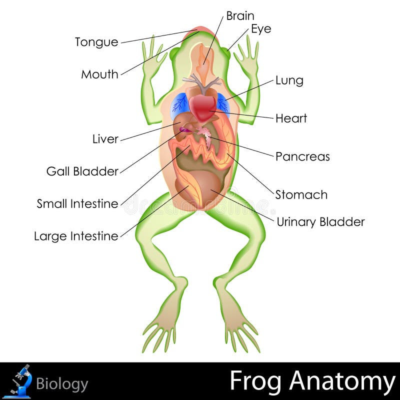
Free with trial Easy to edit vector illustration of frog anatomy. Anatomical model vectors Frog Anatomy
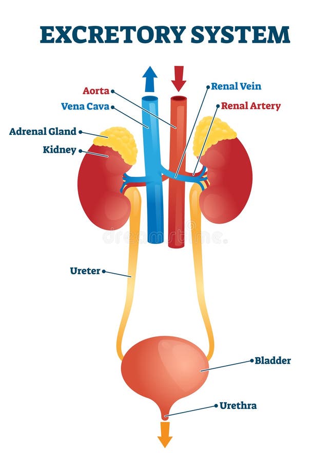
Free with trial Easy to edit vector illustration of human excretory system. Anatomical model vectors Excretory System
Free with trial Realistic human liver. Vector medical illustration. Medicine anatomy, organ human, health and biology. Anatomical model vectors Realistic human liver. Vector medical illustration
Free with trial Side view of man with 3D spine shown. Anatomical model illustrations Man with 3D spine
Free with trial Human respiratory system. Digital illustration. Anatomical model illustrations Respiratory system
Free with trial Heart; abstract blue xray concept. Anatomical model illustrations Heart; xray concept. Heart; abstract blue xray concept
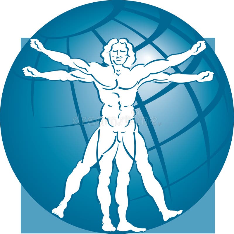
Free with trial An illustration of the vitruvian man with a globe in the background. Anatomical model vectors Vitruvian Man Model & Globe. An illustration of the vitruvian man with a globe in the background.
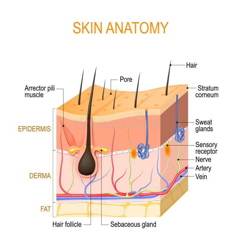
Free with trial Skin anatomy. Layers: epidermis with hair follicle, sweat and sebaceous glands, derma and fat hypodermis. Vector diagram for educational, medical, biological, and scientific use. Anatomical model vectors Skin anatomy. Layers: epidermis with hair follicle, sweat and sebaceous glands, derma and fat hypodermis
Free with trial Human Duodenum isolated on white - 3d illustration. Anatomical model illustrations Human Duodenum isolated on white
Free with trial Shoulder anatomy vector illustration. Labeled inner skeleton and muscle structure scheme. Physiological educational posterior or anterior view with bones titles and location. Healthy organ description. Anatomical model vectors Shoulder anatomy vector illustration. Labeled skeleton and muscle scheme. Shoulder anatomy vector illustration. Labeled inner skeleton and muscle structure scheme. Physiological educational posterior or anterior view with bones titles and location. Healthy organ description
Free with trial Human body vector icon of vitruvian man. Famous leonardo da vinci image vitruvian man, classic proportion form man illustration. Anatomical model vectors Human body vector icon of vitruvian man
Free with trial Dental tooth anatomy. Cross section of human tooth isolated on white. 3d illustration. Anatomical model illustrations Dental tooth anatomy. Cross section of human tooth isolated on white
Free with trial Abstract 3d polygonal wireframe DNA molecule helix spiral. Medical science, genetic biotechnology, chemistry biology, gene cell concept vector illustration. Anatomical model vectors Abstract 3d polygonal wireframe DNA molecule helix spiral. Medical science, genetic biotechnology, chemistry biology, gene cell
Free with trial A man hand muscles and muscle groups and bone shown blue colour body on Isolated background with clipping path. Anatomical model illustrations Body muscles and bone. A man hand muscles and muscle groups and bone shown blue colour body on Isolated background with clipping path
Free with trial Cartoon Silhouette Vitruvian Man Proportion, Human Anatomy. Flat Design Style. Vector illustration. Anatomical model vectors Cartoon Silhouette Vitruvian Man. Vector. Cartoon Silhouette Vitruvian Man Proportion, Human Anatomy. Flat Design Style. Vector illustration
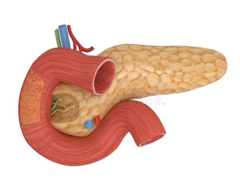
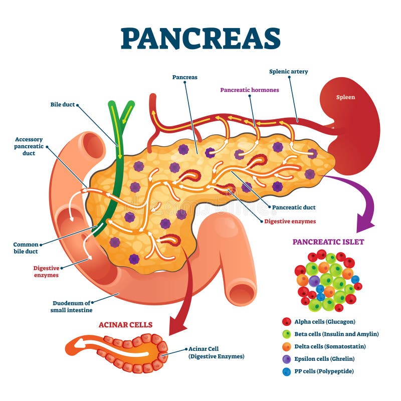
Free with trial Pancreas anatomical cross section model, vector illustration medical example. Blood flow process, cell structure and hormone functions. Digestive enzymes, pancreatic islet and other internal elements. Anatomical model vectors Pancreas anatomical cross section model, vector illustration medical example
Free with trial Rotator cuff muscle with anatomical posterior and anterior view expample. Educational labeled scheme with supraspinatus, infraspinatus, teres minor and subscapularis location vector illustration. Anatomical model vectors Rotator cuff muscle with anatomical posterior and anterior view expample
Free with trial Anterior pelvic tilt model compared with posterior labeled outline diagram. Educational scheme with skeletal bone location and movement angle vector illustration. Anatomical right posture explanation. Anatomical model vectors Anterior pelvic tilt model compared with posterior in labeled outline diagram. Anterior pelvic tilt model compared with posterior labeled outline diagram. Educational scheme with skeletal bone location and movement angle vector illustration. Anatomical right posture explanation.
Free with trial Three-dimensional anatomical model of human legs, on a white background. Anatomical model illustrations Anatomical model of human legs
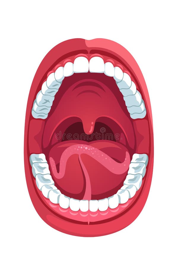
Free with trial Oral cavity. Human open mouth anatomy model. Infographic design for educational poster. Open mouth anatomy and dentistry. Flat style isolated vector visual aid illustration. Anatomical model vectors Oral cavity. Human open mouth anatomy model
Free with trial Science of hair. Anatomical training poster. Hair structure. Detailed medical vector illustration. Anatomical model vectors Science of hair. Anatomical training poster. Hair structure. Medical vector illustration
Free with trial Human muscles anatomy model vector, strong man. Anatomical model vectors Human muscles anatomy model vector
Free with trial Human muscles anatomy model vector, strong man. Anatomical model vectors Human muscles anatomy model vector
Free with trial Human muscles anatomy model vector, strong man. Anatomical model vectors Human muscles anatomy model vector
Free with trial Lungs alveoli schematic, anatomical vector illustration diagram with capillary network. Medical information poster. Anatomical model vectors Lungs alveoli schematic, anatomical vector illustration diagram with capillary network.
Free with trial Dental implants and tooth set model closeup side view realistic image vector illustration. Anatomical model vectors Dental Implant Tooth Set Closeup Model. Dental implants and tooth set model closeup side view realistic image vector illustration
Free with trial Realistic anatomical model of healthy human liver with gallbladder isolated on white. Anatomical model illustrations Realistic human liver illustration. Realistic anatomical model of healthy human liver with gallbladder isolated on white
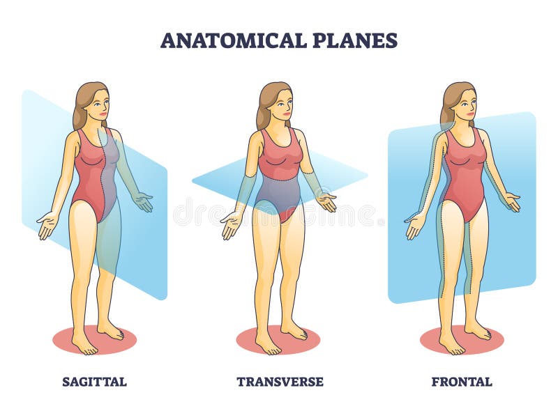
Free with trial Anatomical planes examples for medical human body transection outline diagram. Labeled educational scheme with anatomical sagittal, transverse and frontal person division types vector illustration. Anatomical model vectors Anatomical planes examples for medical human body transection outline diagram
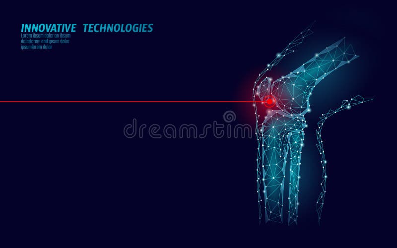
Free with trial Human knee joint 3d model vector illustration. Low poly design future technology cure pain treatment. Blue background and red injury man body leg medicine template art. Anatomical model vectors Human knee joint 3d model vector illustration. Low poly design future technology cure pain treatment. Blue background
Free with trial Dental implant and real tooth anatomy closeup cut away section model side view realistic vector illustration. Anatomical model vectors Dental Implants Anatomy Closeup Model. Dental implant and real tooth anatomy closeup cut away section model side view realistic vector illustration
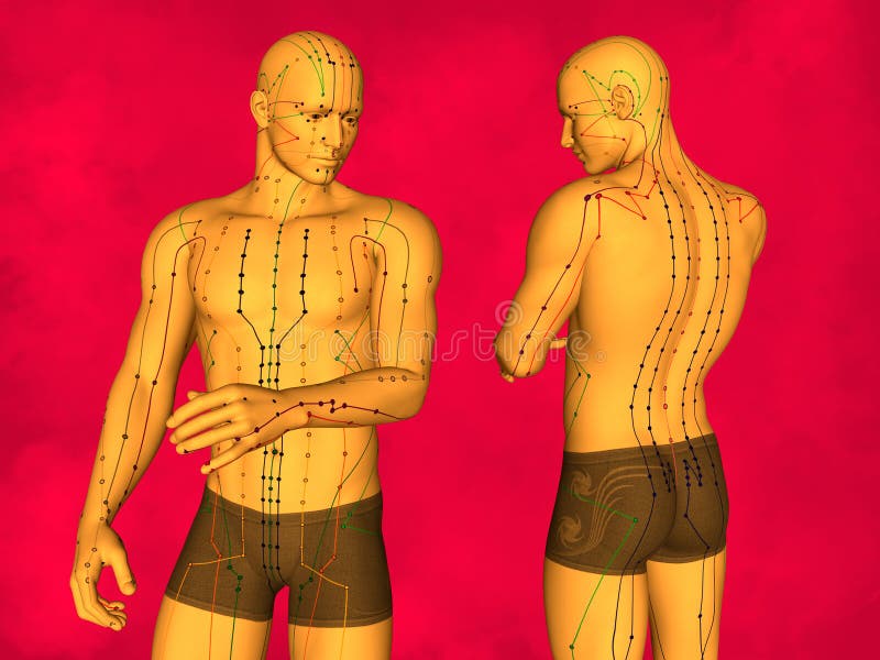
Free with trial Human Body, Acupuncture Model, Acupuncture Points and Meridians, 3D Model, 3D Illustration, Red Background. Anatomical model illustrations Acupuncture model
Free with trial Pneumonia. The anatomical structure of the human lung. Vector illustration on background. Anatomical model vectors Pneumonia. The anatomical structure of the human
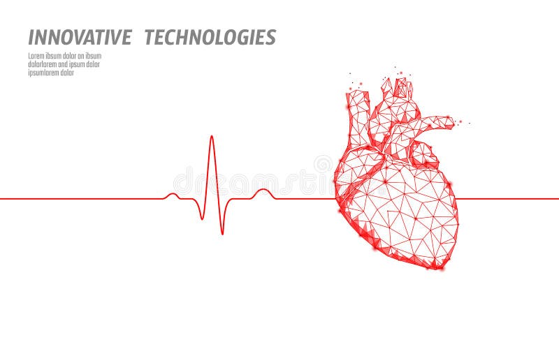
Free with trial Healthy human heart beats 3d medicine model low poly. Triangle connected dots glow point red background. Pulse internal body modern anatomical shape technology render vector illustration. Anatomical model vectors Healthy human heart beats 3d medicine model low poly. Triangle connected dots glow point red background. Pulse internal
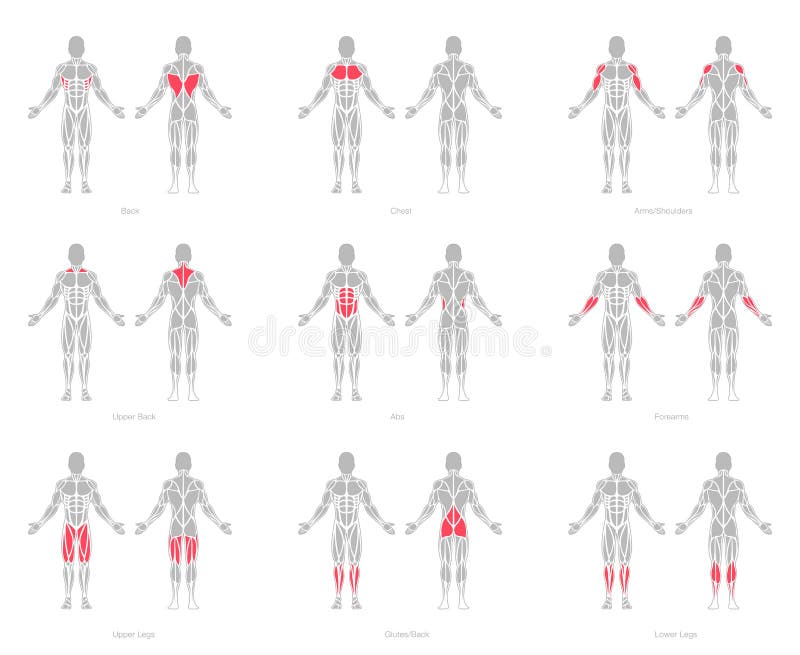
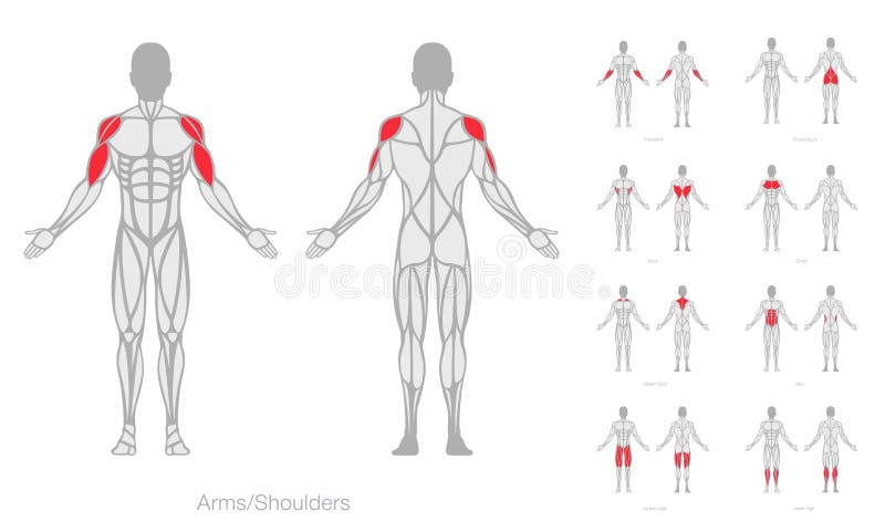
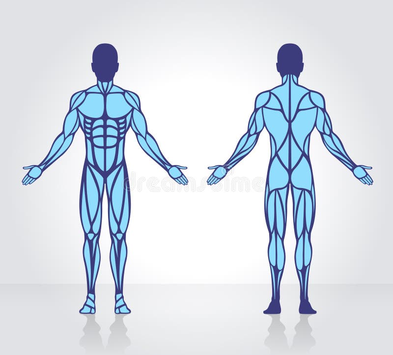
Free with trial Male and female figure with anatomical muscles front and back view set. Red silhouette of muscle structure with biological outline of structure for medical and training vector design. Anatomical model vectors Male and female figure with anatomical muscles front and back view set. Red silhouette of muscle structure with biological outline of structure for medical and
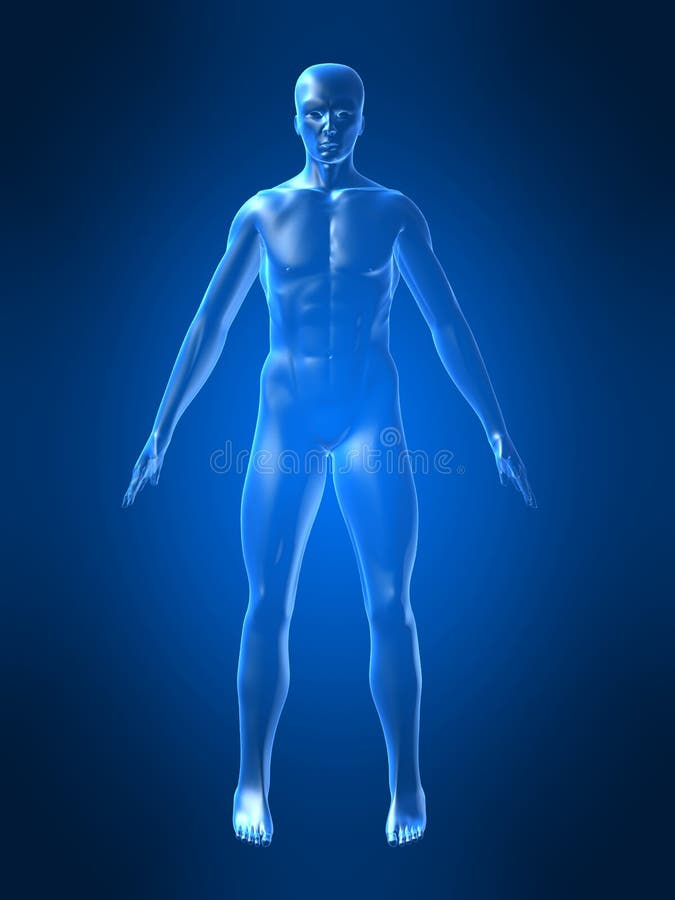
Free with trial A sketch or transparent model of a human body with a notepad and colored pencils in the foreground. Black background. Anatomical model illustrations Sketch of human body. A sketch or transparent model of a human body with a notepad and colored pencils in the foreground. Black background
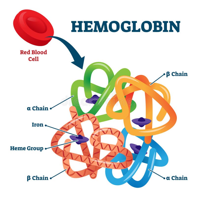
Free with trial Hemoglobin in red blood cells as oxygen transport metalloprotein educational scheme. Medical element structure with closeup iron, heme group, alpha and beta chain. Scientific anatomical inner element. Anatomical model vectors Hemoglobin in red blood cells as oxygen transport metalloprotein scheme. Hemoglobin in red blood cells as oxygen transport metalloprotein educational scheme. Medical element structure with closeup iron, heme group, alpha and beta chain. Scientific anatomical inner element
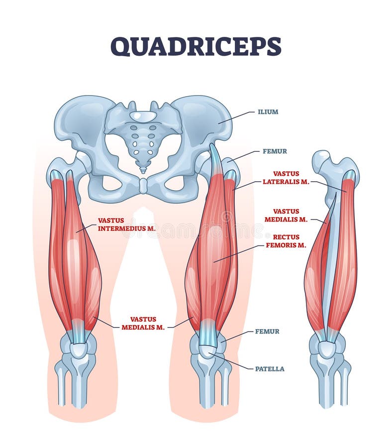
Free with trial Quadriceps muscle and quads leg muscular or bone anatomy outline diagram. Labeled educational medical scheme with vastus intermedius, medialis, lateralis or rectus femoris location vector illustration. Anatomical model vectors Quadriceps muscle or quads leg muscular anatomical structure outline diagram. Quadriceps muscle and quads leg muscular or bone anatomy outline diagram. Labeled educational medical scheme with vastus intermedius, medialis, lateralis or rectus femoris location vector illustration
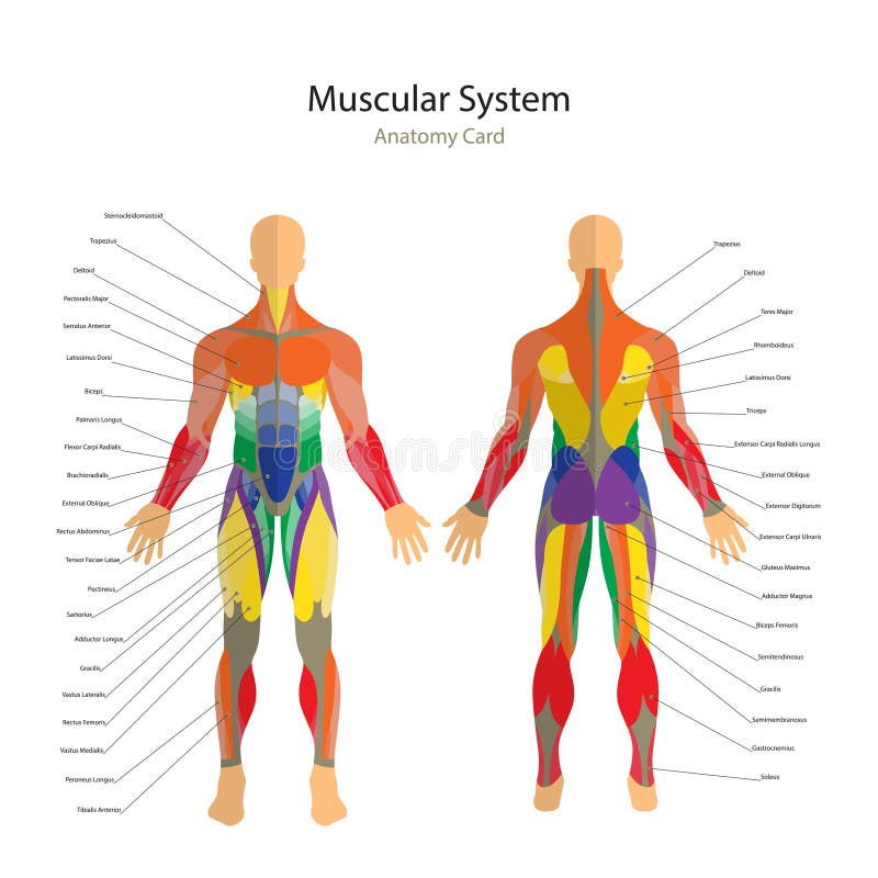
Free with trial Detailed illustration of human muscles. Male fitness model. Exercise and muscle guide. Gym training. Muscle man anatomy. Front and rear view. Anatomical model vectors Illustration of human muscles. Exercise and muscle guide. Gym training. Front and rear view. Muscle man anatomy. Detailed illustration of human muscles. Male fitness model. Exercise and muscle guide. Gym training. Muscle man anatomy. Front and rear view.
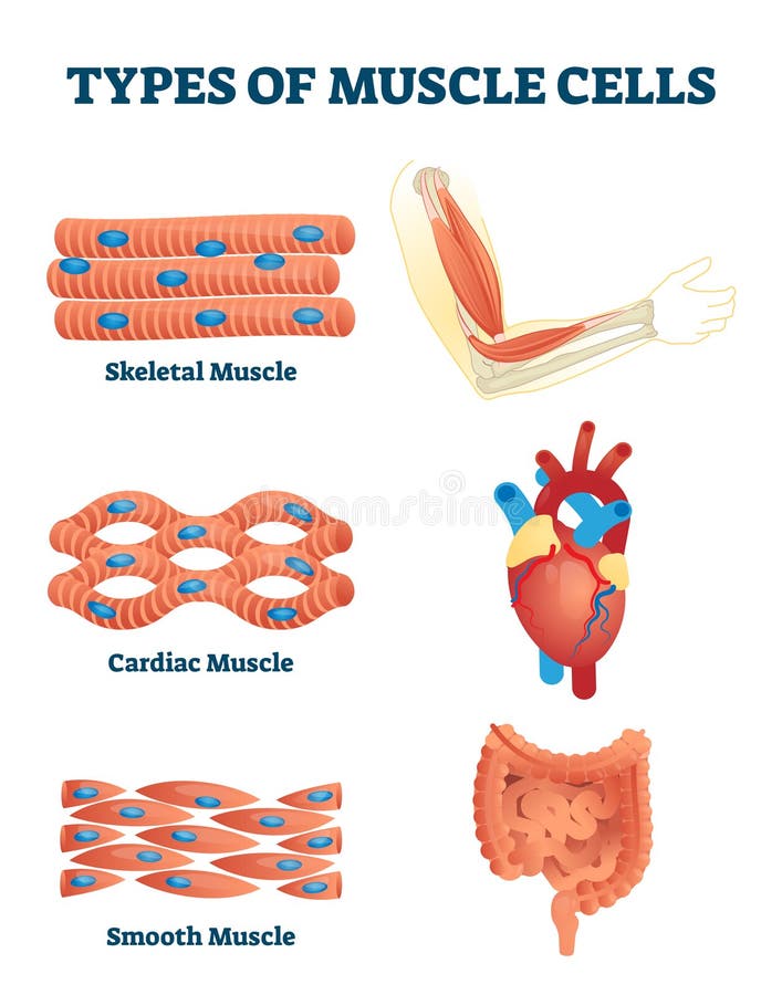
Free with trial Types of muscle cells vector illustration. Labeled various soft tissues differences explanation. Educational skeletal, cardiac and smooth contraction. Anatomical body parts example for biology handout. Anatomical model vectors Types of muscle cells vector illustration. Labeled soft tissues explanation. Types of muscle cells vector illustration. Labeled various soft tissues differences explanation. Educational skeletal, cardiac and smooth contraction. Anatomical body parts example for biology handout
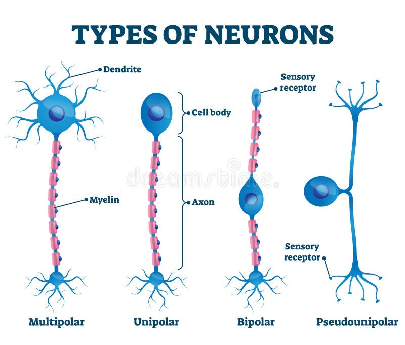
Free with trial Types of neurons vector illustration. Labeled anatomical nerve parts comparison scheme. Synapses receptors educational closeup with multipolar, unipolar, bipolar and pseudounipola sensory receptors. Anatomical model vectors Types of neurons vector illustration. Labeled nerve parts comparison scheme. Types of neurons vector illustration. Labeled anatomical nerve parts comparison scheme. Synapses receptors educational closeup with multipolar, unipolar, bipolar and pseudounipola sensory receptors.
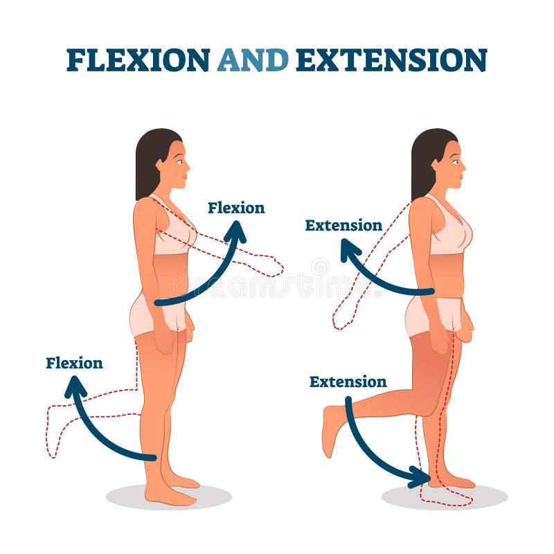
Free with trial Flexion and extension vector illustration. Anatomical movement description. Educational arm or leg exercise to bend or straighten body parts. Normal healthy patient as biological kinesiology example. Anatomical model vectors Flexion and extension vector illustration. Anatomical movement description.
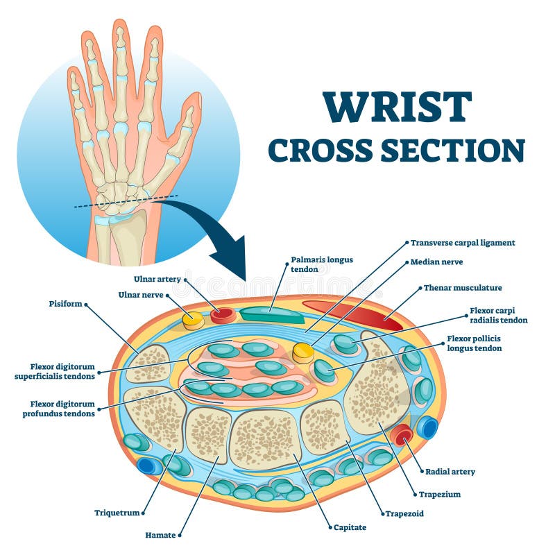
Free with trial Wrist cross section educational structure scheme vector illustration. Anatomical labeled arm research with physiological description and explanation. Nerve, musculature, artery and bone location. Anatomical model vectors Wrist cross section educational anatomy structure scheme vector illustration. Wrist cross section educational structure scheme vector illustration. Anatomical labeled arm research with physiological description and explanation. Nerve, musculature, artery and bone location.
Free with trial Excretory system vector illustration. Labeled educational human organs location scheme. Diagram with isolated bladder, ureter, kidney, aorta and adrenal gland. Anatomical body parts graphic handout. Anatomical model vectors Excretory system vector illustration. Labeled educational organs diagram. Excretory system vector illustration. Labeled educational human organs location scheme. Diagram with isolated bladder, ureter, kidney, aorta and adrenal gland. Anatomical body parts graphic handout.
Free with trial Spinal cord vector icon illustration. Anatomical model vectors Spinal cord vector icon
Free with trial Detailed diagram of the human lungs. Anatomical model vectors Anatomy of the human lungs. Detailed diagram of the human lungs.
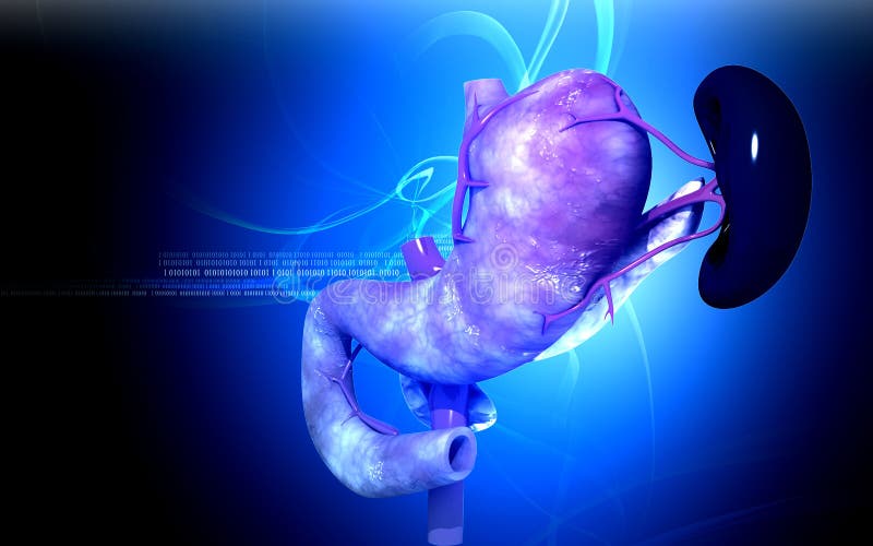
Free with trial Digital illustration of pancreas and spleen in colour background. Anatomical model illustrations Pancreas and spleen
Free with trial The human rib cage is made up of 12 paired rib bones; each are symmetrically paired on a right and left side. Of all 24 ribs, the first seven pairs are often labeled as `true. ` These bones are connected to the costal cartilage, while the five other `false` sets are not. The ribcage also encloses the thoracic cavity and helps protect the heart and lungs from damage. There are 24 ribs in the human body, divided into two sets of 12 curved, flat bones. Each one is attached by cartilage at the back to the thoracic vertebrae. MEN and women have 12 pairs of ribs a few individuals have 13 or 11 pairs. The idea that men have fewer ribs than women is widespread but wrong, perhaps deriving from the biblical story of Eve being made from one of Adam`s ribs. Both men and women have 24 ribs, twelve on each side. Floating rib: One of the last two ribs. A rib is said to be `floating` if it does not attach to the sternum the breast bone or to another rib. There are usually 12 pairs of ribs in all. Each pair of ribs is attached to the building blocks of the spine the vertebrae in the back. The ribs partially enclose and protect the chest cavity, where many vital organs including the heart and the lungs are located. The rib cage is collectively made up of long, curved individual bones with joint-connections to the spinal vertebrae. Anatomical model illustrations Ribs with Ligments anterior view. The human rib cage is made up of 12 paired rib bones; each are symmetrically paired on a right and left side. Of all 24 ribs, the first seven pairs are often labeled as `true.` These bones are connected to the costal cartilage, while the five other `false` sets are not. The ribcage also encloses the thoracic cavity and helps protect the heart and lungs from damage. There are 24 ribs in the human body, divided into two sets of 12 curved, flat bones. Each one is attached by cartilage at the back to the thoracic vertebrae. MEN and women have 12 pairs of ribs a few individuals have 13 or 11 pairs. The idea that men have fewer ribs than women is widespread but wrong, perhaps deriving from the biblical story of Eve being made from one of Adam`s ribs. Both men and women have 24 ribs, twelve on each side. Floating rib: One of the last two ribs. A rib is said to be `floating` if it does not attach to the sternum the breast bone or to another rib. There are usually 12 pairs of ribs in all. Each pair of ribs is attached to the building blocks of the spine the vertebrae in the back. The ribs partially enclose and protect the chest cavity, where many vital organs including the heart and the lungs are located. The rib cage is collectively made up of long, curved individual bones with joint-connections to the spinal vertebrae.
Free with trial Structure of the lungs. Healthcare, and pleura, diaphragm and breath and thorax. Vector illustration. Anatomical model vectors Structure of the lungs