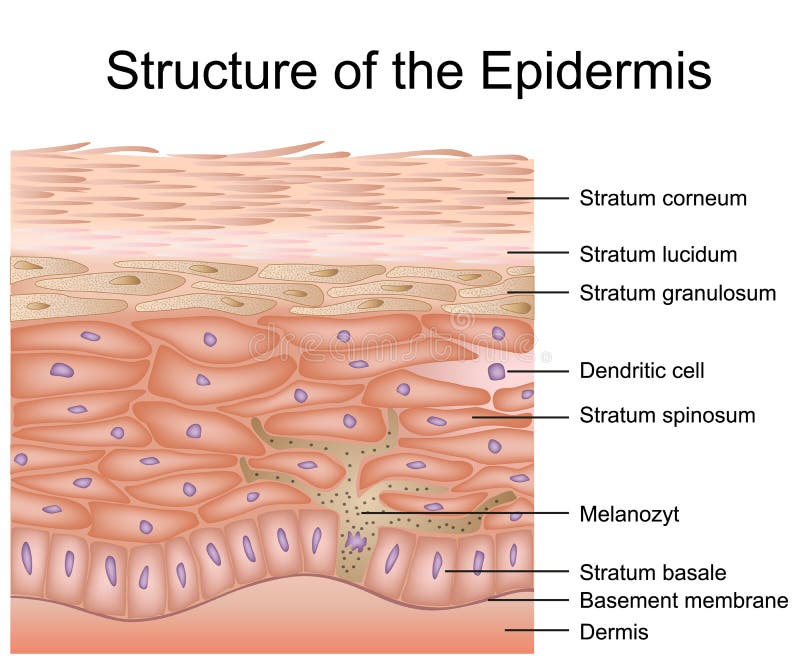
Free with trial Structure of the epidermis medical vector illustration, dermis anatomy eps 10. Anatomy dermis vectors Structure of the epidermis medical vector illustration, dermis anatomy
Free with trial Skin anatomy, detailed illustration. Beautiful bright colors. Anatomy dermis vectors Skin anatomy
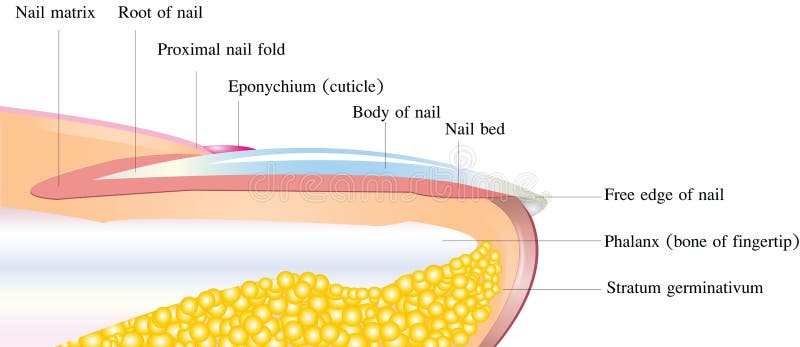
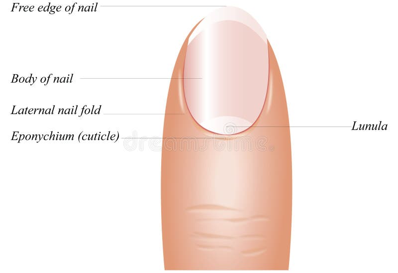
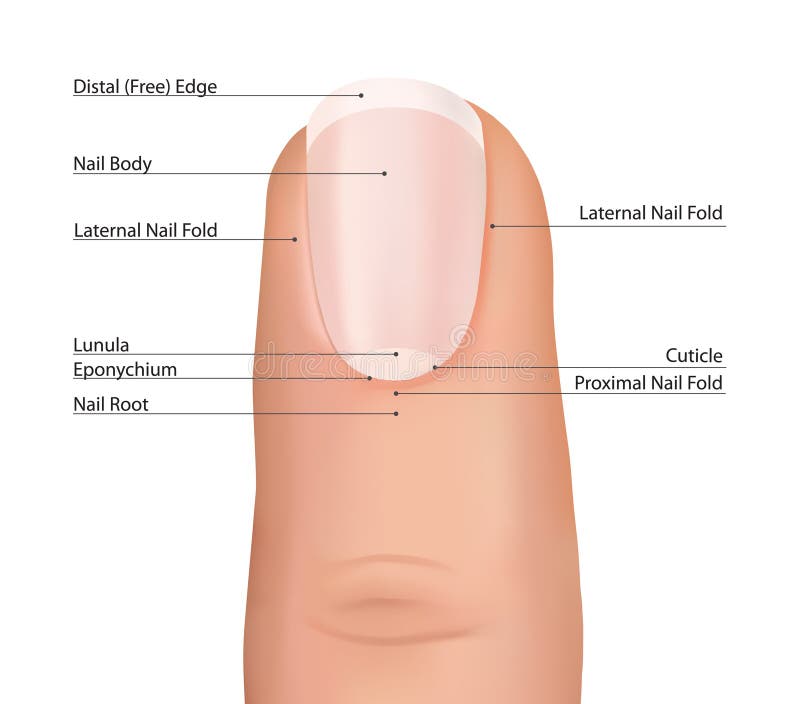
Free with trial Finger Nail detailed anatomy on a white background. Anatomy dermis vectors Detailed nail anatomy. Finger Nail detailed anatomy on a white background
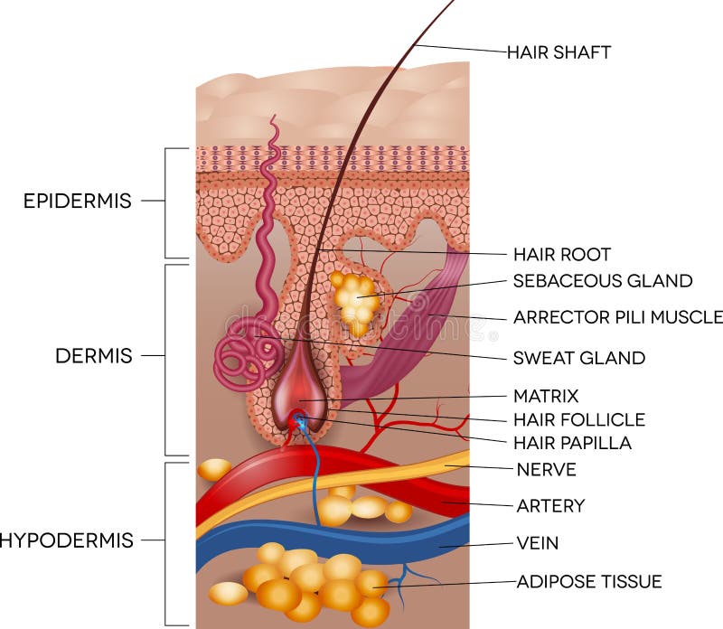
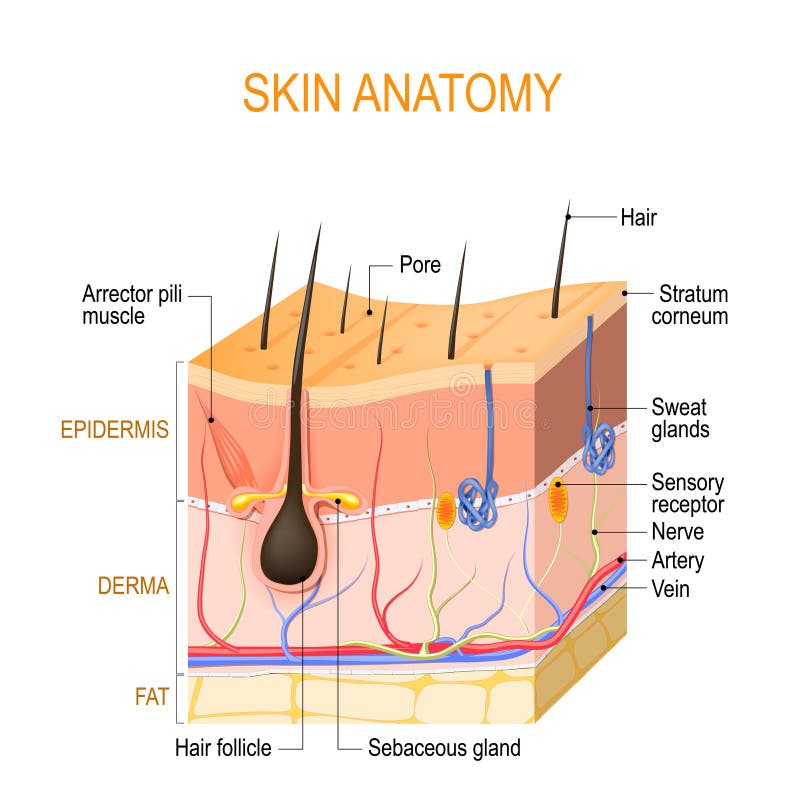
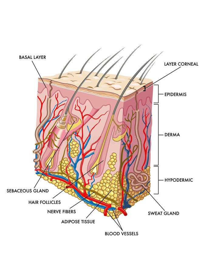
Free with trial Labeled Skin and hair anatomy. Detailed medical illustration. Anatomy dermis vectors Labeled Skin and hair anatomy
Free with trial Easy to edit illustration of Skin Anatomy diagram. Anatomy dermis illustrations Skin Anatomy
Free with trial Skin anatomy. Detailed medical illustration, beautiful bright colors. Anatomy dermis vectors Skin anatomy
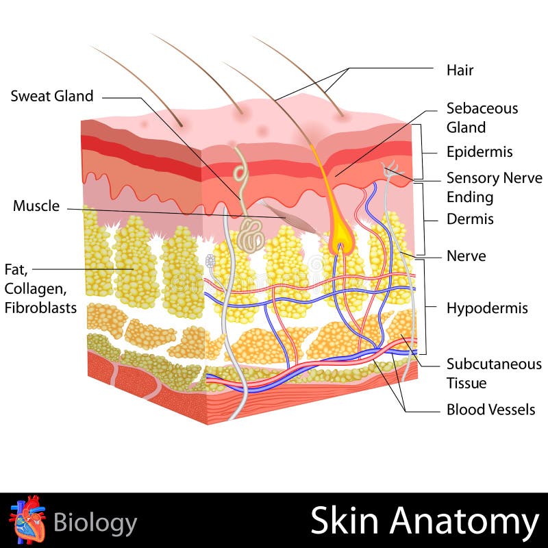
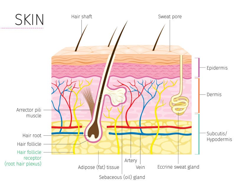
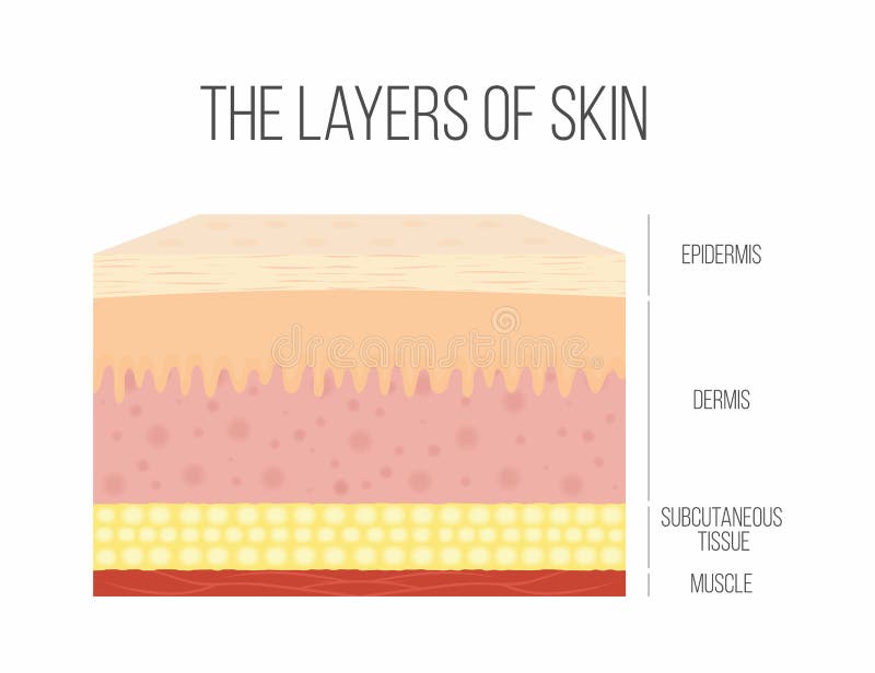
Free with trial The skin is the largest organ of the body, with a total area of about 20 square feet. The skin protects us from microbes and the elements, helps regulate body temperature, and permits the sensations of touch, heat, and cold. Skin has three layers: The epidermis, the outermost layer of skin, provides a waterproof barrier and creates our skin tone. The dermis, beneath the epidermis, contains tough connective tissue, hair follicles, and sweat glands. The deeper subcutaneous tissue hypodermis is made of fat and connective tissue. The skin’s color is created by special cells called melanocytes, which produce the pigment melanin. Melanocytes are located in the epidermis. Anatomy dermis illustrations Skin Anatomy. The skin is the largest organ of the body, with a total area of about 20 square feet. The skin protects us from microbes and the elements, helps regulate body temperature, and permits the sensations of touch, heat, and cold. Skin has three layers: The epidermis, the outermost layer of skin, provides a waterproof barrier and creates our skin tone. The dermis, beneath the epidermis, contains tough connective tissue, hair follicles, and sweat glands. The deeper subcutaneous tissue hypodermis is made of fat and connective tissue. The skin’s color is created by special cells called melanocytes, which produce the pigment melanin. Melanocytes are located in the epidermis.
Free with trial Vector illustration of diagram of human skin anatomy. Anatomy dermis vectors Human Skin Anatomy
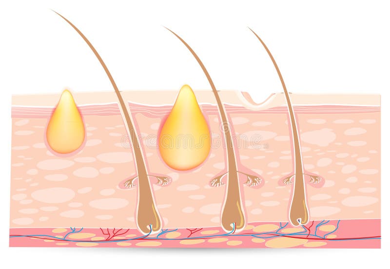
Free with trial Acne types. Pimple skin diseases anatomy medical vector diagram. Illustration of follicle and pimple, medicine anatomy, papule and pustule. Anatomy dermis vectors Acne types. Pimple skin diseases anatomy medical vector diagram
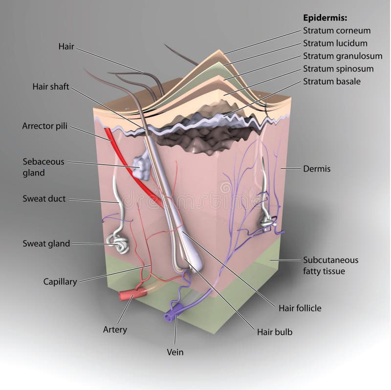
Free with trial Detailed and accurate skin anatomy. Eps8, gradient and mesh printing compatible. Anatomy dermis vectors Human skin anatomy, labeled version. Detailed and accurate skin anatomy. Eps8, gradient and mesh printing compatible
Free with trial Skin anatomy diagram concept with a cross section of the human body surface organ with hair follicle and red and blue blood vessels as a health care and medical symbol of anatomical function. Anatomy dermis illustrations Skin Anatomy
Free with trial Finger Nail detailed anatomy on a white background. Anatomy dermis vectors Finger nail anatomy. Finger Nail detailed anatomy on a white background
Free with trial Detailed skin and hair anatomy on a white background. Anatomy dermis vectors Skin and hair anatomy
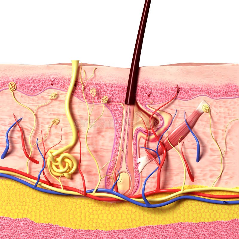
Free with trial 3D art illustration of anatomy of hair follicles. Anatomy dermis illustrations Anatomy of hair follicles
Free with trial Skin anatomy. Layers: epidermis with hair follicle, sweat and sebaceous glands, derma and fat hypodermis. Vector diagram for educational, medical, biological, and scientific use. Anatomy dermis vectors Skin anatomy. Layers: epidermis with hair follicle, sweat and sebaceous glands, derma and fat hypodermis. Vector diagram for educational, medical, biological
Free with trial Detailed and accurate skin anatomy. Eps8, gradient and mesh printing compatible. Anatomy dermis vectors Human skin anatomy, non-labeled version. Detailed and accurate skin anatomy. Eps8, gradient and mesh printing compatible
Free with trial Anatomical training poster. Growth and structure of human hair. Skin and hair anatomy. Cross section of the skin layers. Detailed medical vector illustration. Anatomy dermis vectors Anatomical training poster. Growth and structure of human hair. Skin and hair anatomy. Cross section of the skin layers
Free with trial Nail finger anatomy. Female finger with french manicure. Anatomy dermis vectors Finger Detailed nail anatomy on a white background. Fingernail vector. Nail finger anatomy. Female finger with french manicure .
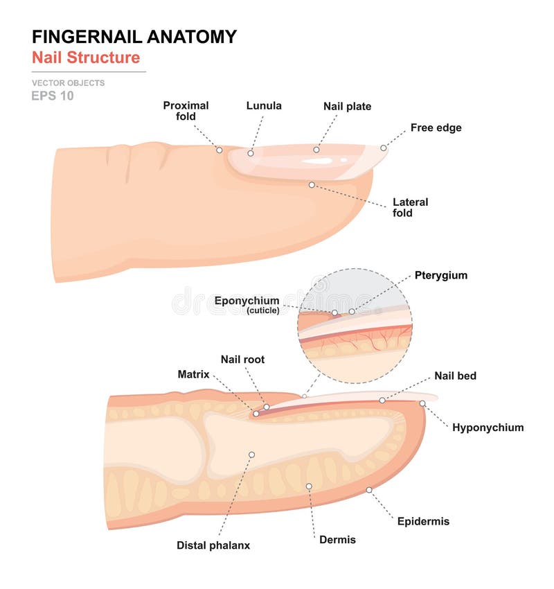
Free with trial Science of human body. Anatomical training poster. Fingernail Anatomy. Structure of human nail. Cross-section of the finger. e view. Detailed medical vector illustration. Anatomy dermis vectors Science of human body. Anatomical training poster. Fingernail Anatomy. Structure of human nail. Cross-section of the finger
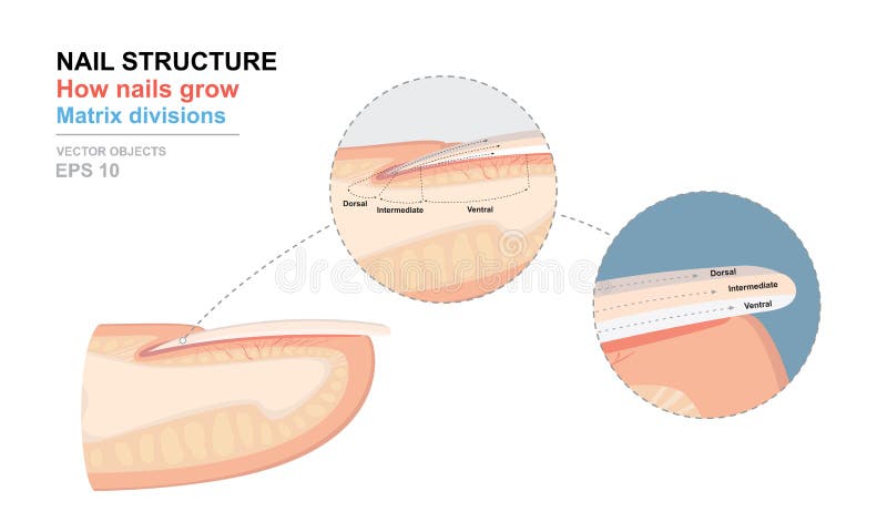
Free with trial Science of human body. Anatomical training poster. Fingernail Anatomy. Structure of human nail. How nails grow. Matrix divisions. Cross-section of the finger. Detailed medical vector illustration. Anatomy dermis vectors Science of human body. Anatomical training poster. Fingernail Anatomy. Structure of human nail. How nails grow. Matrix divisions
Free with trial Layers Of Human Skin. Epidermis (horny layer and granular layer), Dermis (connective tissue) and Subcutaneous fat (adipose tissue). Anatomy dermis vectors Skin layers. Layers Of Human Skin. Epidermis (horny layer and granular layer), Dermis (connective tissue) and Subcutaneous fat (adipose tissue)
Free with trial Anatomy of the epidermis, the outmost layer of human skin, eps8. Anatomy dermis vectors Epidermis of the skin. Anatomy of the epidermis, the outmost layer of human skin, eps8
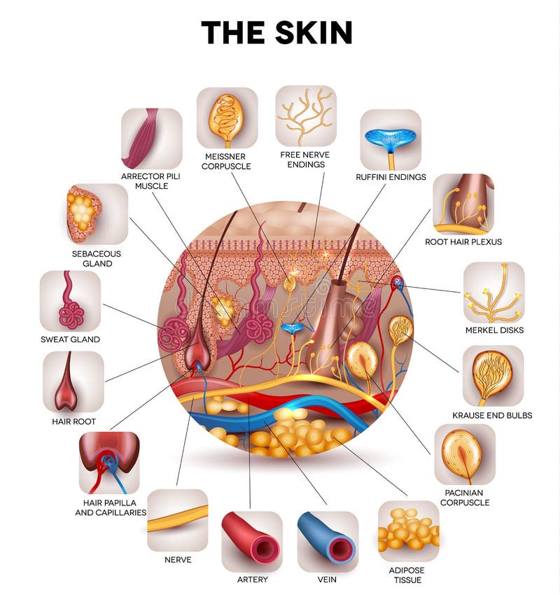
Free with trial Skin anatomy in the round shape, detailed illustration. Beautiful bright colors. Anatomy dermis vectors The skin. Skin anatomy in the round shape, detailed illustration. Beautiful bright colors.
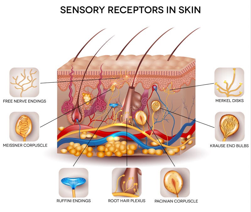
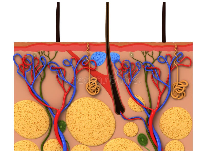
Free with trial Sensory receptors in the skin. Detailed skin anatomy, beautiful bright colors. Anatomy dermis vectors Sensory receptors in the skin
Free with trial Visual representation of skin changes over a lifetime. Collagen and elastin form the structure of the dermis making it tight and plump. Fibroblasts synthesize collagen and elastin. Anatomy dermis vectors Skin aging. Visual representation of skin changes over a lifetime. Collagen and elastin form the structure of the dermis making it tight and plump. Fibroblasts synthesize collagen and elastin.
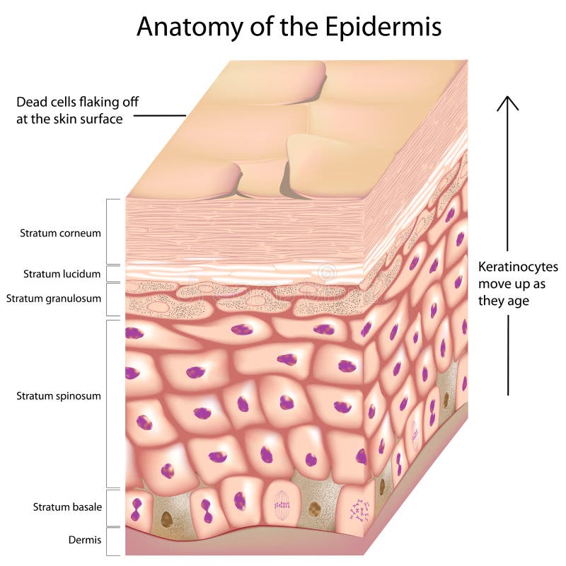
Free with trial Layers of the skin epidermis with keratinocytes moving up as they age, eps8. Anatomy dermis vectors 3d anatomy of the epidermis. Layers of the skin epidermis with keratinocytes moving up as they age, eps8
Free with trial Anatomy of human epidermis with stratum corneum flaking off in dry skin. Anatomy dermis vectors Dry skin
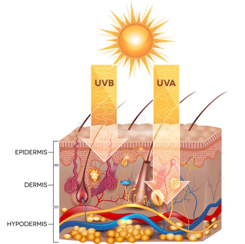
Free with trial UVB and UVA radiation penetrate into skin. Detailed skin anatomy. Anatomy dermis vectors UVB and UVA radiation
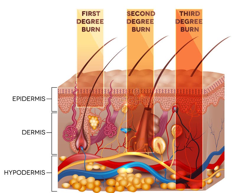
Free with trial Skin burn classification. First, second and third degree skin burns. Detailed skin anatomy. Anatomy dermis vectors Skin burn classification
Free with trial Old skin with age spots and wrinkles caused by atrophy of the dermis (loss of collagen fibers), epidermis and blood vessels leaking. Eps8, gradient and mesh printing compatible. Anatomy dermis vectors Wrinkled versus smooth skin. Old skin with age spots and wrinkles caused by atrophy of the dermis (loss of collagen fibers), epidermis and blood vessels leaking. Eps8, gradient and mesh printing compatible
Free with trial Anatomy skin Hair Cycles of hair follicle. Anatomy dermis vectors Anagen catagen telogen. Anatomy skin Hair Cycles of hair follicle
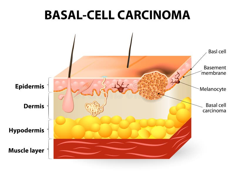
Free with trial Skin cancer. Basal-cell carcinoma or basal cell cancer (BCC). Schematic representation of skin. The separation between epidermis and dermis occurs at the basement membrane zone. Anatomy dermis vectors Basal-cell carcinoma or basal cell cancer
Free with trial Skin cancer melanoma disease concept as a medical symbol of the human epidermis anatomy being attacked by malignant cancerous dark mole shaped as text on the body. Anatomy dermis illustrations Skin Cancer Melanoma
Free with trial Structure of the Human skin. Anatomy diagram. different cell types populating the skin. Anatomy dermis vectors Different cell types populating the skin
Free with trial Various types of skin wound shown as a 3d illustration of the epidermis and dermis of the skin. Anatomy dermis illustrations Skin wounds. Various types of skin wound shown as a 3d illustration of the epidermis and dermis of the skin
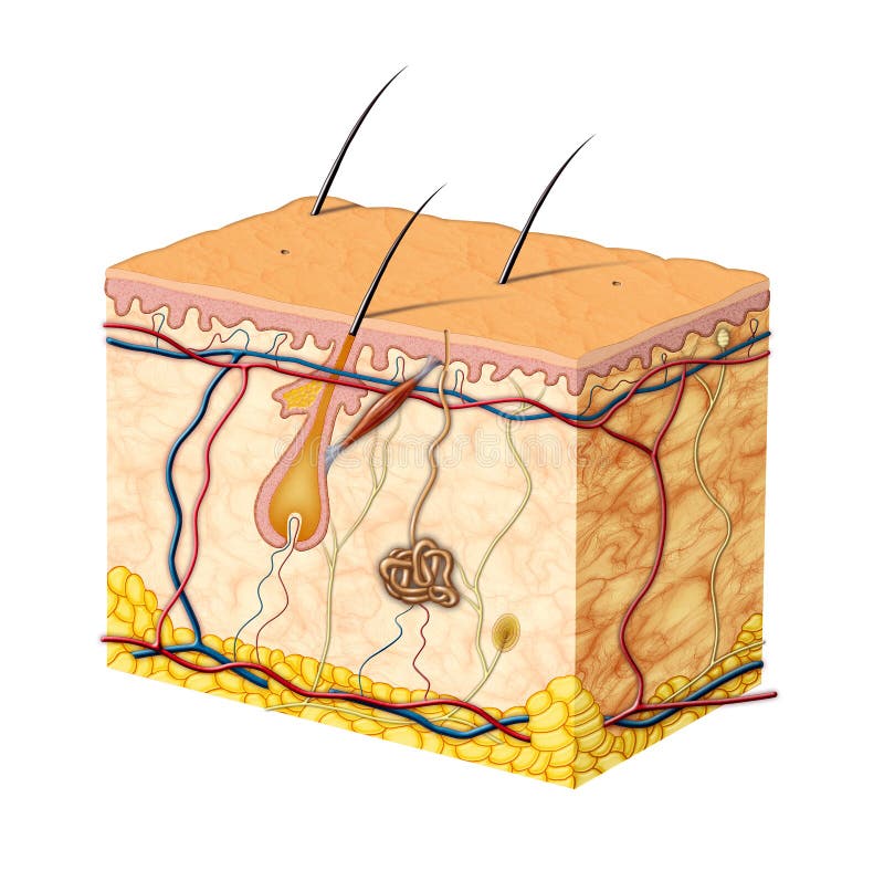
Free with trial The skin and skin structure components, detailed illustration. Skin sensory receptors, vessels, hair, muscle, etc. Beautiful bright colors. Anatomy dermis vectors Skin anatomy. The skin and skin structure components, detailed illustration. Skin sensory receptors, vessels, hair, muscle, etc. Beautiful bright colors.
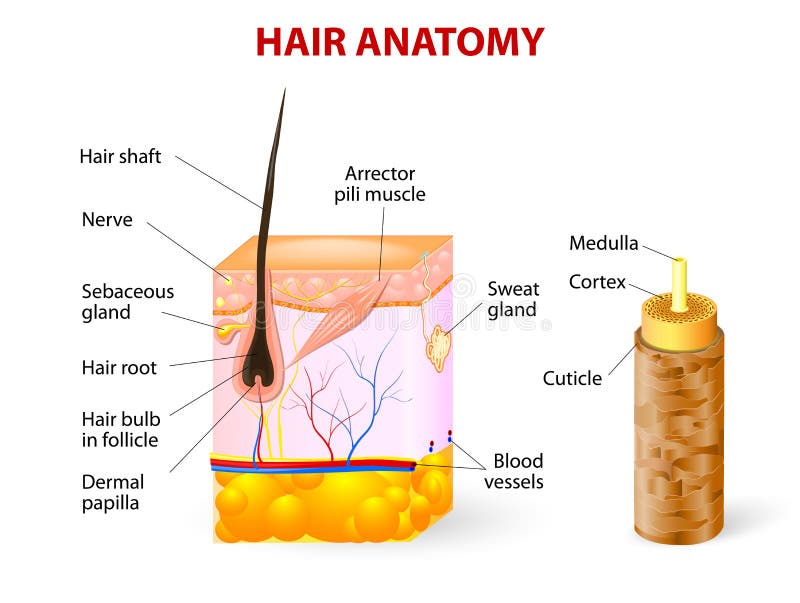
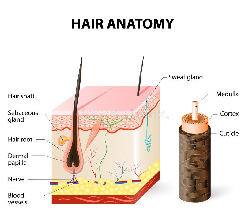
Free with trial Hair anatomy. Vector diagram. The hair shaft grows from the hair follicle consisting of transformed skin tissue. The epidermal cells transform at the command of the dermal papilla cells and generate the hair shaft. Anatomy dermis vectors Diagram of a hair follicle in a cross section of s. Hair anatomy. Vector diagram. The hair shaft grows from the hair follicle consisting of transformed skin tissue. The epidermal cells transform at the command of the dermal papilla cells and generate the hair shaft.
Free with trial Hair anatomy. The hair shaft grows from the hair follicle consisting of transformed skin tissue. The epidermal cells transform at the command of the dermal papilla cells and generate the hair shaft. Anatomy dermis vectors Diagram of a hair follicle in a cross section of skin layers. Hair anatomy. The hair shaft grows from the hair follicle consisting of transformed skin tissue. The epidermal cells transform at the command of the dermal papilla cells and generate the hair shaft.
Free with trial Complexion Physiology Integumentary System Medical Healthy Beauty Cosmetic Makeup Treatment. Anatomy dermis vectors Human Anatomy, Skin And Hair Diagram. Complexion Physiology Integumentary System Medical Healthy Beauty Cosmetic Makeup Treatment
Free with trial Human skin and hair structure. Vector illustration. 3d rendered human skin anatomy. Anatomy dermis vectors Human skin and hair structure. Vector illustration
Free with trial Anatomical training poster. Hair growth phase step by step. Stages of the hair growth cycle. Anagen, telogen, catagen. Skin anatomy. Cross section of the skin layers. Medical vector illustration. Anatomy dermis vectors Anatomical training poster. Hair growth phase step by step. Stages of the hair growth cycle. Anagen, telogen, catagen
Free with trial The cycle of hair growth in human with anagen, catagen and telogen phases, explaining the basis of hair loss and hair removal. Anatomy dermis vectors Hair growth cycle. The cycle of hair growth in human with anagen, catagen and telogen phases, explaining the basis of hair loss and hair removal
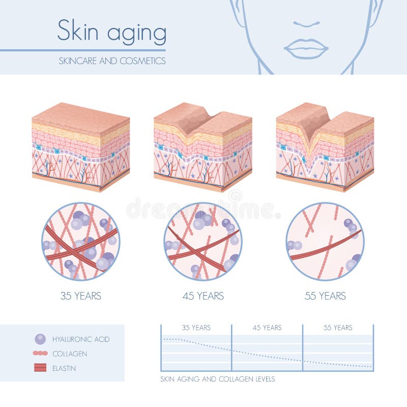
Free with trial Skin aging stages diagrams, collagen and elastin progessive decrease close up, skincare infographics. Anatomy dermis vectors Skin aging
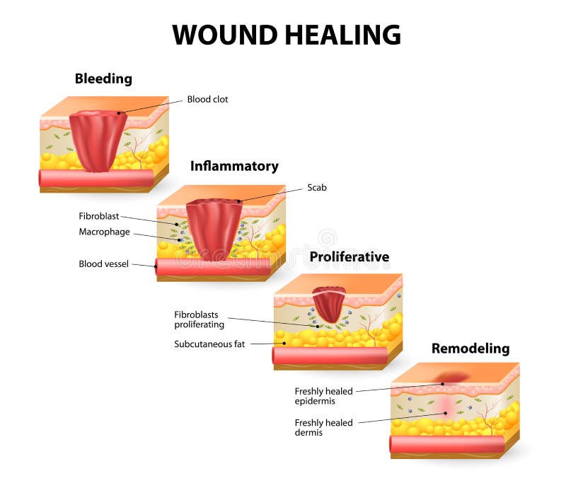
Free with trial Phases of the wound healing process. Hemostasis, Inflammatory, Proliferative, Maturation and remodeling phase. Anatomy dermis vectors Wound healing
Free with trial Types of acne pimples. Healthy skin, Whiteheads and Blackheads, Papules and Pustules. Anatomy dermis vectors Types of acne pimples
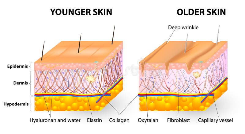
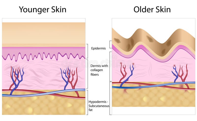
Free with trial Younger skin and aging skin. elastin and collagen. A diagram of younger skin and aging skin showing the decrease in collagen and broken elastin in older skin. Anatomy dermis vectors Younger skin and older skin. Younger skin and aging skin. elastin and collagen. A diagram of younger skin and aging skin showing the decrease in collagen and broken elastin in older skin.
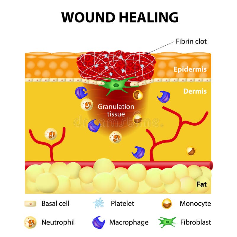
Free with trial The wound healing process. Cutaneous wound after injury. Anatomy dermis vectors Process of wound healing. The wound healing process. Cutaneous wound after injury
Free with trial Overactive melanocyte produces more pigment and is responsible for age spot, eps8. Anatomy dermis vectors Formation of uneven skin tone. Overactive melanocyte produces more pigment and is responsible for age spot, eps8
Free with trial Normal hair tissue and Alopecia areata hair tissue with lymphocytes attacking cells of hair follicles. Anatomy dermis vectors Normal hair and Alopecia areata. Normal hair tissue and Alopecia areata hair tissue with lymphocytes attacking cells of hair follicles
Free with trial Skin layers. Healthy normal human skin. Vector. Anatomy dermis vectors Skin layers. Healthy, normal human skin. Skin layers. Healthy normal human skin. Vector
Free with trial Cross section of skin showing the various layers and structural elements. Anatomy dermis vectors Skin
Free with trial Cross section of skin showing the various layers and structural elements and a variety of nerve endings. Anatomy dermis vectors Skin
Free with trial 3d rendered illustration from a profile of human skin. Anatomy dermis illustrations Skin cut. 3d rendered illustration from a profile of human skin
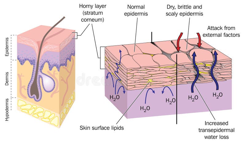
Free with trial Section through skin showing normal epidermis and skin surface structure resulting in water loss and dry, brittle, scaly skin. Created in Adobe Illustrator. EPS 10. Anatomy dermis vectors Dry skin. Section through skin showing normal epidermis and skin surface structure resulting in water loss and dry, brittle, scaly skin. Created in Adobe Illustrator. EPS 10.
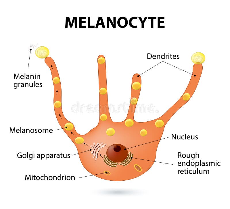
Free with trial Melanocyte, melanin and melanogenesis. Melanocyte - melanin producing cells. Melanin is the pigment responsible for skin color. Anatomy dermis vectors Melanocyte
Free with trial The process of wound healing. Illustration showing skin after injury, appears blood, then blood clot, then scab. Anatomy dermis vectors Wound healing
Free with trial Chemical peeling or procedure chemexfoliation. Human skin layers. Anatomy dermis vectors Chemical peels. Chemical peeling or procedure chemexfoliation. Human skin layers
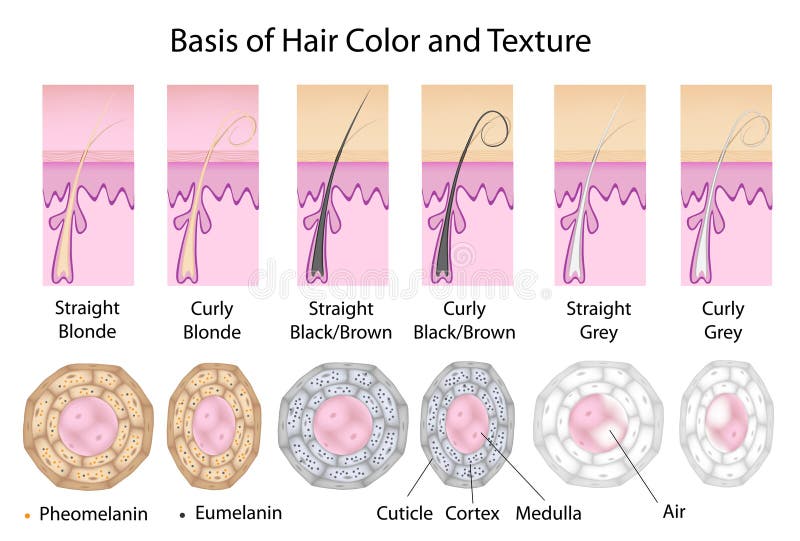
Free with trial In cross section straight hair is round and curly is flatten. Black hair has mostly eumelanin, blonde has more pheomelanin. Grey hair has no pigment and has air in the medulla. Eps8, gradient and mesh printing compatible. Anatomy dermis vectors Cross section of different hair texture and color. In cross section straight hair is round and curly is flatten. Black hair has mostly eumelanin, blonde has more pheomelanin. Grey hair has no pigment and has air in the medulla. Eps8, gradient and mesh printing compatible
Free with trial Hair-follicle cycling. anagen is the growth phase; catagen is the regressing phase; and telogen, the resting or quiescent phase. Vector diagram. Anatomy dermis vectors Human hair growth. Hair-follicle cycling. anagen is the growth phase; catagen is the regressing phase; and telogen, the resting or quiescent phase. Vector diagram
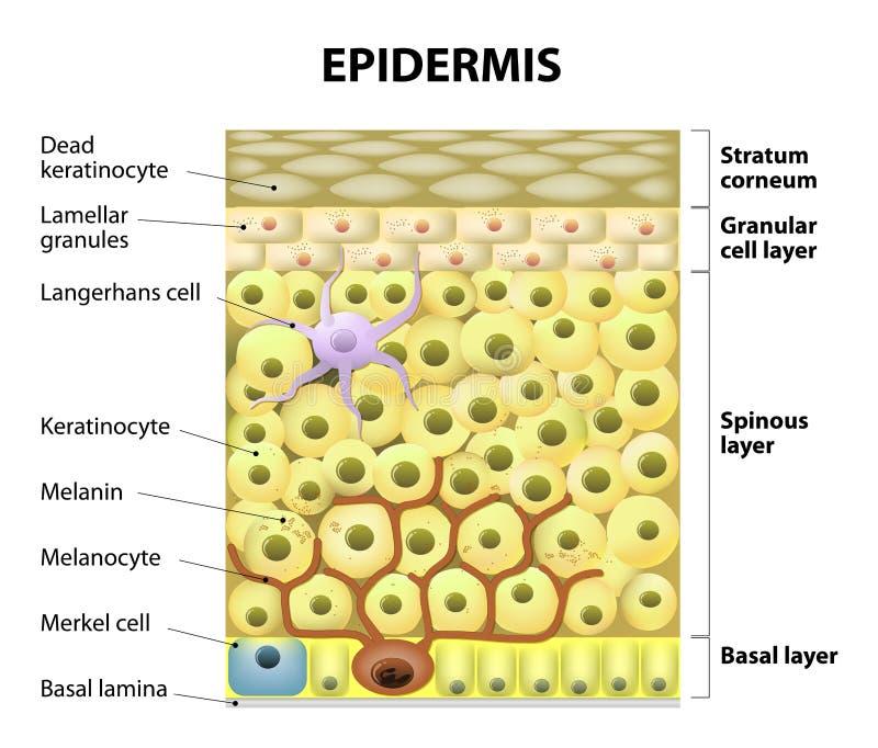
Free with trial Epidermis structure. Cell, and layers of a human skin. vector illustration for medical, educational, biologycal and science use. Skin care. Anatomy dermis vectors Epidermis structure. Cell, and layers of a human skin.
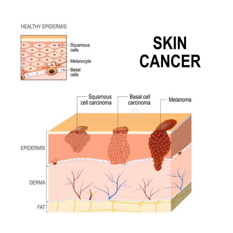
Free with trial Skin cancer: Squamous cell carcinoma disease of older cells on the surface skin, basal-cell cancer begins in the basal cells and Melanoma arises in the pigment cells - melanocytes. layers of human skin and healthy epidermis. Medical diagram. Anatomy dermis vectors Skin cancer. Squamous cell carcinoma, basal-cell cancer and Melanoma. Skin cancer: Squamous cell carcinoma disease of older cells on the surface skin, basal-cell cancer begins in the basal cells and Melanoma arises in the pigment cells - melanocytes. layers of human skin and healthy epidermis. Medical diagram
Free with trial Types of acne pimples. Healthy skin, Whiteheads and Blackheads, Papules and Pustules. Anatomy dermis vectors Types of acne pimples
Free with trial Cell in the epidermis. layers of epidermis. Structure of the human skin. Anatomy dermis vectors Cell in the epidermis
Free with trial Melanoma or skin cancer. This rare type of skin cancer originates from melanocytes. layers of the human skin. Anatomy dermis vectors Melanoma or skin cancer
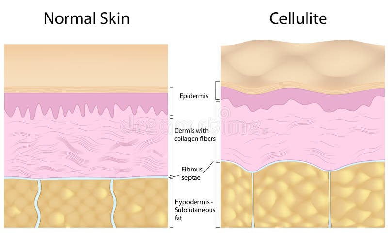
Free with trial Overgrowing fat cells pushing up the fibrous septaes creating dimple appearance in cellulite. Eps8, gradient and mesh printing compatible. Anatomy dermis vectors Cellulite versus smooth skin. Overgrowing fat cells pushing up the fibrous septaes creating dimple appearance in cellulite. Eps8, gradient and mesh printing compatible
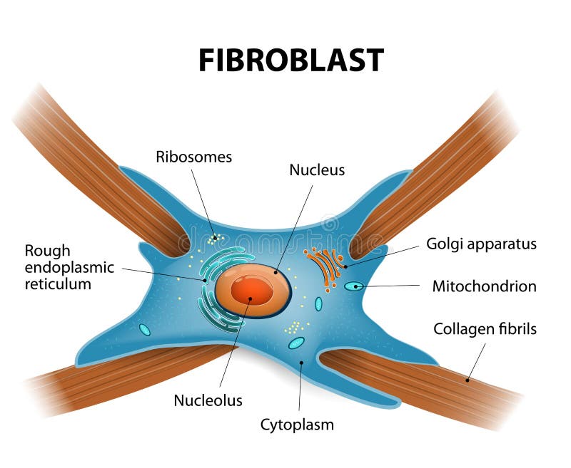
Free with trial Structure of fibroblast cells. These cell are vital to the skin's strength and elasticity. The fibroblasts also synthesise the ground substance of the dermal matrix. Anatomy dermis vectors Structure of fibroblast cells
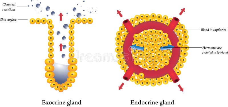
Free with trial Exocrine and endocrine secretion glands on a white background. Anatomy dermis vectors Exocrine and endocrine glands. Exocrine and endocrine secretion glands on a white background
Free with trial Skin showing the dermo-epidermal junction, capillaries and deeper blood vessels. Created in Adobe Illustrator. Contains transparencies. EPS 10. Anatomy dermis vectors Skin epidermal dermal junction. Skin showing the dermo-epidermal junction, capillaries and deeper blood vessels. Created in Adobe Illustrator. Contains transparencies. EPS 10.
Free with trial Collagen is the main structural protein in the: connective tissues, cartilages, bones, nails, derma and hair. Synthesis and types of collagen. Vector illustration for medical, science, and educational use. skincare. Anatomy dermis vectors Collagen is the main structural protein in the: connective tissues, cartilages, bones, nails, derma and hair.
Free with trial Eps8, gradient and mesh printing compatible. Anatomy dermis vectors Formation of skin acnes and pimples. Eps8, gradient and mesh printing compatible
Free with trial Diagram showing skin inflammation, eps8, gradient and mesh printing compatible. Anatomy dermis vectors Eczema. Diagram showing skin inflammation, eps8, gradient and mesh printing compatible
Free with trial 3-dimentional image of the skin, showing all major layers and appendages. Anatomy dermis illustrations Skin 3D. 3-dimentional image of the skin, showing all major layers and appendages
Free with trial #D cross section of skin showing the major layers and structures. Anatomy dermis illustrations 3D Skin. #D cross section of skin showing the major layers and structures