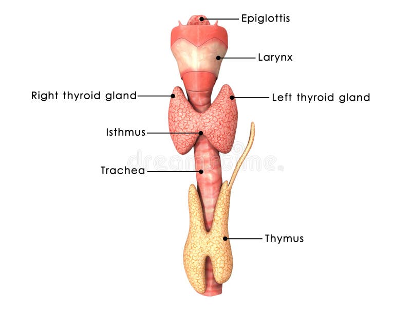
Free with trial Location and relative anatomy of the thyroid gland, showing the lobes, blood supply and situation in the neck. Anatomy neck illustrations The thyroid gland
Free with trial 3d rendered medically accurate illustration of the equine muscle anatomy - Serratus Ventralis. Anatomy neck illustrations Serratus Ventralis
Free with trial 3d rendered medically accurate illustration of the equine muscle anatomy - Rhomboideus Cervicis. Anatomy neck illustrations Rhomboideus Cervicis
Free with trial A sketch in black and white of human anatomy. Anatomy neck illustrations Human anatomy - seventh cervical vertebra. A sketch in black and white of human anatomy
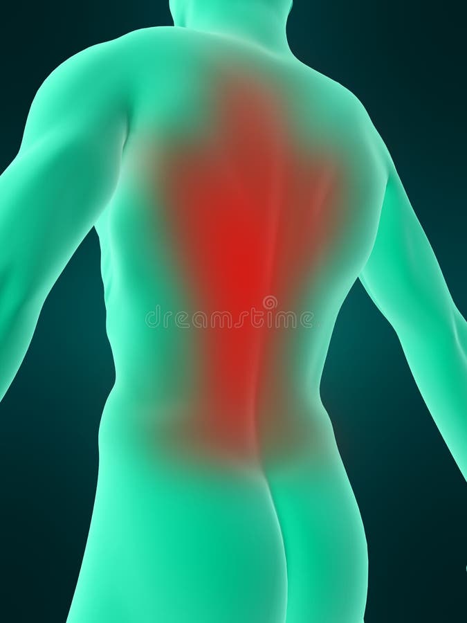
Free with trial 3d rendered illustration of a man having a painful back and neck. Anatomy neck illustrations Highlighted back and neck. 3d rendered illustration of a man having a painful back and neck
Free with trial Frogs are amphibians, living both on land and in water. Their anatomy is very unique. Their bodies are similar to humans in that they have skin, bones, muscles, and organs. The body of a frog can be divided into a head, a short neck, and a trunk. The head contains the brain, mouth, eyes, ears and nose. The frogs head movement is limited due to the short, almost rigid neck. The trunk of a frog forms walls for a single body cavity known as the coelom. The coelom holds all of the frogs internal organs. Frogs have the same kinds of organs as humans and the same organ systems. For example, frogs have a long, sticky tongue which they use to capture food. They also have teeth, which unfortunately are very weak and rather useless. Humans have tongues and teeth as well (and a mouth of course). Anatomy neck illustrations Frog anatomy labelled. Frogs are amphibians, living both on land and in water. Their anatomy is very unique. Their bodies are similar to humans in that they have skin, bones, muscles, and organs. The body of a frog can be divided into a head, a short neck, and a trunk. The head contains the brain, mouth, eyes, ears and nose. The frogs head movement is limited due to the short, almost rigid neck. The trunk of a frog forms walls for a single body cavity known as the coelom. The coelom holds all of the frogs internal organs. Frogs have the same kinds of organs as humans and the same organ systems. For example, frogs have a long, sticky tongue which they use to capture food. They also have teeth, which unfortunately are very weak and rather useless. Humans have tongues and teeth as well (and a mouth of course).
Free with trial 3D illustration, neck painful - cervica spine skeleton x-ray, medical concept. Anatomy neck illustrations Neck painful - cervica spine skeleton x-ray, 3D illustration. 3D illustration, neck painful - cervica spine skeleton x-ray, medical concept.
Free with trial Human head and neck, shown from the nose to the shoulder in cross section. Included are the nasal cavity, mouth, tongue, epiglottis, larynx, vocal cords, esophagus, and trachea (windpipe). Anatomy neck illustrations Head and Neck - Cutaway View. Human head and neck, shown from the nose to the shoulder in cross section. Included are the nasal cavity, mouth, tongue, epiglottis, larynx, vocal cords, esophagus, and trachea (windpipe).
Free with trial Woman having a painful neck - visible spine. Anatomy neck illustrations Painful neck - visible spine
Free with trial Throat anatomy vector illustration diagram, educational medical scheme. Anatomy neck vectors Throat anatomy vector illustration diagram. Throat anatomy vector illustration diagram, educational medical scheme.
Free with trial Woman having a painful neck - visible muscles. Anatomy neck illustrations Painful neck
Free with trial Anatomy Body - Iso View - Blue concept - Medical imaging. Anatomy neck illustrations Anatomy Body - Iso View - Blue concept
Free with trial Anatomy of a thyroid gland. Close-up of thyroid Follicle structure with Follicular cell, C-cell, and Connective tissue. Vector illustration. Anatomy neck vectors Anatomy of a thyroid gland
Free with trial Woman having a painful neck - visible spine. Anatomy neck illustrations Painful neck - visible spine
Free with trial 3d rendered medically accurate illustration of a woman having a painful neck. Anatomy neck illustrations A woman having a painful neck
Free with trial Lever systems in human body for neck, leg and arm movement outline diagram. Labeled educational scheme with biomechanics examples as effort, fulcrum or load force motion principle vector illustration. Anatomy neck vectors Lever systems in human body for neck, leg and arm movement outline diagram. Labeled educational scheme with biomechanics examples as effort, fulcrum or load
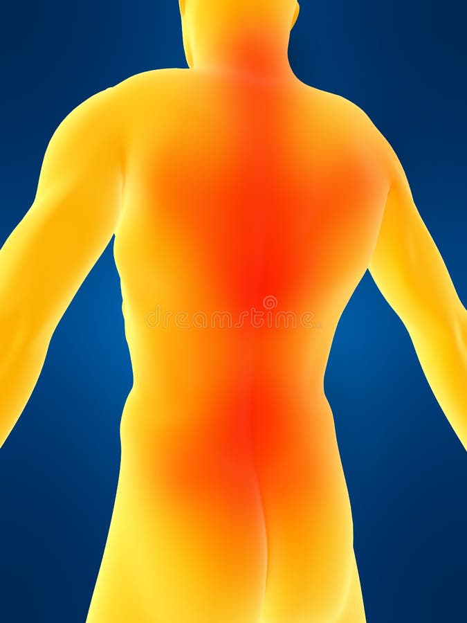
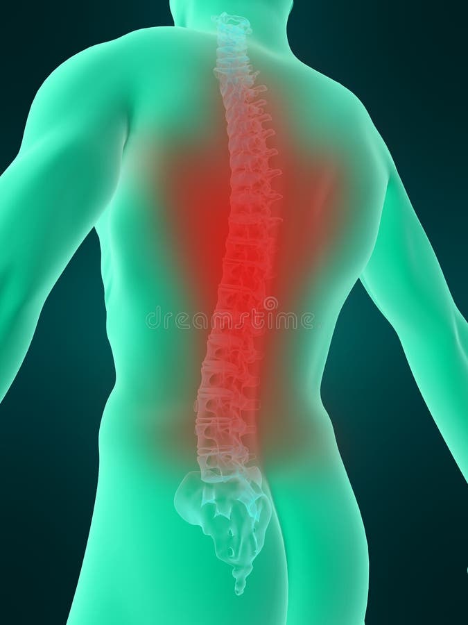
Free with trial 3d rendered illustration of a man having a painful back and neck. Anatomy neck illustrations Highlighted neck and back. 3d rendered illustration of a man having a painful back and neck
Free with trial A vector image illustration Gonstead Chiropractic for acute neck pain. Anatomy neck vectors Gonstead Chiropractic for acute neck pain
Free with trial 3D illustration, neck painful - cervical spine skeleton x-ray, medical concept. Anatomy neck illustrations Neck painful - cervical spine skeleton x-ray, 3D illustration. 3D illustration, neck painful - cervical spine skeleton x-ray, medical concept.
Free with trial 3D illustration, neck painful - cervical spine skeleton x-ray, medical concept. Anatomy neck illustrations Neck painful - cervical spine skeleton x-ray, 3D illustration. 3D illustration, neck painful - cervical spine skeleton x-ray, medical concept.
Free with trial Human Thyroid Gland Anatomy Illustration. 3D render. Anatomy neck illustrations Human Thyroid Gland Anatomy Illustration
Free with trial 3D illustration, neck painful - cervical spine skeleton x-ray, medical concept. Anatomy neck illustrations Neck painful - cervical spine skeleton x-ray, 3D illustration. 3D illustration, neck painful - cervical spine skeleton x-ray, medical concept.
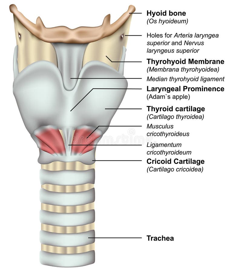
Free with trial Anatomy of the larynx 3d medical illustration on white background eps 10. Anatomy neck vectors Anatomy of the larynx 3d medical illustration on white background
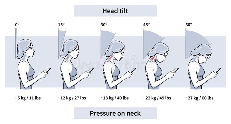
Free with trial Load on neck and back when posture tilting head with phone, pain of weight, outline. Angle of bending head related to pressure on spine. Stage text neck syndrome. Vector illustration. Anatomy neck vectors Load pressure neck head tilt angle vector illustration. Load on neck and back when posture tilting head with phone, pain of weight, outline. Angle of bending head related to pressure on spine. Stage text neck syndrome. Vector illustration
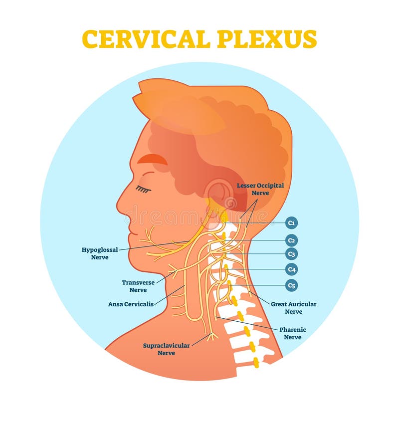
Free with trial Cervical Plexus anatomical nerve diagram, vector illustration medical scheme with human head and neck cross section. Anatomy neck vectors Cervical Plexus anatomical nerve diagram, vector illustration scheme with neck cross section. Cervical Plexus anatomical nerve diagram, vector illustration medical scheme with human head and neck cross section.
Free with trial Femoral neck fractures medical illustration on white background eps 10. Anatomy neck vectors Femoral neck fractures medical illustration on white background
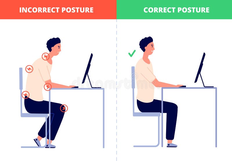
Free with trial Correct sitting. Computer posture, office ergonomics at work desk. Proper position without neck or back pain for healthy spine utter vector concept. Illustration spine body, back orthopedic health. Anatomy neck vectors Correct sitting. Computer posture, office ergonomics at work desk. Proper position without neck or back pain for healthy
Free with trial Improper posture symptoms. Text Neck Syndrome. Spinal curvature, kyphosis, lordosis, scoliosis, arthrosis. Improper posture and stoop. Infographics Vector illustration. Anatomy neck vectors Improper posture symptoms. Text Neck Syndrome. Spinal curvature, kyphosis, lordosis, scoliosis, arthrosis. Improper
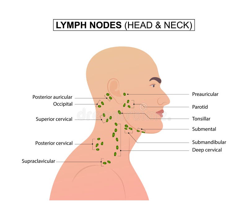
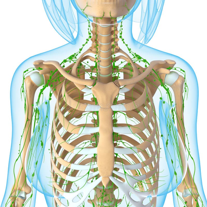
Free with trial Diagram of Lymph nodes of the head and neck. vector illustration. Anatomy neck vectors Lymph nodes of the head and neck
Free with trial Human larynx anatomy with parts. Flat stock illustration. Anatomy neck illustrations Human larynx anatomy
Free with trial 3D Illustration of Human Body Muscles Anatomy (Flexor Hallucis Longus). Anatomy neck illustrations Human Body Muscles Anatomy (Flexor Hallucis Longus)
Free with trial 3D Illustration of Spinal cord Anatomy (Lumber vertebrae). Anatomy neck illustrations Spinal cord Anatomy (Lumber vertebrae)
Free with trial Human body seen from behind with dermatitis on the neck. Anatomy neck illustrations Neck dermatitis, skin, burning inflammation. Human body seen from behind with dermatitis on the neck
Free with trial Vitruvian human or man as a concept or conceptual 3d proportion anatomy body isolated on background. Anatomy neck illustrations Vitruvian human or man conceptual 3d proportion anatomy body. Vitruvian human or man as a concept or conceptual 3d proportion anatomy body isolated on background
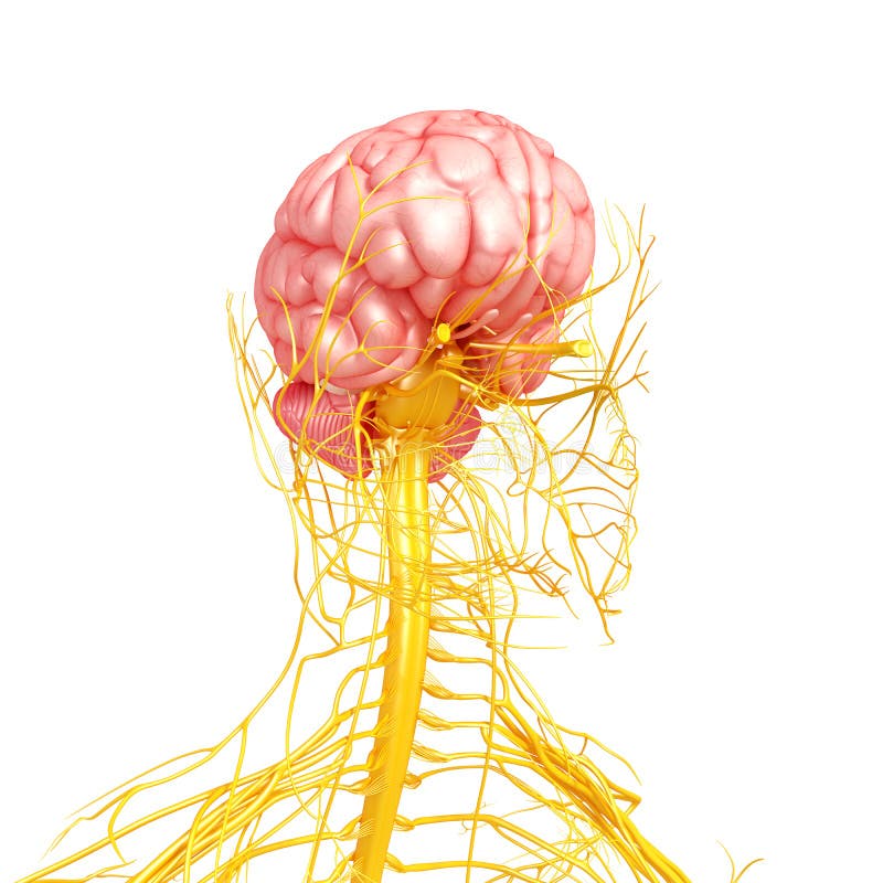
Free with trial Brain Anatomy - Limbic System. 3d rendering illustration. Anatomy neck illustrations Brain Anatomy - Limbic System. 3d rendering
Free with trial Neck pain from using smartphone or texting too much. infographic. right and wrong position for good health - vector illustration. Anatomy neck vectors Neck pain from using smartphone or texting too much. infographic. right and wrong position for good health - vector
Free with trial Neck icon. Cartoon illustration of neck vector icon for web design. Anatomy neck vectors Neck icon, cartoon style. Neck icon. Cartoon illustration of neck vector icon for web design
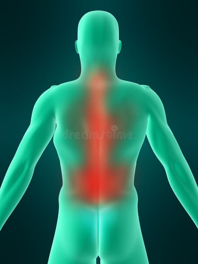
Free with trial 3d rendered anatomy illustration - painful back. Anatomy neck illustrations Highlighted spine. 3d rendered anatomy illustration - painful back
Free with trial 3d rendered medically accurate illustration of the equine muscle anatomy - Brachiocephalicus. Anatomy neck illustrations Brachiocephalicus
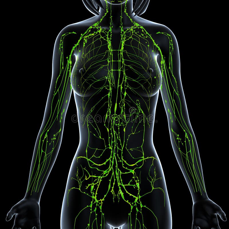
Free with trial Female anatomy illustration of the lymphatic system of male half body. Anatomy neck illustrations Female Lymphatic system of half body. Female anatomy illustration of the lymphatic system of male half body
Free with trial 3d rendered anatomy illustration of a male back with backache. Anatomy neck illustrations Backache
Free with trial 3d rendered anatomy illustration of a male back with backache. Anatomy neck illustrations Backache
Free with trial 3d rendered anatomy illustration of a male back with backache. Anatomy neck illustrations Backache
Free with trial 3d rendered medically accurate illustration of the equine muscle anatomy - Longus Colli. Anatomy neck illustrations Longus Colli
Free with trial Vagus nerve. Branches of the vagus nerve in the neck. Anatomical relationships between superior thyroid artery and external branch of superior laryngeal nerve. Anatomy neck vectors Vagus nerve
Free with trial 3d rendered medically accurate illustration of the equine muscle anatomy - Sternocephalicus. Anatomy neck illustrations Sternocephalicus
Free with trial 3d rendered anatomy illustration of a human spine. Anatomy neck illustrations Human spine
Free with trial The thyroid gland, in vertebrate anatomy, is one of the largest endocrine glands and consists of two connected lobes. Anatomy neck illustrations Thyroid
Free with trial 3d rendered medically accurate illustration of the equine muscle anatomy - Omohyoid. Anatomy neck illustrations Omohyoid
Free with trial 3d rendered illustration of a painful neck. Anatomy neck illustrations Highlighted nerve system. 3d rendered illustration of a painful neck
Free with trial 3d rendered anatomy illustration of a human spine with backache. Anatomy neck illustrations Human spine with backache
Free with trial 3d rendered anatomy illustration of a human spine with backache. Anatomy neck illustrations Human spine with backache
Free with trial 3d rendered medically accurate illustration of the equine muscle anatomy - Sternohyoideus. Anatomy neck illustrations Sternohyoideus
Free with trial Human anatomy illustration of the side view of male nervous system. Anatomy neck illustrations Side view of male nervous system
Free with trial 3d rendered medically accurate illustration of the equine muscle anatomy - Brachiocephalicus. Anatomy neck illustrations Brachiocephalicus
Free with trial The cervical vertebrae of the spine consist of seven bony rings that reside in the neck between the base of the skull and the thoracic vertebrae in the trunk. Among the vertebrae of the spinal column, the cervical vertebrae are the thinnest and most delicate bones. Yet, in spite of their size, the cervical vertebrae have the huge jobs of supporting the head, protecting the spinal cord, and providing mobility to the head and neck. Anatomy neck illustrations Cervical Vertebrae C2. The cervical vertebrae of the spine consist of seven bony rings that reside in the neck between the base of the skull and the thoracic vertebrae in the trunk. Among the vertebrae of the spinal column, the cervical vertebrae are the thinnest and most delicate bones. Yet, in spite of their size, the cervical vertebrae have the huge jobs of supporting the head, protecting the spinal cord, and providing mobility to the head and neck.
Free with trial Female anatomy illustration of the lymphatic system of male half body. Anatomy neck illustrations Female Lymphatic system of half body. Female anatomy illustration of the lymphatic system of male half body
Free with trial The cervical vertebrae of the spine consist of seven bony rings that reside in the neck between the base of the skull and the thoracic vertebrae in the trunk. Among the vertebrae of the spinal column, the cervical vertebrae are the thinnest and most delicate bones. Yet, in spite of their size, the cervical vertebrae have the huge jobs of supporting the head, protecting the spinal cord, and providing mobility to the head and neck. Anatomy neck illustrations Cervical Vertebrae C7. The cervical vertebrae of the spine consist of seven bony rings that reside in the neck between the base of the skull and the thoracic vertebrae in the trunk. Among the vertebrae of the spinal column, the cervical vertebrae are the thinnest and most delicate bones. Yet, in spite of their size, the cervical vertebrae have the huge jobs of supporting the head, protecting the spinal cord, and providing mobility to the head and neck.
Free with trial Human anatomy illustration of the nervous system of male front side view. Anatomy neck illustrations Nervous system of human front side view. Human anatomy illustration of the nervous system of male front side view
Free with trial Female Anatomy Medical Imaging by @ decade3d. Anatomy neck illustrations Full Female Body - Front View - Blue concept. Female Anatomy Medical Imaging by @ decade3d
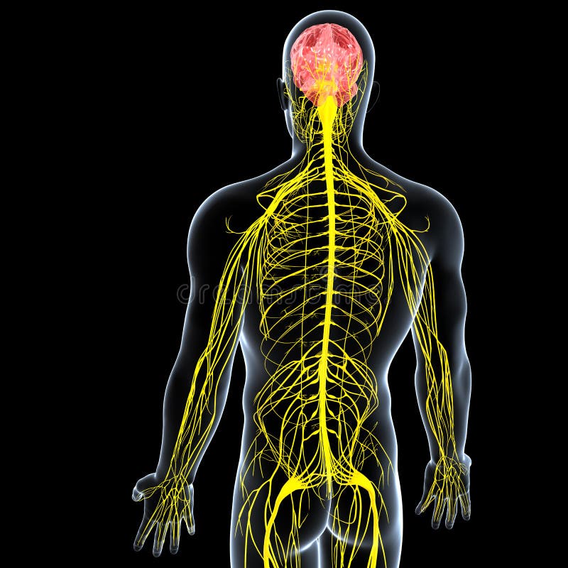
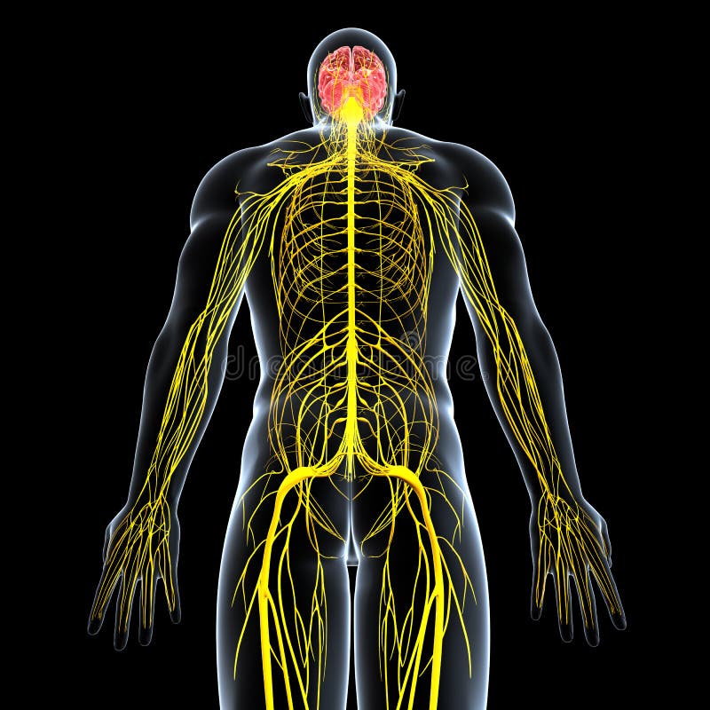
Free with trial Human anatomy illustration of the nervous system of male with full back body. Anatomy neck illustrations Nervous system of male with full back body
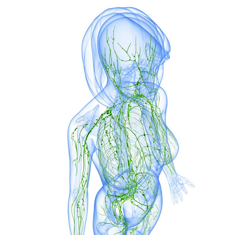
Free with trial Female anatomy illustration of the Lymphatic system & x ray. Anatomy neck illustrations Female Lymphatic system x ray. Female anatomy illustration of the Lymphatic system & x ray
Free with trial 3d rendered medically accurate illustration of the equine muscle anatomy - Sternocephalicus. Anatomy neck illustrations Sternocephalicus
Free with trial 3d rendered medically accurate illustration of the equine muscle anatomy - Cleidobrachialis. Anatomy neck illustrations Cleidobrachialis
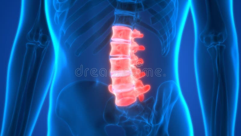
Free with trial The lumbar vertebrae are, in human anatomy, the five vertebrae between the rib cage and the pelvis. They are the largest segments of the vertebral column and are characterized by the absence of the foramen transversarium within the transverse process as it is only found in the cervical region, and by the absence of facets on the sides of the body. They are designated L1 to L5, starting at the top. The lumbar vertebrae help support the weight of the body, and permit movement. Anatomy neck illustrations Lumbar vertebrae L3. The lumbar vertebrae are, in human anatomy, the five vertebrae between the rib cage and the pelvis. They are the largest segments of the vertebral column and are characterized by the absence of the foramen transversarium within the transverse process as it is only found in the cervical region, and by the absence of facets on the sides of the body. They are designated L1 to L5, starting at the top. The lumbar vertebrae help support the weight of the body, and permit movement.
Free with trial The lumbar vertebrae are, in human anatomy, the five vertebrae between the rib cage and the pelvis. They are the largest segments of the vertebral column and are characterized by the absence of the foramen transversarium within the transverse process as it is only found in the cervical region, and by the absence of facets on the sides of the body. They are designated L1 to L5, starting at the top. The lumbar vertebrae help support the weight of the body, and permit movement. Anatomy neck illustrations Lumbar vertebrae L1. The lumbar vertebrae are, in human anatomy, the five vertebrae between the rib cage and the pelvis. They are the largest segments of the vertebral column and are characterized by the absence of the foramen transversarium within the transverse process as it is only found in the cervical region, and by the absence of facets on the sides of the body. They are designated L1 to L5, starting at the top. The lumbar vertebrae help support the weight of the body, and permit movement.
Free with trial The lumbar vertebrae are, in human anatomy, the five vertebrae between the rib cage and the pelvis. They are the largest segments of the vertebral column and are characterized by the absence of the foramen transversarium within the transverse process as it is only found in the cervical region, and by the absence of facets on the sides of the body. They are designated L1 to L5, starting at the top. The lumbar vertebrae help support the weight of the body, and permit movement. Anatomy neck illustrations Lumbar vertebrae L1. The lumbar vertebrae are, in human anatomy, the five vertebrae between the rib cage and the pelvis. They are the largest segments of the vertebral column and are characterized by the absence of the foramen transversarium within the transverse process as it is only found in the cervical region, and by the absence of facets on the sides of the body. They are designated L1 to L5, starting at the top. The lumbar vertebrae help support the weight of the body, and permit movement.
Free with trial Female anatomy illustration of the lymphatic system of male half body. Anatomy neck illustrations Female Lymphatic system of half body. Female anatomy illustration of the lymphatic system of male half body
Free with trial The cervical vertebrae of the spine consist of seven bony rings that reside in the neck between the base of the skull and the thoracic vertebrae in the trunk. Among the vertebrae of the spinal column, the cervical vertebrae are the thinnest and most delicate bones. Yet, in spite of their size, the cervical vertebrae have the huge jobs of supporting the head, protecting the spinal cord, and providing mobility to the head and neck. Anatomy neck illustrations Cervical Vertebrae C4. The cervical vertebrae of the spine consist of seven bony rings that reside in the neck between the base of the skull and the thoracic vertebrae in the trunk. Among the vertebrae of the spinal column, the cervical vertebrae are the thinnest and most delicate bones. Yet, in spite of their size, the cervical vertebrae have the huge jobs of supporting the head, protecting the spinal cord, and providing mobility to the head and neck.
Free with trial Femoral neck fracture. Major types. Anatomy neck vectors Fracture neck of femur. Femoral neck fracture. Major types.
Free with trial The cervical vertebrae of the spine consist of seven bony rings that reside in the neck between the base of the skull and the thoracic vertebrae in the trunk. Among the vertebrae of the spinal column, the cervical vertebrae are the thinnest and most delicate bones. Yet, in spite of their size, the cervical vertebrae have the huge jobs of supporting the head, protecting the spinal cord, and providing mobility to the head and neck. Anatomy neck illustrations Cervical spine with Nerves lateral view. The cervical vertebrae of the spine consist of seven bony rings that reside in the neck between the base of the skull and the thoracic vertebrae in the trunk. Among the vertebrae of the spinal column, the cervical vertebrae are the thinnest and most delicate bones. Yet, in spite of their size, the cervical vertebrae have the huge jobs of supporting the head, protecting the spinal cord, and providing mobility to the head and neck.
Free with trial Female anatomy illustration of the Lymphatic system isolated. Anatomy neck illustrations Female Lymphatic system. Female anatomy illustration of the Lymphatic system isolated
Free with trial Face lift before and after treatment. Anatomy neck vectors Neck lift. Face lift before and after treatment
Free with trial 3d rendered anatomy illustration of a human spine with backache. Anatomy neck illustrations Human spine with backache
Free with trial The anatomy of a Champagne bottle. Hand drawn Illustration of Infographic in vintage engraved style. Isolated on white background. Anatomy neck vectors Hand drawn Illustration of Champagne bottle. The anatomy of a Champagne bottle. Hand drawn Illustration of Infographic in vintage engraved style. Isolated on white background.
Free with trial The thyroid gland, in vertebrate anatomy, is one of the largest endocrine glands and consists of two connected lobes. Anatomy neck illustrations Thyroid gland
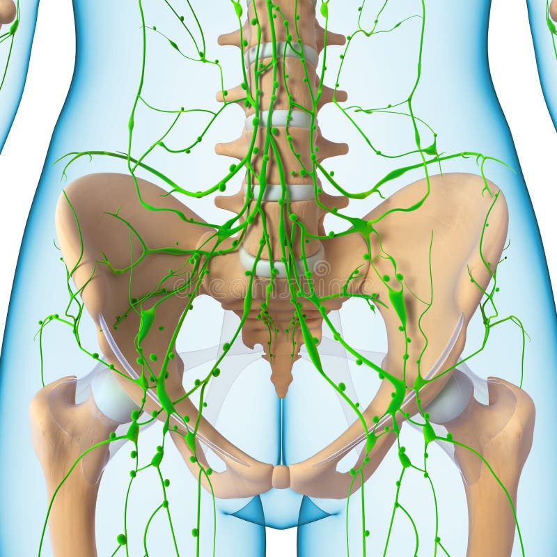
Free with trial Female anatomy illustration of the Lymphatic system. Anatomy neck illustrations Lymphatic system
Free with trial 3d rendered medically accurate illustration of the equine muscle anatomy - Brachiocephalicus. Anatomy neck illustrations Brachiocephalicus