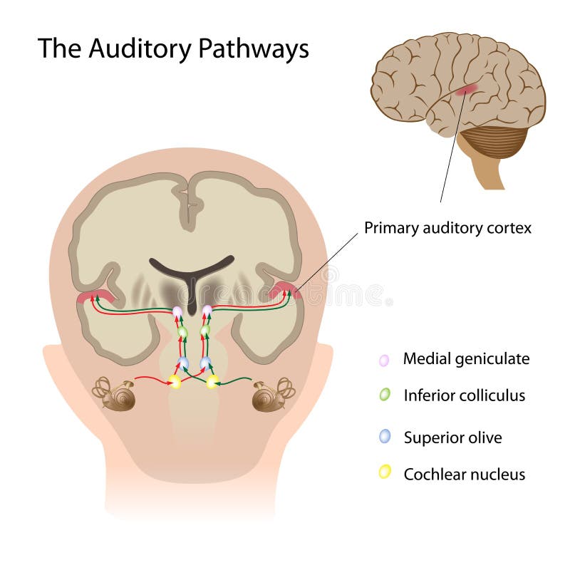
Free with trial Diagram of the auditory pathway in the brain, eps10. Auditory pathway vectors The auditory pathways. Diagram of the auditory pathway in the brain, eps10
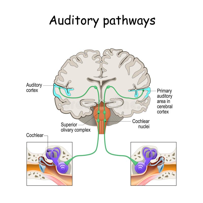
Free with trial The auditory pathway medical illustration on white background eps 10 cross view of the human brain. Auditory pathway vectors The auditory pathway medical illustration on white background. The auditory pathway medical illustration on white background eps 10 cross view of the human brain
Free with trial Auditory pathways from cochlea in ear to cortex in brain. Sound Localization. anatomy of the auditory system. Structures of the Ear. Auditory pathway vectors Auditory pathways from cochlea in ear to cortex in brain
Free with trial Auditory system from Tympanic membrane and Cochlear in the ear to Auditory cortex on the brain. sensory system for the sense of hearing. Anatomy of the human ear. Vector poster. Auditory pathway vectors Auditory system. sensory system. human ear anatomy. Auditory system from Tympanic membrane and Cochlear in the ear to Auditory cortex on the brain. sensory system for the sense of hearing. Anatomy of the human ear. Vector poster
Free with trial Auditory pathway from the receptors in the organ of Corti of the inner ear to the central nervous system. Primary auditory cortex. Vestibulocochlear nerve function medical flat vector illustration. Auditory pathway vectors Auditory pathway diagram. Auditory pathway from the receptors in the organ of Corti of the inner ear to the central nervous system. Primary auditory cortex. Vestibulocochlear nerve function medical flat vector illustration.
Free with trial Auditory pathway from the receptors in the organ of Corti of the inner ear to the central nervous system. Primary auditory cortex. Vestibulocochlear nerve function medical flat vector illustration. Auditory pathway vectors Auditory pathway diagram. Auditory pathway from the receptors in the organ of Corti of the inner ear to the central nervous system. Primary auditory cortex. Vestibulocochlear nerve function medical flat vector illustration.
Free with trial Auditory pathway from the receptors in the organ of Corti of the inner ear to the central nervous system. Primary auditory cortex. Vestibulocochlear nerve function medical flat vector illustration. Auditory pathway vectors Auditory pathway diagram. Auditory pathway from the receptors in the organ of Corti of the inner ear to the central nervous system. Primary auditory cortex. Vestibulocochlear nerve function medical flat vector illustration.
Free with trial The Inner Ear Cochlea. Cross-section of one spiral of cochlea. Organ of Corti, the sensory organ of hearing. Spiral ganglion, Osseous Spiral Lamina. Auditory Pathway. Auditory pathway vectors The Inner Ear Cochlea. Cross-section of one spiral of cochlea. Organ of Corti, the sensory organ of hearing. Spiral
Free with trial The cell bodies of the three neurons in a typical somatosensory pathway are located in the dorsal root ganglion, the spinal cord, and the thalamus. A major target of somatosensory pathways is the postcentral gyrus in the parietal lobe of the cerebral cortex. Auditory pathway illustrations Somatosensory association area of human brain. The cell bodies of the three neurons in a typical somatosensory pathway are located in the dorsal root ganglion, the spinal cord, and the thalamus. A major target of somatosensory pathways is the postcentral gyrus in the parietal lobe of the cerebral cortex.
Free with trial Anatomy of Inner Ear and cross-section of Cochlea. Organ of Corti, the sensory organ of hearing. Spiral ganglion, Osseous Spiral Lamina. Structural diagram of the Ear Anatomy. Auditory pathway vectors Anatomy of Inner Ear and cross-section of Cochlea. Organ of Corti, the sensory organ of hearing. Spiral ganglion
Free with trial Auditory ossicles brief diagram shows the middle ear sound pathway, malleus, incus, and stapes amplify vibrations from eardrum to cochlea. Outline diagram. Auditory pathway vectors Auditory ossicles brief diagram shows the middle ear sound pathway, ... Auditory ossicles brief diagram shows the middle ear sound pathway, malleus, incus, and stapes amplify vibrations from eardrum to cochlea. Outline diagram
Free with trial The auditory pathway conveys the special sense of hearing. Information travels from the receptors in the organ of Corti of the inner ear (cochlear hair cells) to the central nervous system, carried by the vestibulocochlear nerve (CN VIII). Auditory pathway illustrations Auditory Pathway Transmits Sensory Signals for Hearing. The auditory pathway conveys the special sense of hearing. Information travels from the receptors in the organ of Corti of the inner ear (cochlear hair cells) to the central nervous system, carried by the vestibulocochlear nerve (CN VIII).
Free with trial The auditory pathway conveys the special sense of hearing. Information travels from the receptors in the organ of Corti of the inner ear (cochlear hair cells) to the central nervous system, carried by the vestibulocochlear nerve (CN VIII). Auditory pathway illustrations Auditory Pathway Transmits Sensory Signals for Hearing. The auditory pathway conveys the special sense of hearing. Information travels from the receptors in the organ of Corti of the inner ear (cochlear hair cells) to the central nervous system, carried by the vestibulocochlear nerve (CN VIII).
Free with trial The auditory pathway conveys the special sense of hearing. Information travels from the receptors in the organ of Corti of the inner ear (cochlear hair cells) to the central nervous system, carried by the vestibulocochlear nerve (CN VIII). Auditory pathway illustrations Auditory Pathway Transmits Sensory Signals for Hearing. The auditory pathway conveys the special sense of hearing. Information travels from the receptors in the organ of Corti of the inner ear (cochlear hair cells) to the central nervous system, carried by the vestibulocochlear nerve (CN VIII).
Free with trial The auditory pathway conveys the special sense of hearing. Information travels from the receptors in the organ of Corti of the inner ear (cochlear hair cells) to the central nervous system, carried by the vestibulocochlear nerve (CN VIII). Auditory pathway illustrations The Auditory Pathway Carries Sensory Hearing Information. The auditory pathway conveys the special sense of hearing. Information travels from the receptors in the organ of Corti of the inner ear (cochlear hair cells) to the central nervous system, carried by the vestibulocochlear nerve (CN VIII).
Free with trial The auditory pathway conveys the special sense of hearing. Information travels from the receptors in the organ of Corti of the inner ear (cochlear hair cells) to the central nervous system, carried by the vestibulocochlear nerve (CN VIII). Auditory pathway illustrations The Auditory Pathway Carries Sensory Hearing Information. The auditory pathway conveys the special sense of hearing. Information travels from the receptors in the organ of Corti of the inner ear (cochlear hair cells) to the central nervous system, carried by the vestibulocochlear nerve (CN VIII).
Free with trial The auditory pathway conveys the special sense of hearing. Information travels from the receptors in the organ of Corti of the inner ear (cochlear hair cells) to the central nervous system, carried by the vestibulocochlear nerve (CN VIII). Auditory pathway illustrations The Auditory Pathway Carries Sensory Hearing Information. The auditory pathway conveys the special sense of hearing. Information travels from the receptors in the organ of Corti of the inner ear (cochlear hair cells) to the central nervous system, carried by the vestibulocochlear nerve (CN VIII).
Free with trial This vibrant illustration displays a detailed depiction of the human ear connected by a red neural pathway to the brain, with sound waves visually represented around them it can be used for educational purposes or in medical publications explaining auditory processing. Generative AI. Auditory pathway illustrations Human Ear to Brain Connection Illustration - Auditory Pathway and Neural Signal Transmission Diagram. This vibrant illustration displays a detailed depiction of the human ear connected by a red neural pathway to the brain, with sound waves visually represented around them it can be used for educational purposes or in medical publications explaining auditory processing. Generative AI
Free with trial Detailed anatomy illustration of human ear outer middle, inner ear structures. Shows auditory system pathway with cochlea, eardrum. Visual representation of ear anatomy emphasizes ear. Auditory pathway illustrations Detailed anatomy illustration of human ear outer middle, inner ear structures. Shows auditory system pathway with cochlea
Free with trial A detailed vector illustration of the human ear's auditory anatomy, showing the pathway of sound perception. This medical diagram is perfect for audiology education, scientific presentations, health awareness campaigns, or biological studies. Its intricate details and accurate depiction provide a valuable and versatile visual asset for various professional uses, vector design Generative AI. Auditory pathway vectors Human Ear Auditory Anatomy, Medical Science Hearing Vector Art, vector design Generative AI. A detailed vector illustration of the human ear's auditory anatomy, showing the pathway of sound perception. This medical diagram is perfect for audiology education, scientific presentations, health awareness campaigns, or biological studies. Its intricate details and accurate depiction provide a valuable and versatile visual asset for various professional uses, vector design Generative AI
Free with trial This detailed illustration showcases a cross-section of the human ear connected to the brain via auditory nerves, highlighting sound wave transmission and neurological pathways. Educators can utilize this graphic as a visual aid to explain hearing physiology in biology classes or medical training programs. Generative AI. Auditory pathway illustrations Human Ear Anatomy Illustration with Auditory Pathway to Brain - Medical Diagram for Education. This detailed illustration showcases a cross-section of the human ear connected to the brain via auditory nerves, highlighting sound wave transmission and neurological pathways. Educators can utilize this graphic as a visual aid to explain hearing physiology in biology classes or medical training programs. Generative AI
Free with trial Anatomical illustration of the cochlea featuring a detailed cross-section. The cochlea is a spiral-shaped organ in the inner ear, essential for hearing. Visible structures include the scala vestibuli, scala tympani, and cochlear duct, with intricate details of the bony and membranous labyrinth. The image highlights the organ of Corti, essential for transmitting sound vibrations into nerve impulses. The section shows the cochlea's coiled nature and intricate network of fluid-filled channels important for auditory processing. Auditory pathway illustrations Detailed anatomical illustration of the cochlea, inner ear, cross-section, and auditory system. Anatomical illustration of the cochlea featuring a detailed cross-section. The cochlea is a spiral-shaped organ in the inner ear, essential for hearing. Visible structures include the scala vestibuli, scala tympani, and cochlear duct, with intricate details of the bony and membranous labyrinth. The image highlights the organ of Corti, essential for transmitting sound vibrations into nerve impulses. The section shows the cochlea's coiled nature and intricate network of fluid-filled channels important for auditory processing.
Free with trial Cross-sectional diagram of a human ear illustrating external, middle, and internal sections. The external ear includes labeled parts like the pinna and auditory canal. The middle ear shows the malleus, incus, and stapes. The internal ear highlights the semicircular canals and cochlea. Notations indicate redness, swelling, and itchiness in the external auditory canal, while wax, shed skin, and pus are present. Key components like the tympanic cavity, eardrum, eustachian tube, and auditory pathway are marked, providing a detailed overview of ear anatomy. Auditory pathway vectors Swimmer\'s Ear Cross section illustration. Cross-sectional diagram of a human ear illustrating external, middle, and internal sections. The external ear includes labeled parts like the pinna and auditory canal. The middle ear shows the malleus, incus, and stapes. The internal ear highlights the semicircular canals and cochlea. Notations indicate redness, swelling, and itchiness in the external auditory canal, while wax, shed skin, and pus are present. Key components like the tympanic cavity, eardrum, eustachian tube, and auditory pathway are marked, providing a detailed overview of ear anatomy.
Free with trial A modern bus station featuring visual and auditory guidance systems. The design emphasizes accessibility and user-friendly travel options with ample seating and information displays Generated By AI. Auditory pathway illustrations Modern Bus Station with Visual and Auditory Guidance. A modern bus station featuring visual and auditory guidance systems. The design emphasizes accessibility and user-friendly travel options with ample seating and information displays Generated By AI
Free with trial This digital illustration depicts a detailed view of the human ear, showcasing sound waves entering and stimulating the auditory nerve. It can be used for educational materials, medical presentations, or informative graphics about hearing health. Generative AI. Auditory pathway illustrations Human Ear Anatomy Illustration: Sound Waves, Auditory Nerve & Hearing Process Explained. This digital illustration depicts a detailed view of the human ear, showcasing sound waves entering and stimulating the auditory nerve. It can be used for educational materials, medical presentations, or informative graphics about hearing health. Generative AI
Free with trial This vibrant, detailed illustration showcases the inner workings of a human cochlea connected to the auditory nerve against a backdrop of a rainy city night. The diagram can be utilized for educational resources about hearing, audiology, or medical explanations relating to sound perception. Generative AI. Auditory pathway illustrations Detailed 3D Render of Human Cochlea and Auditory Nerve Functioning in Rainy Cityscape. This vibrant, detailed illustration showcases the inner workings of a human cochlea connected to the auditory nerve against a backdrop of a rainy city night. The diagram can be utilized for educational resources about hearing, audiology, or medical explanations relating to sound perception. Generative AI
Free with trial This detailed illustration showcases a cross-section of the human inner ear, highlighting structures like the auditory nerve and cochlea alongside labeled components it can be utilized for medical education materials or scientific publications to explain auditory function. Generative AI. Auditory pathway illustrations Detailed Anatomy of the Human Inner Ear with Auditory Nerve and Structures Illustration. This detailed illustration showcases a cross-section of the human inner ear, highlighting structures like the auditory nerve and cochlea alongside labeled components it can be utilized for medical education materials or scientific publications to explain auditory function. Generative AI
Free with trial This detailed illustration showcases the human ear, depicting sound waves traveling through the auditory canal and connecting to the brain via a nerve pathway. It serves as a useful visual aid for educational materials explaining hearing physiology and neurological processes. Generative AI. Auditory pathway illustrations Human Ear Anatomy Illustration: Sound Waves to Brain Connection - Medical Diagram for Education. This detailed illustration showcases the human ear, depicting sound waves traveling through the auditory canal and connecting to the brain via a nerve pathway. It serves as a useful visual aid for educational materials explaining hearing physiology and neurological processes. Generative AI
Free with trial This digital illustration depicts a human ear connected to a detailed brain model via flowing blue sound waves and a vibrant neurological pathway. It can be utilized for educational materials on auditory processing, neuroscience diagrams or illustrating how our senses impact cognitive function. Generative AI. Auditory pathway illustrations Human Ear to Brain Connection Illustration - Sound Waves, Neurological Pathways & Cognitive Function. This digital illustration depicts a human ear connected to a detailed brain model via flowing blue sound waves and a vibrant neurological pathway. It can be utilized for educational materials on auditory processing, neuroscience diagrams or illustrating how our senses impact cognitive function. Generative AI
Free with trial This digital illustration depicts a detailed, cross-section of the human ear alongside a visual representation of sound waves impacting it, highlighting the auditory cortex. It can be utilized for educational materials concerning hearing mechanics, medical illustrations, or informative science presentations. Generative AI. Auditory pathway illustrations Detailed Human Ear Anatomy Illustration with Sound Waves and Auditory Cortex Diagram - Science Visual. This digital illustration depicts a detailed, cross-section of the human ear alongside a visual representation of sound waves impacting it, highlighting the auditory cortex. It can be utilized for educational materials concerning hearing mechanics, medical illustrations, or informative science presentations. Generative AI
Free with trial This detailed diagram showcases the anatomy of the human ear, tracing the pathway from sound waves entering the outer ear through the middle and inner ear structures to stimulation of the auditory cortex in the brain. It can serve as a valuable visual aid for educational materials related to audiology, neuroscience, or medical studies exploring hearing processes. Generative AI. Auditory pathway illustrations Human Ear Anatomy Diagram Illustrating Sound Transmission to the Brain - Medical Illustration. This detailed diagram showcases the anatomy of the human ear, tracing the pathway from sound waves entering the outer ear through the middle and inner ear structures to stimulation of the auditory cortex in the brain. It can serve as a valuable visual aid for educational materials related to audiology, neuroscience, or medical studies exploring hearing processes. Generative AI
Free with trial Detailed anatomy of human ear outer, middle, inner structures with auditory system. Highlighted canal, vestibule, frequency pathways, eardrum, balance labyrinth. Acoustic details worked. Auditory pathway illustrations Detailed anatomy of human ear outer, middle, inner structures with auditory system. Highlighted canal, vestibule, frequency
Free with trial A transparent, glowing 3D anatomical diagram showcases the intricate structures of the human inner ear, including the cochlea, semicircular canals, and vestibular system, set against a dark gradient background with subtle highlights for emphasis. The image is designed for educational or medical use, highlighting the complexity and functionality of the auditory system. Auditory pathway illustrations Detailed Anatomical Illustration of Human Inner Ear Structure. A transparent, glowing 3D anatomical diagram showcases the intricate structures of the human inner ear, including the cochlea, semicircular canals, and vestibular system, set against a dark gradient background with subtle highlights for emphasis. The image is designed for educational or medical use, highlighting the complexity and functionality of the auditory system
Free with trial Detailed medical illustration of the human cochlea, the auditory portion of the inner ear. The image presents a cross-sectional view, showcasing the intricate structure with labeled components. The cochlea is depicted in vibrant colors, highlighting the different sections and their relationships. This educational diagram is suitable for medical textbooks, educational materials, or presentations on human anatomy and the auditory system. Auditory pathway illustrations Anatomy of the Human Cochlea - Medical Illustration. Detailed medical illustration of the human cochlea, the auditory portion of the inner ear. The image presents a cross-sectional view, showcasing the intricate structure with labeled components. The cochlea is depicted in vibrant colors, highlighting the different sections and their relationships. This educational diagram is suitable for medical textbooks, educational materials, or presentations on human anatomy and the auditory system.
Free with trial A flat a white car emitting orange and yellow sound waves towards a man. The man, with a red flushed face and closed eyes, holds his hands over his ears, indicating discomfort. Text labels point to a diagram of a brain labeled "Cour Inestnges" and a cross-section of an ear with a pathway labeled "Sound sensitivity pathway. " The overall style is clean and with a light blue background. Auditory pathway illustrations Man with noise induced headache from car sound waves. A flat a white car emitting orange and yellow sound waves towards a man. The man, with a red flushed face and closed eyes, holds his hands over his ears, indicating discomfort. Text labels point to a diagram of a brain labeled "Cour Inestnges" and a cross-section of an ear with a pathway labeled "Sound sensitivity pathway." The overall style is clean and with a light blue background
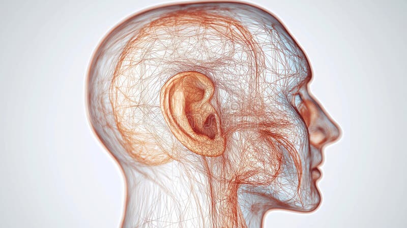
Free with trial A highly detailed and scientifically accurate illustration of the human ear anatomy showcasing the intricate nerve connections leading to the brain This educational image highlights the outer middle and inner ear structures including the cochlea auditory nerve and brainstem pathways Perfect for medical textbooks healthcare presentations biology lectures or educational websites The clean labeled. Auditory pathway illustrations Detailed Anatomy of Human Ear with Nerve Connections and Brain Illustration. A highly detailed and scientifically accurate illustration of the human ear anatomy showcasing the intricate nerve connections leading to the brain This educational image highlights the outer middle and inner ear structures including the cochlea auditory nerve and brainstem pathways Perfect for medical textbooks healthcare presentations biology lectures or educational websites The clean labeled
Free with trial A comprehensive medical illustration showcasing the intricate internal and external structures of the human ear, detailing the path of sound waves from the outer ear to the inner ear, highlighting the cochlea and auditory nerve. Auditory pathway illustrations Detailed Cross-Sectional View of Human Ear Anatomy with Sound Wave Illustration. A comprehensive medical illustration showcasing the intricate internal and external structures of the human ear, detailing the path of sound waves from the outer ear to the inner ear, highlighting the cochlea and auditory nerve
Free with trial A modern bus station featuring clear visual and auditory guidance. The design emphasizes accessibility, safety, and comfort for commuters in an urban environment Generated By AI. Auditory pathway illustrations Modern Bus Station with Clear Guidance Features. A modern bus station featuring clear visual and auditory guidance. The design emphasizes accessibility, safety, and comfort for commuters in an urban environment Generated By AI
Free with trial Detailed anatomy illustration shows human ear structures. Outer ear canal, eardrum highlighted. Middle ear ossicles, inner ear labyrinth displayed. Auditory pathway balance system. Auditory pathway illustrations Detailed anatomy illustration shows human ear structures. Outer ear canal, eardrum highlighted. Middle ear ossicles, inner ear
Free with trial This digital illustration depicts a human ear receiving sound waves, with a connection visualized through neural pathways leading to the brain and its vascular system. It can be utilized as an educational tool or visual aid in medical presentations concerning auditory processing and neurological function. Generative AI. Auditory pathway illustrations Sound Waves to Brain Illustration - Human Ear & Neural Pathways Diagram for Medical Use. This digital illustration depicts a human ear receiving sound waves, with a connection visualized through neural pathways leading to the brain and its vascular system. It can be utilized as an educational tool or visual aid in medical presentations concerning auditory processing and neurological function. Generative AI
Free with trial This detailed illustration depicts the inner workings of a human ear, showing sound waves traveling through the canal and stimulating auditory receptors before transmitting signals via the auditory nerve to the brain. It can be utilized as an educational visual aid in medical textbooks or presentations concerning hearing physiology and anatomy. Generative AI. Auditory pathway illustrations Human Ear Anatomy Illustration: Sound Waves to Brain Processing - Medical Diagram for Education. This detailed illustration depicts the inner workings of a human ear, showing sound waves traveling through the canal and stimulating auditory receptors before transmitting signals via the auditory nerve to the brain. It can be utilized as an educational visual aid in medical textbooks or presentations concerning hearing physiology and anatomy. Generative AI
Free with trial This illustration depicts a detailed human ear connected via glowing red neural pathways to a vibrant, textured brain, set against a dark blue and black backdrop. It can be used in educational materials or medical presentations to visually explain auditory processing and the neurological system. Generative AI. Auditory pathway illustrations Human Ear to Brain Connection Diagram - Neuroscience Illustration for Medical & Educational Purposes. This illustration depicts a detailed human ear connected via glowing red neural pathways to a vibrant, textured brain, set against a dark blue and black backdrop. It can be used in educational materials or medical presentations to visually explain auditory processing and the neurological system. Generative AI
Free with trial This detailed medical illustration depicts the human ear and how sound waves travel through it, showing nerve pathways leading to the auditory cortex of the brain. Educators or healthcare professionals could use this visual aid to explain hearing processes and anatomy in a clear, understandable way. Generative AI. Auditory pathway illustrations Human Ear Anatomy Diagram Illustrating Sound Transmission to the Brain - Medical Illustration. This detailed medical illustration depicts the human ear and how sound waves travel through it, showing nerve pathways leading to the auditory cortex of the brain. Educators or healthcare professionals could use this visual aid to explain hearing processes and anatomy in a clear, understandable way. Generative AI
Free with trial Cross-sectional anatomical illustration of a dog's ear, including internal structures. Highlights the external ear canal, inner ear, cochlea, and related auditory components with labels. Detailed depiction of the Eustachian tube, tympanic membrane, and ossicles. The intricate layout showcases the auditory pathway and its connection with the cranial region, emphasizing the complexity of canine ear anatomy. The diagram also provides a visual of the surrounding structures, such as the eye and nose, aiding in spatial understanding. Auditory pathway illustrations Anatomy of a dogs ear with labels. Cross-sectional anatomical illustration of a dog's ear, including internal structures. Highlights the external ear canal, inner ear, cochlea, and related auditory components with labels. Detailed depiction of the Eustachian tube, tympanic membrane, and ossicles. The intricate layout showcases the auditory pathway and its connection with the cranial region, emphasizing the complexity of canine ear anatomy. The diagram also provides a visual of the surrounding structures, such as the eye and nose, aiding in spatial understanding.
Free with trial Detailed diagram showing the inner anatomy of a human ear listening to music, with sound waves and musical notes. Auditory pathway illustrations Human Ear Listening to Music Anatomy. Detailed diagram showing the inner anatomy of a human ear listening to music, with sound waves and musical notes.
Free with trial Human Ear Anatomy Cross Section Diagram. Detailed illustration of human ear anatomy. Auditory pathway illustrations Human Ear Anatomy Cross Section Diagram. Detailed illustration of human ear anatomy
Free with trial A winding mountain path is painted with piano keys, creating a surreal landscape where music meets nature's grandeur. Auditory pathway illustrations Surreal mountain road transforms into a piano keyboard under a dramatic sky. A winding mountain path is painted with piano keys, creating a surreal landscape where music meets nature's grandeur
Free with trial A detailed illustration of the human ear, showcasing its anatomical structure and components. Auditory pathway vectors Anatomical Illustration of the Human Ear - Detailed Diagram. A detailed illustration of the human ear, showcasing its anatomical structure and components
Free with trial The temporal lobes of the human brain are highlighted in a 3D anatomical rendering to illustrate their functions related. Auditory pathway illustrations The temporal lobes of the human brain are highlighted in a 3D anatomical rendering to illustrate their functions related
Free with trial Detailed anatomy of the human ear, highlighting the outer, middle, and inner ear structures gerenetive ai illustration. Auditory pathway illustrations Detailed anatomy of the human ear, highlighting the outer, middle, and inner ear structures
Free with trial Cross-sectional diagram of the human ear showing earwax blockage. It labels the external, middle, and internal parts. Key components include the pinna, external auditory canal, earwax, eardrum, malleus, incus, stapes, tympanic cavity, semicircular canals, cochlea, auditory pathway, and Eustachian tube. The diagram illustrates the placement of earwax within the external auditory canal, highlighting its potential to block sound transmission. Auditory pathway vectors Earwax Blockage (Cerumen Impaction). Cross-sectional diagram of the human ear showing earwax blockage. It labels the external, middle, and internal parts. Key components include the pinna, external auditory canal, earwax, eardrum, malleus, incus, stapes, tympanic cavity, semicircular canals, cochlea, auditory pathway, and Eustachian tube. The diagram illustrates the placement of earwax within the external auditory canal, highlighting its potential to block sound transmission.
Free with trial This striking 3D render depicts a human ear with vibrant blue and red energy flows, showcasing the conversion of sound waves into electrical signals. The image could be utilized for educational materials on audiology, neuroscience, or explaining how hearing functions in scientific publications. Generative AI. Auditory pathway illustrations Futuristic Ear Diagram Illustrating Sound Waves and Neural Transmission - 3D Render. This striking 3D render depicts a human ear with vibrant blue and red energy flows, showcasing the conversion of sound waves into electrical signals. The image could be utilized for educational materials on audiology, neuroscience, or explaining how hearing functions in scientific publications. Generative AI
Free with trial This captivating close-up image showcases the intricate anatomy of a healthy human ear. Visible are the delicate structures of the outer ear, including the pinna and its unique folds, which help collect sound waves. The ear canal, a crucial pathway for sound transmission, is clearly depicted, revealing its smooth, slightly curved interior. The eardrum, a thin, translucent membrane, is. Auditory pathway illustrations Detailed Anatomy of a Healthy Human Ear Outer Ear Ear Canal and Eardrum CloseUp. This captivating close-up image showcases the intricate anatomy of a healthy human ear. Visible are the delicate structures of the outer ear, including the pinna and its unique folds, which help collect sound waves. The ear canal, a crucial pathway for sound transmission, is clearly depicted, revealing its smooth, slightly curved interior. The eardrum, a thin, translucent membrane, is
Free with trial This digital illustration depicts a detailed cross-section of the human ear, showcasing its internal structures alongside vibrant sound waves radiating outwards. It can be utilized as an educational tool in biology classes or for creating informative medical presentations regarding hearing physiology. Generative AI. Auditory pathway illustrations Human Ear Anatomy Illustration with Sound Waves, Medical Visualization for Education and Healthcare. This digital illustration depicts a detailed cross-section of the human ear, showcasing its internal structures alongside vibrant sound waves radiating outwards. It can be utilized as an educational tool in biology classes or for creating informative medical presentations regarding hearing physiology. Generative AI
Free with trial This vibrant illustration depicts an ear connected by glowing pathways to a human brain set against a backdrop of planets and stars, resembling a psychedelic universe. It could be utilized for educational materials on neuroscience, music appreciation, or as striking visual content in creative projects. Generative AI. Auditory pathway illustrations Psychedelic Illustration of Ear to Brain Connection Within a Cosmic, Starry Sky Background - Visual Representation. This vibrant illustration depicts an ear connected by glowing pathways to a human brain set against a backdrop of planets and stars, resembling a psychedelic universe. It could be utilized for educational materials on neuroscience, music appreciation, or as striking visual content in creative projects. Generative AI
Free with trial Auditory pathway from the receptors in the organ of Corti of the inner ear to the central nervous system. Primary auditory cortex. Vestibulocochlear nerve function medical flat vector illustration. Auditory pathway vectors Auditory pathway diagram. Auditory pathway from the receptors in the organ of Corti of the inner ear to the central nervous system. Primary auditory cortex. Vestibulocochlear nerve function medical flat vector illustration.
Free with trial Binaural hearing. Human ability to hear in two ears. Auditory pathways from ear to auditory cortex. Anatomy of the auditory system. Structure of the hearing system. Flat vector illustration. Auditory pathway vectors Binaural hearing. Human ability to hear in two ears. Auditory pathways
Free with trial The cell bodies of the three neurons in a typical somatosensory pathway are located in the dorsal root ganglion, the spinal cord, and the thalamus. A major target of somatosensory pathways is the postcentral gyrus in the parietal lobe of the cerebral cortex. Auditory pathway illustrations Somatosensory association area of human brain. The cell bodies of the three neurons in a typical somatosensory pathway are located in the dorsal root ganglion, the spinal cord, and the thalamus. A major target of somatosensory pathways is the postcentral gyrus in the parietal lobe of the cerebral cortex.
Free with trial The cell bodies of the three neurons in a typical somatosensory pathway are located in the dorsal root ganglion, the spinal cord, and the thalamus. A major target of somatosensory pathways is the postcentral gyrus in the parietal lobe of the cerebral cortex. Auditory pathway illustrations Somatosensory association area of human brain. The cell bodies of the three neurons in a typical somatosensory pathway are located in the dorsal root ganglion, the spinal cord, and the thalamus. A major target of somatosensory pathways is the postcentral gyrus in the parietal lobe of the cerebral cortex.
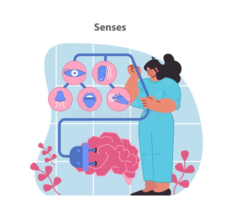
Free with trial Senses concept illustration. A visual exploration of the five senses connected to brain function. Insights into sensory perception. Flat vector illustration. Auditory pathway vectors Senses concept illustration. A visual exploration of the five senses connected to brain function.
Free with trial Injury to head and ear concept icon. Acquired hearing loss cause idea thin line illustration. Damage to auditory pathway. Blunt head trauma. Vector isolated outline RGB color drawing. Editable stroke. Auditory pathway vectors Injury to head and ear concept icon