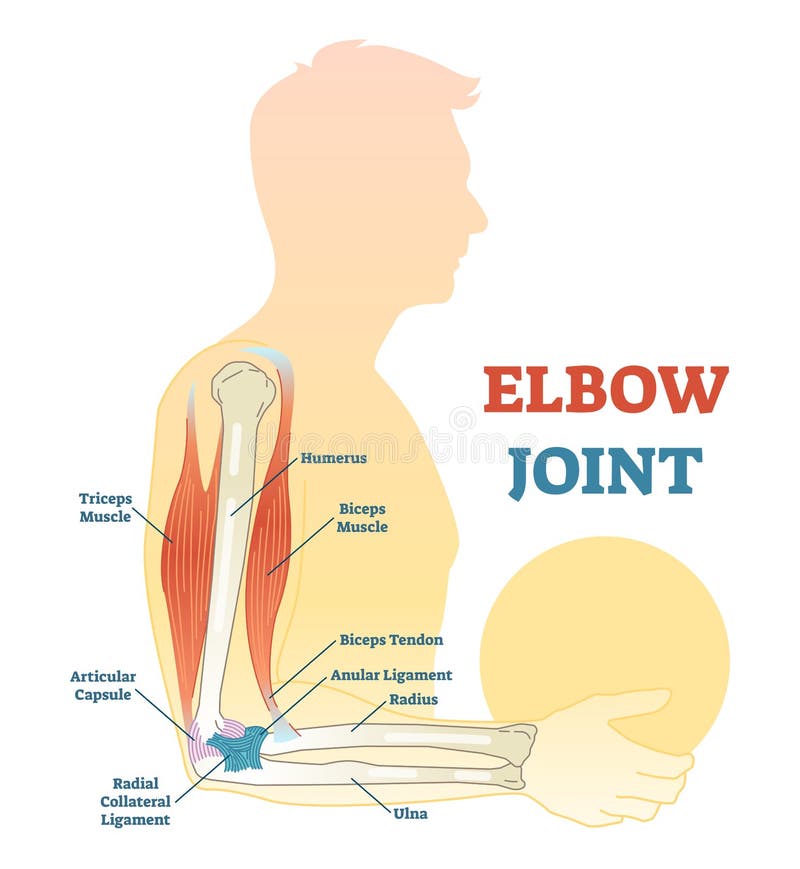
Free with trial Anatomy of a elbow joint. parts of the arm. bones humerus, radius, ulna Muscle Triceps, Biceps and Distal tendon of triceps. Biceps triceps movement arm vectors Anatomy of a elbow joint
Free with trial Muscle is a soft tissue found in most animals. Muscle cells contain protein filaments of actin and myosin that slide past one another, producing a contraction that changes both the length and the shape of the cell. Muscles function to produce force and motion. They are primarily responsible for maintaining and changing posture, locomotion, as well as movement of internal organs, such as the contraction of the heart and the movement of food through the digestive system via peristalsis. Biceps triceps movement arm illustrations Muscle Anatomy. Muscle is a soft tissue found in most animals. Muscle cells contain protein filaments of actin and myosin that slide past one another, producing a contraction that changes both the length and the shape of the cell. Muscles function to produce force and motion. They are primarily responsible for maintaining and changing posture, locomotion, as well as movement of internal organs, such as the contraction of the heart and the movement of food through the digestive system via peristalsis.
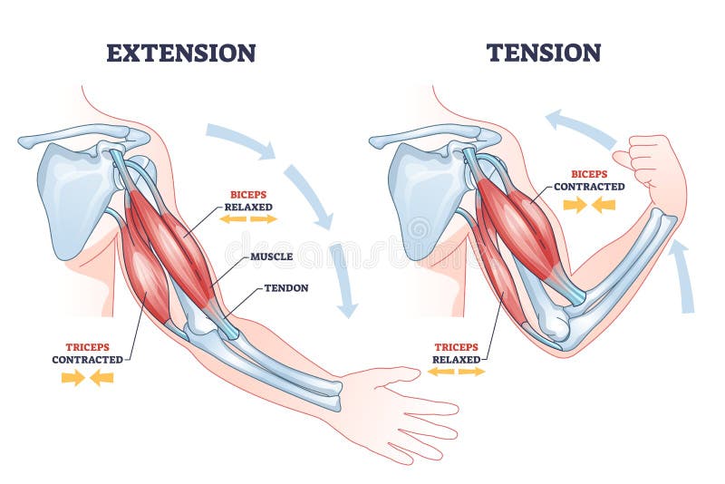
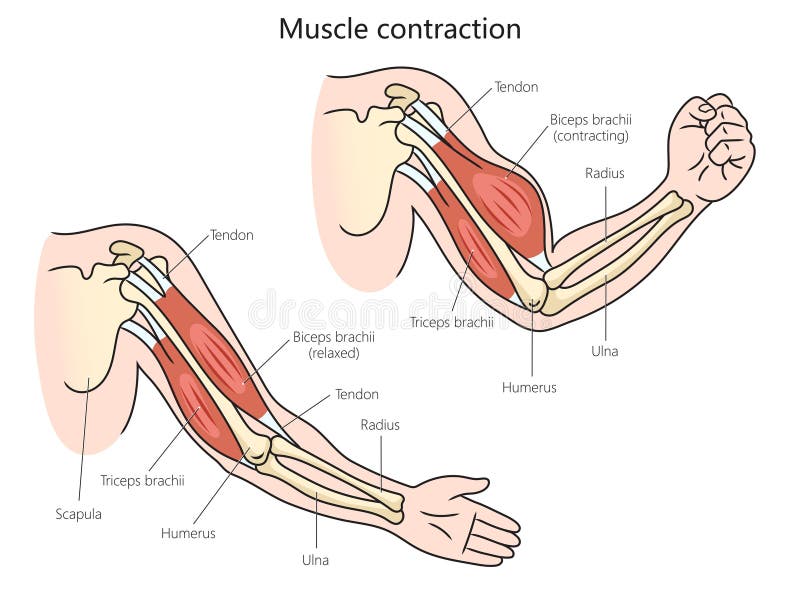
Free with trial Contracting and relaxing of arms biceps and triceps muscles outline diagram. Labeled educational scheme with anatomical contracted and relaxed muscular system structure description vector illustration. Biceps triceps movement arm vectors Contracting and relaxing of arms biceps and triceps muscles outline diagram
Free with trial Elbow joint vector illustrated diagram, medical scheme. Educational sports injury information. Biceps triceps movement arm vectors Elbow joint vector illustrated diagram, medical scheme.
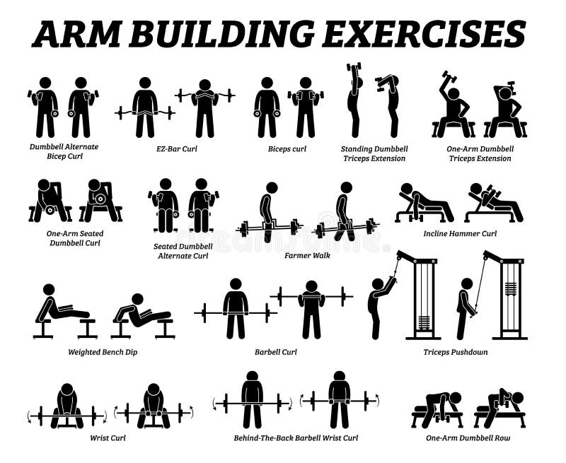
Free with trial Artworks depict a set of weight training reps workout for arm hand muscle by gym machine and tools with step by step instructions. Biceps triceps movement arm vectors Arm building exercises and muscle building stick figure pictograms. Artworks depict a set of weight training reps workout for arm hand muscle by gym machine and tools with step by step instructions
Free with trial Muscles of the hand and arm beautiful bright illustration on a white background. Biceps triceps movement arm vectors Muscles of the hand and arm
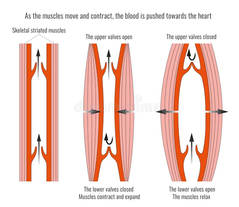
Free with trial Muscle contraction mechanism. Muscles work principle scheme. Anatomical structure and physical movement process example with contracted and relaxed biceps. Flat vector illustration. Biceps triceps movement arm vectors Muscle contraction mechanism. Muscles work principle scheme
Free with trial Different muscles in human body and muscular classification outline diagram. Labeled educational physiological parts scheme with anatomic skeletal, smooth and cardiac division vector illustration. Biceps triceps movement arm vectors Different muscles in human body and muscular classification outline diagram
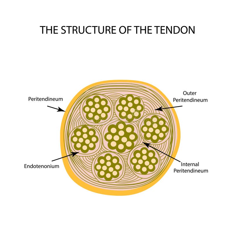
Free with trial Structure of tendon. Infographics. Vector illustration on isolated background. Biceps triceps movement arm vectors Structure of tendon. Infographics. Vector illustration on isolated background
Free with trial Human muscle contraction structure diagram hand drawn schematic vector illustration. Medical science educational illustration. Biceps triceps movement arm vectors Muscle contraction diagram medical science. Human muscle contraction structure diagram hand drawn schematic vector illustration. Medical science educational illustration
Free with trial Detailed illustration showcasing the human arm muscular anatomy including the biceps triceps deltoids and forearm muscles ideal for medical educational and fitness related content This image was generated with AI. Biceps triceps movement arm illustrations Human Arm Muscles Anatomy Diagram Biceps Triceps Deltoids Forearm Medical Illustration. Detailed illustration showcasing the human arm muscular anatomy including the biceps triceps deltoids and forearm muscles ideal for medical educational and fitness related content This image was generated with AI.
Free with trial Diagram showing human arm with triceps contracted and biceps relaxed, in flat medical graphic style, isolated on light background. Concept of muscle movement. Vector illustration. Biceps triceps movement arm vectors Arm muscles contraction diagram. Vector illustration. Diagram showing human arm with triceps contracted and biceps relaxed, in flat medical graphic style, isolated on light background. Concept of muscle movement. Vector illustration
Free with trial Diagram showing arm muscles during contraction and relaxation, flat anatomical style on light background. Concept of muscle movement and bone interaction. Vector illustration. Biceps triceps movement arm vectors Biceps and triceps muscle movement diagram. Vector illustration. Diagram showing arm muscles during contraction and relaxation, flat anatomical style on light background. Concept of muscle movement and bone interaction. Vector illustration
Free with trial Anatomical illustration showcasing the human arm muscles, highlighting the biceps and triceps with a detailed view. Perfect for medical or educational purposes. Biceps triceps movement arm illustrations Anatomical view of the human arm, showcasing the muscles involved in movement and function. Anatomical illustration showcasing the human arm muscles, highlighting the biceps and triceps with a detailed view. Perfect for medical or educational purposes.
Free with trial A detailed and labeled illustration showcasing the anatomy of the arm muscles. The image highlights the deltoid, biceps, triceps, brachialis, and other key muscle groups, providing a clear visual representation of their structure and location. Ideal for educational materials, medical publications, and fitness resources. The artwork is clean, informative, and suitable for a variety of applications. Biceps triceps movement arm illustrations Anatomy of the Arm Muscles: Detailed Illustration. A detailed and labeled illustration showcasing the anatomy of the arm muscles. The image highlights the deltoid, biceps, triceps, brachialis, and other key muscle groups, providing a clear visual representation of their structure and location. Ideal for educational materials, medical publications, and fitness resources. The artwork is clean, informative, and suitable for a variety of applications.
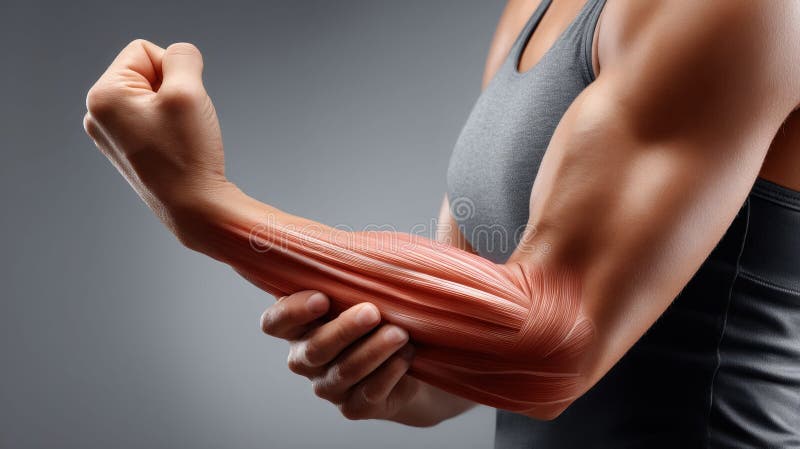
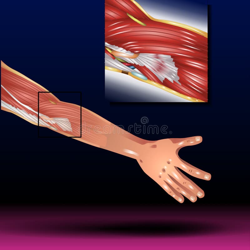
Free with trial Detailed illustration of biceps and triceps anatomy showcasing muscle structure, tendons, and muscle fibers. Useful for medical education, healthcare research, and biological studies. Ideal for. Biceps triceps movement arm illustrations Illustration of Biceps and Triceps Anatomy with Muscles in Red and White Background Featuring Tendons and Muscle Fibers. Detailed illustration of biceps and triceps anatomy showcasing muscle structure, tendons, and muscle fibers. Useful for medical education, healthcare research, and biological studies. Ideal for
Free with trial A detailed anatomical illustration of the human arm's muscular system, showcasing the biceps, triceps, and forearm muscles. This educational image is perfect for fitness, sports science, and biology. Ideal for understanding muscle function, strength training, and anatomical studies, explaining movement, vector design Generative AI. Biceps triceps movement arm vectors Detailed Human Arm Muscular System Anatomy, Bicep, Tricep, vector design Generative AI. A detailed anatomical illustration of the human arm's muscular system, showcasing the biceps, triceps, and forearm muscles. This educational image is perfect for fitness, sports science, and biology. Ideal for understanding muscle function, strength training, and anatomical studies, explaining movement, vector design Generative AI
Free with trial A 3D illustration image of contracted biceps muscle, demonstrating muscle contraction during arm flexion. Biceps triceps movement arm illustrations A 3D illustration image of contracted biceps muscle. A 3D illustration image of contracted biceps muscle, demonstrating muscle contraction during arm flexion.
Free with trial A 3D illustration image of contracted biceps muscle, demonstrating muscle contraction during arm flexion. Biceps triceps movement arm illustrations A 3D illustration image of contracted biceps muscle. A 3D illustration image of contracted biceps muscle, demonstrating muscle contraction during arm flexion.
Free with trial A 3D illustration image of contracted biceps muscle, demonstrating muscle contraction during arm flexion. Biceps triceps movement arm illustrations A 3D illustration image of contracted biceps muscle. A 3D illustration image of contracted biceps muscle, demonstrating muscle contraction during arm flexion.
Free with trial A black and white illustration of a flexed arm showcasing muscle definition, ideal for use in fitness and health-related marketing materials or educational resources. Biceps triceps movement arm vectors Flexed Arm Muscle Illustration. A black and white illustration of a flexed arm showcasing muscle definition, ideal for use in fitness and health-related. A black and white illustration of a flexed arm showcasing muscle definition, ideal for use in fitness and health-related marketing materials or educational resources
Free with trial Workers strength symbolized by a flexing muscular arm next to text. Power and solidarity for labor movements. Strong workforce concept. Biceps triceps movement arm illustrations Workers strength represented by muscular arm and text. Workers strength symbolized by a flexing muscular arm next to text. Power and solidarity for labor movements. Strong workforce concept
Free with trial A detailed, close-up view of a human arm, showcasing the intricate network of muscles and nerves. The internal structures are highlighted with vibrant, glowing blue, red, and orange light, creating a futuristic and energetic visual representation of the bodys power and complexity. Biceps triceps movement arm illustrations Human Arm Muscles and Nerves Illuminated with Glowing Energy. A detailed, close-up view of a human arm, showcasing the intricate network of muscles and nerves. The internal structures are highlighted with vibrant, glowing blue, red, and orange light, creating a futuristic and energetic visual representation of the bodys power and complexity
Free with trial Detailed 3D render of the human arm musculature, highlighting tendons in red and the body outline in blue against a dark background. Biceps triceps movement arm illustrations Anatomical Illustration of Human Arm Muscles with Glowing Red Tendons and Blue Outline. Detailed 3D render of the human arm musculature, highlighting tendons in red and the body outline in blue against a dark background
Free with trial Part of Tricep muscles anatomy. Medial, Long, and Lateral head of triceps. Flat vector illustration isolated on white background. Biceps triceps movement arm vectors Part of Tricep muscles anatomy. Medial, Long, and Lateral head of triceps
Free with trial Tricep muscles anatomy. Medial, Long, and Lateral head of triceps. Flat vector illustration isolated on white background. Biceps triceps movement arm illustrations Tricep muscles anatomy. Medial, Long, and Lateral head of triceps
Free with trial Black silhouette of a flexing arm, symbolizing strength, fitness and a healthy lifestyle. Biceps triceps movement arm vectors A strong muscular arm silhouette flexing represents power and physical fitness. Black silhouette of a flexing arm, symbolizing strength, fitness and a healthy lifestyle.
Free with trial A 3D illustration image of contracted biceps muscle, demonstrating muscle contraction during arm flexion. Biceps triceps movement arm illustrations A 3D illustration image of contracted biceps muscle. A 3D illustration image of contracted biceps muscle, demonstrating muscle contraction during arm flexion.
Free with trial Illustration of human arm muscle anatomy. Composite of muscle contraction close up view for medical textbook health care visuals or science catalog. Bodybuilding arm muscles. Biceps triceps movement arm illustrations Illustration of human arm muscle anatomy. Composite of muscle contraction close up view for medical textbook, health care visuals. Illustration of human arm muscle anatomy. Composite of muscle contraction close up view for medical textbook health care visuals or science catalog. Bodybuilding arm muscles.
Free with trial This vibrant illustration showcases a strong, muscular man executing a pull-up on a rustic wooden bar. His defined back and arm muscles highlight dedication to fitness and strength training. Biceps triceps movement arm illustrations Strong man demonstrating powerful back and arm muscles during a pull-up exercise routine. This vibrant illustration showcases a strong, muscular man executing a pull-up on a rustic wooden bar. His defined back and arm muscles highlight dedication to fitness and strength training
Free with trial A person presenting a diagram of arm muscles, encapsulated within a circular flow, suggesting a continuous learning or training process. Biceps triceps movement arm illustrations Muscle Growth Cycle Stick Figure Explains. A person presenting a diagram of arm muscles, encapsulated within a circular flow, suggesting a continuous learning or training process
Free with trial This is an illustration of a very muscular man performing a pull-up on a wooden bar, focusing on his strong back and arm muscles. It represents fitness and strength. Generative AI. Biceps triceps movement arm illustrations Strong muscular man doing pullups on a wooden bar demonstrating powerful back muscles and core. This is an illustration of a very muscular man performing a pull-up on a wooden bar, focusing on his strong back and arm muscles. It represents fitness and strength. Generative AI
Free with trial Strong male silhouette performs a challenging seated cable row exercise with focus and determination, strengthening back and arm muscles effectively as part of a comprehensive fitness regimen. This graphic illustration emphasizes proper form and technique for resistance training, showcasing a dedicated individual committed to their physical fitness journey, achieving peak performance and overall wellness in a gym setting or training studio through consistent powerful workouts to build an impressive physique and improve endurance and overall athletic capability. Biceps triceps movement arm vectors Male silhouette performing seated cable row exercise in gym. Strong male silhouette performs a challenging seated cable row exercise with focus and determination, strengthening back and arm muscles effectively as part of a comprehensive fitness regimen. This graphic illustration emphasizes proper form and technique for resistance training, showcasing a dedicated individual committed to their physical fitness journey, achieving peak performance and overall wellness in a gym setting or training studio through consistent powerful workouts to build an impressive physique and improve endurance and overall athletic capability.
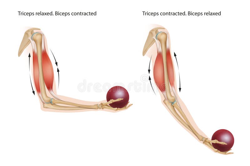
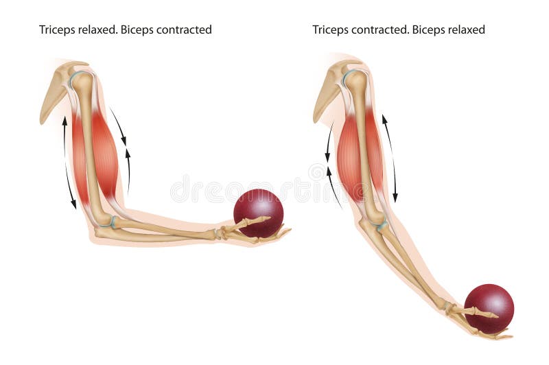
Free with trial An example of an anatomical and physical movement process where the biceps are contracted and the triceps are relaxed, the biceps is relaxed and the triceps are contracted. Biceps triceps movement arm illustrations Contracting and relaxing of biceps and triceps during flexion and extension. An example of an anatomical and physical movement process where the biceps are contracted and the triceps are relaxed, the biceps is relaxed and the triceps are contracted.
Free with trial Muscles transfer force to bones through tendons. They move our bones and associated body parts by pulling on them – this process is called muscle contraction. Biceps triceps movement arm illustrations An example of an anatomical and physical movement process where the biceps are contracted and the triceps are relaxed, the biceps. Muscles transfer force to bones through tendons. They move our bones and associated body parts by pulling on them – this process is called muscle contraction.
Free with trial Muscles transfer force to bones through tendons. They move our bones and associated body parts by pulling on them – this process is called muscle contraction. Biceps triceps movement arm illustrations An example of an anatomical and physical movement process where the biceps are contracted and the triceps are relaxed, the biceps. Muscles transfer force to bones through tendons. They move our bones and associated body parts by pulling on them – this process is called muscle contraction.
Free with trial Overhead Triceps extension Workout Stick Figure reference. Vector Image. Black and white character showing the movement representation. Biceps triceps movement arm illustrations Overhead Triceps extension Workout Stick Figure reference
Free with trial 3D Isometric Flat Vector Illustration of Anatomical Contracted And Relaxed Arm Muscular, Biceps and Triceps Motion Anatomy. Item 2. Biceps triceps movement arm vectors 3D Isometric Flat Vector Illustration of Anatomical Contracted And Relaxed Arm Muscular. Item 2. 3D Isometric Flat Vector Illustration of Anatomical Contracted And Relaxed Arm Muscular, Biceps and Triceps Motion Anatomy. Item 2
Free with trial Vektor ilustration gym icons , arm muscles. Biceps triceps movement arm vectors Arm muscle
Free with trial The femur, also known as the thighbone, is the largest and strongest bone in the human body. It is located in the upper leg, extending from the hip to the knee joint. The femur is surrounded by several muscles that attach to it, allowing for movement of the leg. The muscles that attach to the femur include:Quadriceps femoris muscle: This muscle group includes the rectus femoris, vastus lateralis, vastus medialis, and vastus intermedius muscles. These muscles originate from the pelvis and femur and attach to the patella and tibia via the patellar tendon. They are responsible for extending the knee joint and flexing the hip joint. Hamstring muscles: The hamstring muscle group includes the biceps femoris, semimembranosus, and semitendinosus muscles. These muscles originate from the ischial tuberosity of the pelvis and attach to the tibia and fibula bones of the lower leg. Gluteus maximus muscle: This muscle originates from the ilium bone of the pelvis and attaches to the femur bone. It is responsible for extending the hip joint and rotating the thigh laterally. Biceps triceps movement arm illustrations Male human skeleton femur muscle anatomy. 3d render. The femur, also known as the thighbone, is the largest and strongest bone in the human body. It is located in the upper leg, extending from the hip to the knee joint. The femur is surrounded by several muscles that attach to it, allowing for movement of the leg.The muscles that attach to the femur include:Quadriceps femoris muscle: This muscle group includes the rectus femoris, vastus lateralis, vastus medialis, and vastus intermedius muscles. These muscles originate from the pelvis and femur and attach to the patella and tibia via the patellar tendon. They are responsible for extending the knee joint and flexing the hip joint.Hamstring muscles: The hamstring muscle group includes the biceps femoris, semimembranosus, and semitendinosus muscles. These muscles originate from the ischial tuberosity of the pelvis and attach to the tibia and fibula bones of the lower leg. They are responsible for flexing the knee joint and extending the hip joint.Gluteus maximus muscle: This muscle originates from the ilium bone of the pelvis and attaches to the femur bone. It is responsible for extending the hip joint and rotating the thigh laterally.
Free with trial The femur, also known as the thighbone, is the largest and strongest bone in the human body. It is located in the upper leg, extending from the hip to the knee joint. The femur is surrounded by several muscles that attach to it, allowing for movement of the leg. The muscles that attach to the femur include:Quadriceps femoris muscle: This muscle group includes the rectus femoris, vastus lateralis, vastus medialis, and vastus intermedius muscles. These muscles originate from the pelvis and femur and attach to the patella and tibia via the patellar tendon. They are responsible for extending the knee joint and flexing the hip joint. Hamstring muscles: The hamstring muscle group includes the biceps femoris, semimembranosus, and semitendinosus muscles. These muscles originate from the ischial tuberosity of the pelvis and attach to the tibia and fibula bones of the lower leg. Gluteus maximus muscle: This muscle originates from the ilium bone of the pelvis and attaches to the femur bone. It is responsible for extending the hip joint and rotating the thigh laterally. Biceps triceps movement arm illustrations Male human skeleton femur muscle anatomy. 3d render. The femur, also known as the thighbone, is the largest and strongest bone in the human body. It is located in the upper leg, extending from the hip to the knee joint. The femur is surrounded by several muscles that attach to it, allowing for movement of the leg.The muscles that attach to the femur include:Quadriceps femoris muscle: This muscle group includes the rectus femoris, vastus lateralis, vastus medialis, and vastus intermedius muscles. These muscles originate from the pelvis and femur and attach to the patella and tibia via the patellar tendon. They are responsible for extending the knee joint and flexing the hip joint.Hamstring muscles: The hamstring muscle group includes the biceps femoris, semimembranosus, and semitendinosus muscles. These muscles originate from the ischial tuberosity of the pelvis and attach to the tibia and fibula bones of the lower leg. They are responsible for flexing the knee joint and extending the hip joint.Gluteus maximus muscle: This muscle originates from the ilium bone of the pelvis and attaches to the femur bone. It is responsible for extending the hip joint and rotating the thigh laterally.
Free with trial The femur, also known as the thighbone, is the largest and strongest bone in the human body. It is located in the upper leg, extending from the hip to the knee joint. The femur is surrounded by several muscles that attach to it, allowing for movement of the leg. The muscles that attach to the femur include:Quadriceps femoris muscle: This muscle group includes the rectus femoris, vastus lateralis, vastus medialis, and vastus intermedius muscles. These muscles originate from the pelvis and femur and attach to the patella and tibia via the patellar tendon. They are responsible for extending the knee joint and flexing the hip joint. Hamstring muscles: The hamstring muscle group includes the biceps femoris, semimembranosus, and semitendinosus muscles. These muscles originate from the ischial tuberosity of the pelvis and attach to the tibia and fibula bones of the lower leg. Gluteus maximus muscle: This muscle originates from the ilium bone of the pelvis and attaches to the femur bone. It is responsible for extending the hip joint and rotating the thigh laterally. Biceps triceps movement arm illustrations Male human skeleton femur muscle anatomy. 3d render. The femur, also known as the thighbone, is the largest and strongest bone in the human body. It is located in the upper leg, extending from the hip to the knee joint. The femur is surrounded by several muscles that attach to it, allowing for movement of the leg.The muscles that attach to the femur include:Quadriceps femoris muscle: This muscle group includes the rectus femoris, vastus lateralis, vastus medialis, and vastus intermedius muscles. These muscles originate from the pelvis and femur and attach to the patella and tibia via the patellar tendon. They are responsible for extending the knee joint and flexing the hip joint.Hamstring muscles: The hamstring muscle group includes the biceps femoris, semimembranosus, and semitendinosus muscles. These muscles originate from the ischial tuberosity of the pelvis and attach to the tibia and fibula bones of the lower leg. They are responsible for flexing the knee joint and extending the hip joint.Gluteus maximus muscle: This muscle originates from the ilium bone of the pelvis and attaches to the femur bone. It is responsible for extending the hip joint and rotating the thigh laterally.
Free with trial The femur, also known as the thighbone, is the largest and strongest bone in the human body. It is located in the upper leg, extending from the hip to the knee joint. The femur is surrounded by several muscles that attach to it, allowing for movement of the leg. The muscles that attach to the femur include:Quadriceps femoris muscle: This muscle group includes the rectus femoris, vastus lateralis, vastus medialis, and vastus intermedius muscles. These muscles originate from the pelvis and femur and attach to the patella and tibia via the patellar tendon. They are responsible for extending the knee joint and flexing the hip joint. Hamstring muscles: The hamstring muscle group includes the biceps femoris, semimembranosus, and semitendinosus muscles. These muscles originate from the ischial tuberosity of the pelvis and attach to the tibia and fibula bones of the lower leg. Gluteus maximus muscle: This muscle originates from the ilium bone of the pelvis and attaches to the femur bone. It is responsible for extending the hip joint and rotating the thigh laterally. Biceps triceps movement arm illustrations Male human skeleton femur muscle anatomy. 3d render. The femur, also known as the thighbone, is the largest and strongest bone in the human body. It is located in the upper leg, extending from the hip to the knee joint. The femur is surrounded by several muscles that attach to it, allowing for movement of the leg.The muscles that attach to the femur include:Quadriceps femoris muscle: This muscle group includes the rectus femoris, vastus lateralis, vastus medialis, and vastus intermedius muscles. These muscles originate from the pelvis and femur and attach to the patella and tibia via the patellar tendon. They are responsible for extending the knee joint and flexing the hip joint.Hamstring muscles: The hamstring muscle group includes the biceps femoris, semimembranosus, and semitendinosus muscles. These muscles originate from the ischial tuberosity of the pelvis and attach to the tibia and fibula bones of the lower leg. They are responsible for flexing the knee joint and extending the hip joint.Gluteus maximus muscle: This muscle originates from the ilium bone of the pelvis and attaches to the femur bone. It is responsible for extending the hip joint and rotating the thigh laterally.
Free with trial The femur, also known as the thighbone, is the largest and strongest bone in the human body. It is located in the upper leg, extending from the hip to the knee joint. The femur is surrounded by several muscles that attach to it, allowing for movement of the leg. The muscles that attach to the femur include:Quadriceps femoris muscle: This muscle group includes the rectus femoris, vastus lateralis, vastus medialis, and vastus intermedius muscles. These muscles originate from the pelvis and femur and attach to the patella and tibia via the patellar tendon. They are responsible for extending the knee joint and flexing the hip joint. Hamstring muscles: The hamstring muscle group includes the biceps femoris, semimembranosus, and semitendinosus muscles. These muscles originate from the ischial tuberosity of the pelvis and attach to the tibia and fibula bones of the lower leg. Gluteus maximus muscle: This muscle originates from the ilium bone of the pelvis and attaches to the femur bone. It is responsible for extending the hip joint and rotating the thigh laterally. Biceps triceps movement arm illustrations Male human skeleton femur muscle anatomy. 3d render. The femur, also known as the thighbone, is the largest and strongest bone in the human body. It is located in the upper leg, extending from the hip to the knee joint. The femur is surrounded by several muscles that attach to it, allowing for movement of the leg.The muscles that attach to the femur include:Quadriceps femoris muscle: This muscle group includes the rectus femoris, vastus lateralis, vastus medialis, and vastus intermedius muscles. These muscles originate from the pelvis and femur and attach to the patella and tibia via the patellar tendon. They are responsible for extending the knee joint and flexing the hip joint.Hamstring muscles: The hamstring muscle group includes the biceps femoris, semimembranosus, and semitendinosus muscles. These muscles originate from the ischial tuberosity of the pelvis and attach to the tibia and fibula bones of the lower leg. They are responsible for flexing the knee joint and extending the hip joint.Gluteus maximus muscle: This muscle originates from the ilium bone of the pelvis and attaches to the femur bone. It is responsible for extending the hip joint and rotating the thigh laterally.
Free with trial The femur, also known as the thighbone, is the largest and strongest bone in the human body. It is located in the upper leg, extending from the hip to the knee joint. The femur is surrounded by several muscles that attach to it, allowing for movement of the leg. The muscles that attach to the femur include:Quadriceps femoris muscle: This muscle group includes the rectus femoris, vastus lateralis, vastus medialis, and vastus intermedius muscles. These muscles originate from the pelvis and femur and attach to the patella and tibia via the patellar tendon. They are responsible for extending the knee joint and flexing the hip joint. Hamstring muscles: The hamstring muscle group includes the biceps femoris, semimembranosus, and semitendinosus muscles. These muscles originate from the ischial tuberosity of the pelvis and attach to the tibia and fibula bones of the lower leg. Gluteus maximus muscle: This muscle originates from the ilium bone of the pelvis and attaches to the femur bone. It is responsible for extending the hip joint and rotating the thigh laterally. Biceps triceps movement arm illustrations Male human skeleton femur muscle anatomy. 3d render. The femur, also known as the thighbone, is the largest and strongest bone in the human body. It is located in the upper leg, extending from the hip to the knee joint. The femur is surrounded by several muscles that attach to it, allowing for movement of the leg.The muscles that attach to the femur include:Quadriceps femoris muscle: This muscle group includes the rectus femoris, vastus lateralis, vastus medialis, and vastus intermedius muscles. These muscles originate from the pelvis and femur and attach to the patella and tibia via the patellar tendon. They are responsible for extending the knee joint and flexing the hip joint.Hamstring muscles: The hamstring muscle group includes the biceps femoris, semimembranosus, and semitendinosus muscles. These muscles originate from the ischial tuberosity of the pelvis and attach to the tibia and fibula bones of the lower leg. They are responsible for flexing the knee joint and extending the hip joint.Gluteus maximus muscle: This muscle originates from the ilium bone of the pelvis and attaches to the femur bone. It is responsible for extending the hip joint and rotating the thigh laterally.
Free with trial The femur, also known as the thighbone, is the largest and strongest bone in the human body. It is located in the upper leg, extending from the hip to the knee joint. The femur is surrounded by several muscles that attach to it, allowing for movement of the leg. The muscles that attach to the femur include:Quadriceps femoris muscle: This muscle group includes the rectus femoris, vastus lateralis, vastus medialis, and vastus intermedius muscles. These muscles originate from the pelvis and femur and attach to the patella and tibia via the patellar tendon. They are responsible for extending the knee joint and flexing the hip joint. Hamstring muscles: The hamstring muscle group includes the biceps femoris, semimembranosus, and semitendinosus muscles. These muscles originate from the ischial tuberosity of the pelvis and attach to the tibia and fibula bones of the lower leg. Gluteus maximus muscle: This muscle originates from the ilium bone of the pelvis and attaches to the femur bone. It is responsible for extending the hip joint and rotating the thigh laterally. Biceps triceps movement arm illustrations Male human skeleton femur muscle anatomy. 3d render. The femur, also known as the thighbone, is the largest and strongest bone in the human body. It is located in the upper leg, extending from the hip to the knee joint. The femur is surrounded by several muscles that attach to it, allowing for movement of the leg.The muscles that attach to the femur include:Quadriceps femoris muscle: This muscle group includes the rectus femoris, vastus lateralis, vastus medialis, and vastus intermedius muscles. These muscles originate from the pelvis and femur and attach to the patella and tibia via the patellar tendon. They are responsible for extending the knee joint and flexing the hip joint.Hamstring muscles: The hamstring muscle group includes the biceps femoris, semimembranosus, and semitendinosus muscles. These muscles originate from the ischial tuberosity of the pelvis and attach to the tibia and fibula bones of the lower leg. They are responsible for flexing the knee joint and extending the hip joint.Gluteus maximus muscle: This muscle originates from the ilium bone of the pelvis and attaches to the femur bone. It is responsible for extending the hip joint and rotating the thigh laterally.
Free with trial The femur, also known as the thighbone, is the largest and strongest bone in the human body. It is located in the upper leg, extending from the hip to the knee joint. The femur is surrounded by several muscles that attach to it, allowing for movement of the leg. The muscles that attach to the femur include:Quadriceps femoris muscle: This muscle group includes the rectus femoris, vastus lateralis, vastus medialis, and vastus intermedius muscles. These muscles originate from the pelvis and femur and attach to the patella and tibia via the patellar tendon. They are responsible for extending the knee joint and flexing the hip joint. Hamstring muscles: The hamstring muscle group includes the biceps femoris, semimembranosus, and semitendinosus muscles. These muscles originate from the ischial tuberosity of the pelvis and attach to the tibia and fibula bones of the lower leg. Gluteus maximus muscle: This muscle originates from the ilium bone of the pelvis and attaches to the femur bone. It is responsible for extending the hip joint and rotating the thigh laterally. Biceps triceps movement arm illustrations Male human skeleton femur muscle anatomy. 3d render. The femur, also known as the thighbone, is the largest and strongest bone in the human body. It is located in the upper leg, extending from the hip to the knee joint. The femur is surrounded by several muscles that attach to it, allowing for movement of the leg.The muscles that attach to the femur include:Quadriceps femoris muscle: This muscle group includes the rectus femoris, vastus lateralis, vastus medialis, and vastus intermedius muscles. These muscles originate from the pelvis and femur and attach to the patella and tibia via the patellar tendon. They are responsible for extending the knee joint and flexing the hip joint.Hamstring muscles: The hamstring muscle group includes the biceps femoris, semimembranosus, and semitendinosus muscles. These muscles originate from the ischial tuberosity of the pelvis and attach to the tibia and fibula bones of the lower leg. They are responsible for flexing the knee joint and extending the hip joint.Gluteus maximus muscle: This muscle originates from the ilium bone of the pelvis and attaches to the femur bone. It is responsible for extending the hip joint and rotating the thigh laterally.
Free with trial The femur, also known as the thighbone, is the largest and strongest bone in the human body. It is located in the upper leg, extending from the hip to the knee joint. The femur is surrounded by several muscles that attach to it, allowing for movement of the leg. The muscles that attach to the femur include:Quadriceps femoris muscle: This muscle group includes the rectus femoris, vastus lateralis, vastus medialis, and vastus intermedius muscles. These muscles originate from the pelvis and femur and attach to the patella and tibia via the patellar tendon. They are responsible for extending the knee joint and flexing the hip joint. Hamstring muscles: The hamstring muscle group includes the biceps femoris, semimembranosus, and semitendinosus muscles. These muscles originate from the ischial tuberosity of the pelvis and attach to the tibia and fibula bones of the lower leg. Gluteus maximus muscle: This muscle originates from the ilium bone of the pelvis and attaches to the femur bone. It is responsible for extending the hip joint and rotating the thigh laterally. Biceps triceps movement arm illustrations Male human skeleton femur muscle anatomy. 3d render. The femur, also known as the thighbone, is the largest and strongest bone in the human body. It is located in the upper leg, extending from the hip to the knee joint. The femur is surrounded by several muscles that attach to it, allowing for movement of the leg.The muscles that attach to the femur include:Quadriceps femoris muscle: This muscle group includes the rectus femoris, vastus lateralis, vastus medialis, and vastus intermedius muscles. These muscles originate from the pelvis and femur and attach to the patella and tibia via the patellar tendon. They are responsible for extending the knee joint and flexing the hip joint.Hamstring muscles: The hamstring muscle group includes the biceps femoris, semimembranosus, and semitendinosus muscles. These muscles originate from the ischial tuberosity of the pelvis and attach to the tibia and fibula bones of the lower leg. They are responsible for flexing the knee joint and extending the hip joint.Gluteus maximus muscle: This muscle originates from the ilium bone of the pelvis and attaches to the femur bone. It is responsible for extending the hip joint and rotating the thigh laterally.
Free with trial The femur, also known as the thighbone, is the largest and strongest bone in the human body. It is located in the upper leg, extending from the hip to the knee joint. The femur is surrounded by several muscles that attach to it, allowing for movement of the leg. The muscles that attach to the femur include:Quadriceps femoris muscle: This muscle group includes the rectus femoris, vastus lateralis, vastus medialis, and vastus intermedius muscles. These muscles originate from the pelvis and femur and attach to the patella and tibia via the patellar tendon. They are responsible for extending the knee joint and flexing the hip joint. Hamstring muscles: The hamstring muscle group includes the biceps femoris, semimembranosus, and semitendinosus muscles. These muscles originate from the ischial tuberosity of the pelvis and attach to the tibia and fibula bones of the lower leg. Gluteus maximus muscle: This muscle originates from the ilium bone of the pelvis and attaches to the femur bone. It is responsible for extending the hip joint and rotating the thigh laterally. Biceps triceps movement arm illustrations Male human skeleton femur muscle anatomy. 3d render. The femur, also known as the thighbone, is the largest and strongest bone in the human body. It is located in the upper leg, extending from the hip to the knee joint. The femur is surrounded by several muscles that attach to it, allowing for movement of the leg.The muscles that attach to the femur include:Quadriceps femoris muscle: This muscle group includes the rectus femoris, vastus lateralis, vastus medialis, and vastus intermedius muscles. These muscles originate from the pelvis and femur and attach to the patella and tibia via the patellar tendon. They are responsible for extending the knee joint and flexing the hip joint.Hamstring muscles: The hamstring muscle group includes the biceps femoris, semimembranosus, and semitendinosus muscles. These muscles originate from the ischial tuberosity of the pelvis and attach to the tibia and fibula bones of the lower leg. They are responsible for flexing the knee joint and extending the hip joint.Gluteus maximus muscle: This muscle originates from the ilium bone of the pelvis and attaches to the femur bone. It is responsible for extending the hip joint and rotating the thigh laterally.
Free with trial The femur, also known as the thighbone, is the largest and strongest bone in the human body. It is located in the upper leg, extending from the hip to the knee joint. The femur is surrounded by several muscles that attach to it, allowing for movement of the leg. The muscles that attach to the femur include:Quadriceps femoris muscle: This muscle group includes the rectus femoris, vastus lateralis, vastus medialis, and vastus intermedius muscles. These muscles originate from the pelvis and femur and attach to the patella and tibia via the patellar tendon. They are responsible for extending the knee joint and flexing the hip joint. Hamstring muscles: The hamstring muscle group includes the biceps femoris, semimembranosus, and semitendinosus muscles. These muscles originate from the ischial tuberosity of the pelvis and attach to the tibia and fibula bones of the lower leg. Gluteus maximus muscle: This muscle originates from the ilium bone of the pelvis and attaches to the femur bone. It is responsible for extending the hip joint and rotating the thigh laterally. Biceps triceps movement arm illustrations Male human skeleton femur muscle anatomy. 3d render. The femur, also known as the thighbone, is the largest and strongest bone in the human body. It is located in the upper leg, extending from the hip to the knee joint. The femur is surrounded by several muscles that attach to it, allowing for movement of the leg.The muscles that attach to the femur include:Quadriceps femoris muscle: This muscle group includes the rectus femoris, vastus lateralis, vastus medialis, and vastus intermedius muscles. These muscles originate from the pelvis and femur and attach to the patella and tibia via the patellar tendon. They are responsible for extending the knee joint and flexing the hip joint.Hamstring muscles: The hamstring muscle group includes the biceps femoris, semimembranosus, and semitendinosus muscles. These muscles originate from the ischial tuberosity of the pelvis and attach to the tibia and fibula bones of the lower leg. They are responsible for flexing the knee joint and extending the hip joint.Gluteus maximus muscle: This muscle originates from the ilium bone of the pelvis and attaches to the femur bone. It is responsible for extending the hip joint and rotating the thigh laterally.
Free with trial The femur, also known as the thighbone, is the largest and strongest bone in the human body. It is located in the upper leg, extending from the hip to the knee joint. The femur is surrounded by several muscles that attach to it, allowing for movement of the leg. The muscles that attach to the femur include:Quadriceps femoris muscle: This muscle group includes the rectus femoris, vastus lateralis, vastus medialis, and vastus intermedius muscles. These muscles originate from the pelvis and femur and attach to the patella and tibia via the patellar tendon. They are responsible for extending the knee joint and flexing the hip joint. Hamstring muscles: The hamstring muscle group includes the biceps femoris, semimembranosus, and semitendinosus muscles. These muscles originate from the ischial tuberosity of the pelvis and attach to the tibia and fibula bones of the lower leg. Gluteus maximus muscle: This muscle originates from the ilium bone of the pelvis and attaches to the femur bone. It is responsible for extending the hip joint and rotating the thigh laterally. Biceps triceps movement arm illustrations Male human skeleton femur muscle anatomy. 3d render. The femur, also known as the thighbone, is the largest and strongest bone in the human body. It is located in the upper leg, extending from the hip to the knee joint. The femur is surrounded by several muscles that attach to it, allowing for movement of the leg.The muscles that attach to the femur include:Quadriceps femoris muscle: This muscle group includes the rectus femoris, vastus lateralis, vastus medialis, and vastus intermedius muscles. These muscles originate from the pelvis and femur and attach to the patella and tibia via the patellar tendon. They are responsible for extending the knee joint and flexing the hip joint.Hamstring muscles: The hamstring muscle group includes the biceps femoris, semimembranosus, and semitendinosus muscles. These muscles originate from the ischial tuberosity of the pelvis and attach to the tibia and fibula bones of the lower leg. They are responsible for flexing the knee joint and extending the hip joint.Gluteus maximus muscle: This muscle originates from the ilium bone of the pelvis and attaches to the femur bone. It is responsible for extending the hip joint and rotating the thigh laterally.
Free with trial Lateral raises Dumbell Workout Stick Figure reference. Vector Image. Black and white character showing the movement representation. Biceps triceps movement arm illustrations Lateral raises DBWorkout Stick Figure reference. Lateral raises Dumbell Workout Stick Figure reference. Vector Image. Black and white character showing the movement representation.
Free with trial Front Raises Dumbell Workout Stick Figure reference. Vector Image. Black and white character showing the movement representation. Biceps triceps movement arm illustrations Front Raises DB Workout Stick Figure reference. Front Raises Dumbell Workout Stick Figure reference. Vector Image. Black and white character showing the movement representation.
Free with trial Body Squat Workout Stick Figure reference. Vector Image. Black and white character showing the movement representation. Biceps triceps movement arm illustrations Body Squats Workout Stick Figure reference. Body Squat Workout Stick Figure reference. Vector Image. Black and white character showing the movement representation.
Free with trial Fla source file available - Role of calcium for human. Man suffering from pain in elbow, digital compositing with illustration of arm bone. Biceps triceps movement arm illustrations Elbow Tendonitis, Elbow Dislocation, Elbow Bone Illustration, Pain, Forearm, Cramp, Illustration, Bone, Ache. Fla source file available - Role of calcium for human. Man suffering from pain in elbow, digital compositing with illustration of arm bone
Free with trial Human muscle contraction structure diagram hand drawn schematic raster illustration. Medical science educational illustration. Biceps triceps movement arm illustrations Muscle contraction diagram medical science. Human muscle contraction structure diagram hand drawn schematic raster illustration. Medical science educational illustration
Free with trial Structure of tendon. Infographics. Vector illustration on isolated background. Biceps triceps movement arm vectors Structure of tendon. Infographics. Vector illustration on isolated background
Free with trial Vector illustration of muscle and cells sign. Collection of muscle and anatomy stock symbol for web. Biceps triceps movement arm vectors Vector design of muscle and cells symbol. Set of muscle and anatomy vector icon for stock. Vector illustration of muscle and cells sign. Collection of muscle and anatomy stock symbol for web.
Free with trial Vector design of muscle and cells logo. Set of muscle and anatomy stock symbol for web. Biceps triceps movement arm vectors Isolated object of muscle and cells sign. Collection of muscle and anatomy vector icon for stock. Vector design of muscle and cells logo. Set of muscle and anatomy stock symbol for web.
Free with trial Vector illustration of muscle and cells symbol. Set of muscle and anatomy stock vector illustration. Biceps triceps movement arm vectors Vector design of muscle and cells icon. Collection of muscle and anatomy stock symbol for web. Vector illustration of muscle and cells symbol. Set of muscle and anatomy stock vector illustration.
Free with trial Isolated object of muscle and cells logo. Set of muscle and anatomy stock symbol for web. Biceps triceps movement arm vectors Vector illustration of muscle and cells sign. Collection of muscle and anatomy vector icon for stock. Isolated object of muscle and cells logo. Set of muscle and anatomy stock symbol for web.
Free with trial Human exercising with dumbbells icon, Weightlifting workout pictogram, Vector illustration. Biceps triceps movement arm vectors Human exercising with dumbbells icon, Weightlifting workout pictogram, Vector illustration.
Free with trial Isolated object of muscle and cells symbol. Set of muscle and anatomy stock vector illustration. Biceps triceps movement arm vectors Vector illustration of muscle and cells icon. Collection of muscle and anatomy stock symbol for web. Isolated object of muscle and cells symbol. Set of muscle and anatomy stock vector illustration.
Free with trial Vector illustration of fiber and muscular logo. Set of fiber and body stock symbol for web. Biceps triceps movement arm vectors Vector design of fiber and muscular sign. Collection of fiber and body vector icon for stock. Vector illustration of fiber and muscular logo. Set of fiber and body stock symbol for web.
Free with trial Vector illustration of human and body logo. Collection of human and cells stock symbol for web. Biceps triceps movement arm vectors Vector design of human and body sign. Set of human and cells vector icon for stock. Vector illustration of human and body logo. Collection of human and cells stock symbol for web.
Free with trial Vector design of human and body logo. Set of human and cells vector icon for stock. Biceps triceps movement arm vectors Isolated object of human and body sign. Collection of human and cells stock vector illustration. Vector design of human and body logo. Set of human and cells vector icon for stock.
Free with trial Vector illustration of muscle and cells logo. Collection of muscle and anatomy vector icon for stock. Biceps triceps movement arm vectors Vector design of muscle and cells sign. Set of muscle and anatomy stock vector illustration. Vector illustration of muscle and cells logo. Collection of muscle and anatomy vector icon for stock.
Free with trial Isolated object of human and body logo. Collection of human and cells vector icon for stock. Biceps triceps movement arm vectors Vector illustration of human and body sign. Set of human and cells stock vector illustration. Isolated object of human and body logo. Collection of human and cells vector icon for stock.
Free with trial Isolated object of fiber and muscular symbol. Collection of fiber and body vector icon for stock. Biceps triceps movement arm vectors Vector illustration of fiber and muscular icon. Set of fiber and body stock vector illustration. Isolated object of fiber and muscular symbol. Collection of fiber and body vector icon for stock.
Free with trial Isolated object of fiber and muscular icon. Collection of fiber and body vector icon for stock. Biceps triceps movement arm vectors Vector illustration of fiber and muscular logo. Set of fiber and body stock vector illustration. Isolated object of fiber and muscular icon. Collection of fiber and body vector icon for stock.
Free with trial Isolated object of muscle and cells logo. Collection of muscle and anatomy vector icon for stock. Biceps triceps movement arm vectors Vector illustration of muscle and cells sign. Set of muscle and anatomy stock vector illustration. Isolated object of muscle and cells logo. Collection of muscle and anatomy vector icon for stock.
Free with trial Vector illustration of muscle and cells symbol. Set of muscle and anatomy vector icon for stock. Biceps triceps movement arm vectors Vector design of muscle and cells icon. Collection of muscle and anatomy stock vector illustration. Vector illustration of muscle and cells symbol. Set of muscle and anatomy vector icon for stock.
Free with trial Vector illustration of fiber and muscular logo. Collection of fiber and body stock symbol for web. Biceps triceps movement arm vectors Vector design of fiber and muscular sign. Set of fiber and body vector icon for stock. Vector illustration of fiber and muscular logo. Collection of fiber and body stock symbol for web.
Free with trial Vector illustration of fiber and muscular logo. Collection of fiber and body stock symbol for web. Biceps triceps movement arm vectors Vector design of fiber and muscular sign. Set of fiber and body vector icon for stock. Vector illustration of fiber and muscular logo. Collection of fiber and body stock symbol for web.
Free with trial Vector illustration of fiber and muscular sign. Collection of fiber and body stock symbol for web. Biceps triceps movement arm vectors Vector design of fiber and muscular symbol. Set of fiber and body vector icon for stock. Vector illustration of fiber and muscular sign. Collection of fiber and body stock symbol for web.
Free with trial Vector design of fiber and muscular sign. Collection of fiber and body stock vector illustration. Biceps triceps movement arm vectors Isolated object of fiber and muscular symbol. Set of fiber and body stock symbol for web. Vector design of fiber and muscular sign. Collection of fiber and body stock vector illustration.