Free with trial Biotech/medicine related graph map in light blue. Conceptual. Biotech illustration illustrations Biotech/Medicine Graph Map. Biotech/medicine related graph map in light blue. Conceptual.
Free with trial Polygonal DNA concept. Vector mesh spheres from flying debris. Thin line concept. Blue structure style illustration. Biotech illustration vectors Abstract DNA concept. Polygonal DNA concept. Vector mesh spheres from flying debris. Thin line concept. Blue structure style illustration
Free with trial Biotechnology researcher concept or biotech science, close up of male biologist doing experiment. Biotech illustration illustrations Biotechnology scientist concept. Biotechnology researcher concept or biotech science, close up of male biologist doing experiment.
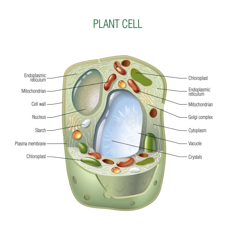
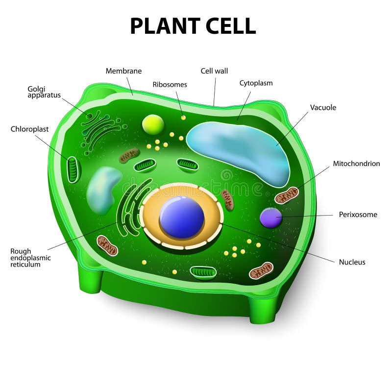
Free with trial Plant cell cut-away - scientifically correct vector illustration for best prints and other uses. Biotech illustration vectors Plant cell
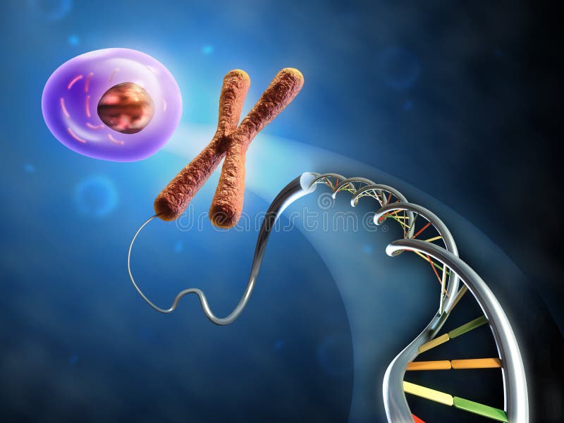
Free with trial Illustration showing the formation of an animal cell from dna and chromosomes. Digital illustration. Biotech illustration illustrations From Dna to cell. Illustration showing the formation of an animal cell from dna and chromosomes. Digital illustration.
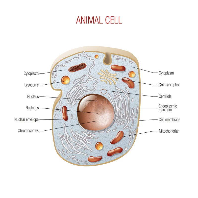
Free with trial Animal cell cut-away - scientifically correct vector illustration for best prints and other uses. Biotech illustration vectors Animal cell
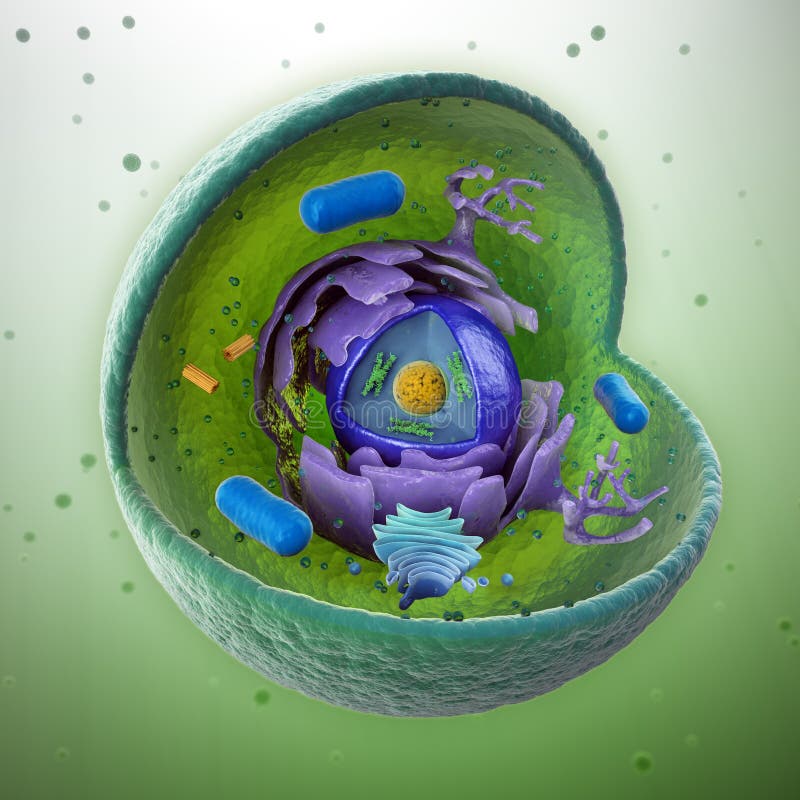
Free with trial Animal cell cut-away - scientifically correct 3d illustration. Biotech illustration illustrations Animal cell cut-away
Free with trial Background suitable for medical and research subjects. Digital illustration. Biotech illustration illustrations Medical Research background. Background suitable for medical and research subjects. Digital illustration.
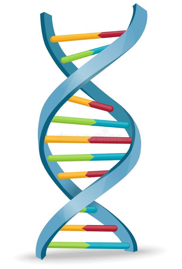
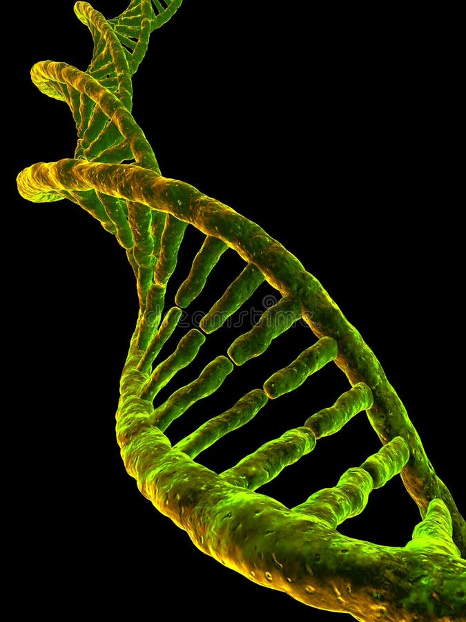
Free with trial Conceptual DNA strands - genetics research concept illustration. Biotech illustration illustrations DNA strands
Free with trial Organic chemistry: model of the DNA molecule - illustration of a biological particle. Biotech illustration illustrations DNA molecule
Free with trial Origins of life: from simple molecules to dna. An human being materialize from dna and holds the Earth between its hands. Digital illustration. Biotech illustration illustrations Life creation. Origins of life: from simple molecules to dna. An human being materialize from dna and holds the Earth between its hands. Digital illustration.
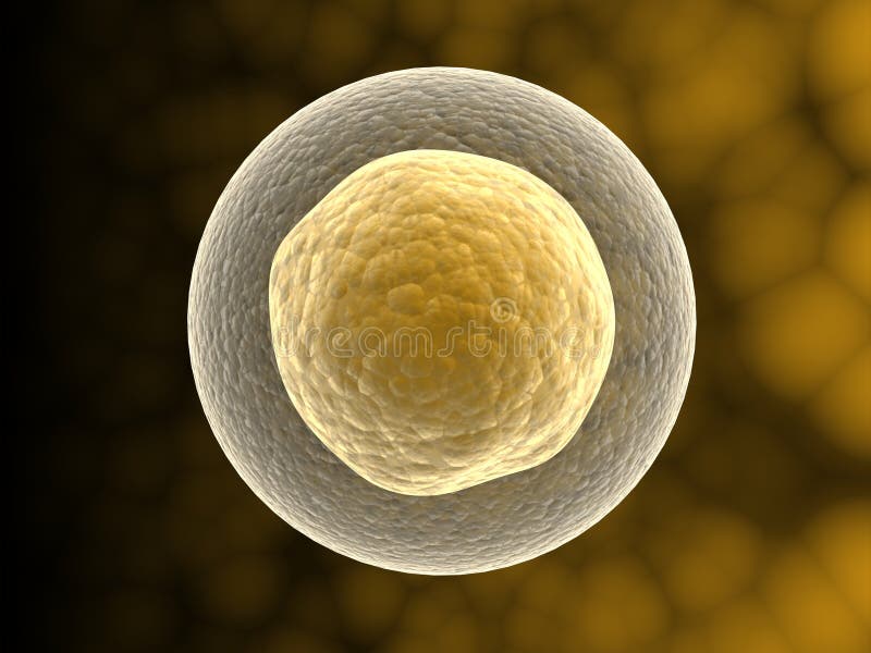
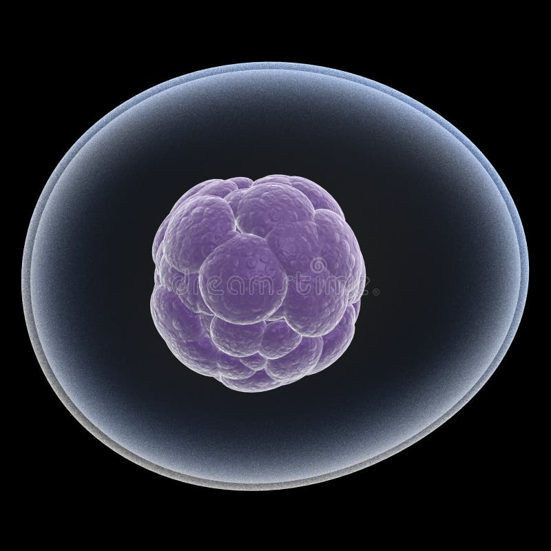
Free with trial Digital illustration of CELL WITH NUCLEUS in 3d on digital background. Biotech illustration illustrations CELL WITH NUCLEUS
Free with trial DNA Medical Science and Biotech Chemistry Genes. Biotech illustration illustrations DNA Medical Science
Free with trial DNA sequencing concept CG illustration. Biotech illustration illustrations DNA sequencing concept
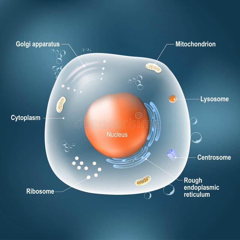
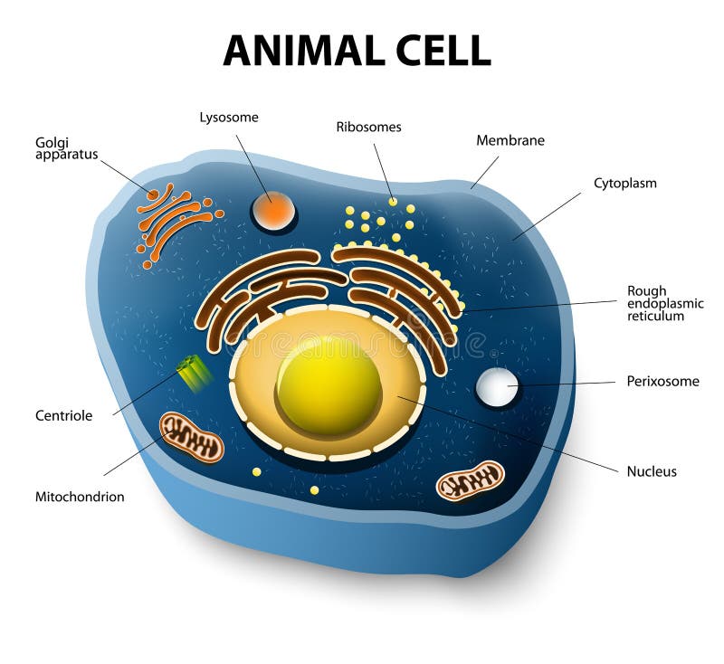
Free with trial Anatomy of cell. All organelles: Nucleus, Ribosome, Rough endoplasmic reticulum, Golgi apparatus, mitochondrion, cytoplasm, lysosome, Centrosome. Animal cell on the dark background. Illustration easy editable for Your color. Biotech illustration vectors Anatomy of animal cell. Anatomy of cell. All organelles: Nucleus, Ribosome, Rough endoplasmic reticulum, Golgi apparatus, mitochondrion, cytoplasm, lysosome, Centrosome. Animal cell on the dark background. Illustration easy editable for Your color
Free with trial Science Biotechnology DNA concept. 3D rendering illustration with glowing lights. Science and technology. Biotech illustration illustrations Science Biotechnology DNA concept. 3D rendering illustration with glowing lights. Science and technology.
Free with trial Cross section diagram of Prokaryotic and Eukaryotic cells. Biotech illustration vectors Cells
Free with trial Plant cell structure. Vector diagram. Biotech illustration vectors Plant cell anatomy. Plant cell structure. Vector diagram
Free with trial A logo that can be used for company branding. Biotech illustration vectors Ecoworld logo. A logo that can be used for company branding
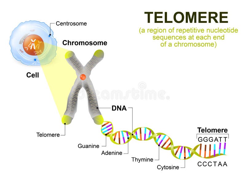
Free with trial A telomere is a repeating sequence of double-stranded DNA located at the ends of chromosomes. Each time a cell divides, the telomeres become shorter. Eventually, the telomeres become so short that the cell can no longer divide. Biotech illustration vectors Human cell, chromosome and telomere. A telomere is a repeating sequence of double-stranded DNA located at the ends of chromosomes. Each time a cell divides, the telomeres become shorter. Eventually, the telomeres become so short that the cell can no longer divide.
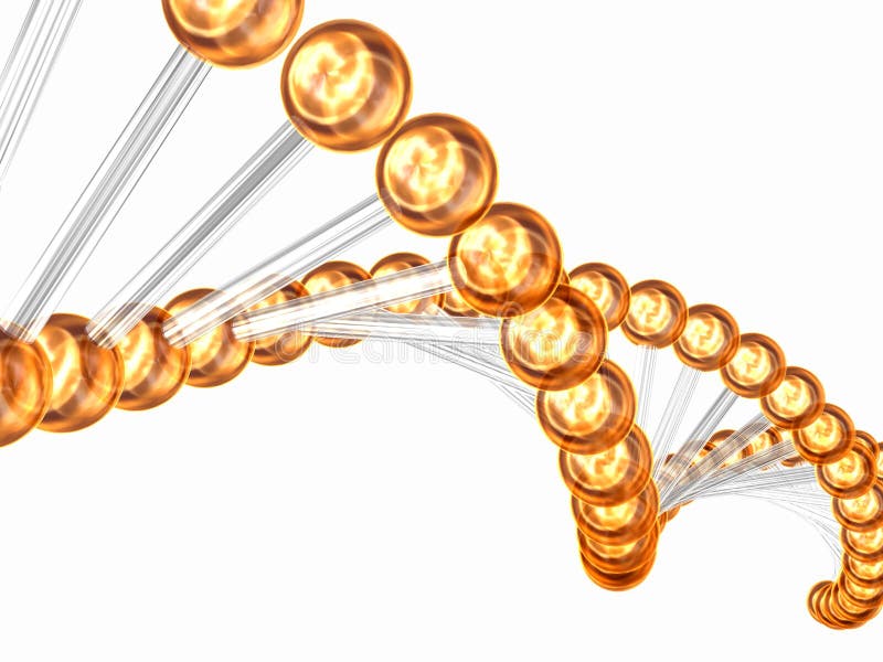
Free with trial 3d rendered illustration of a double helix. Biotech illustration illustrations 3d gene. 3d rendered illustration of a double helix
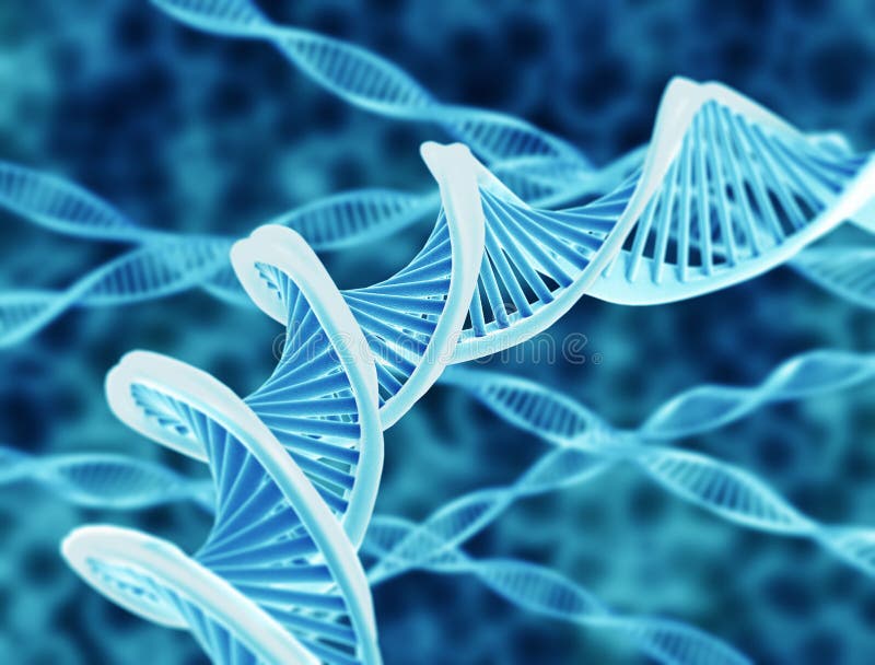
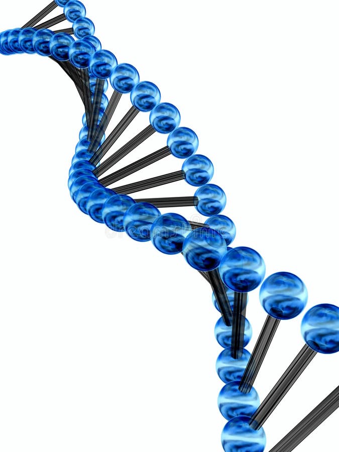
Free with trial DNA spiral with a blue background for genetic research or something else. Biotech illustration vectors DNA
Free with trial 3d rendered illustration of a cell injection. Biotech illustration illustrations Cell injection
Free with trial 3d rendered illustration of a cell injection. Biotech illustration illustrations Cell injection
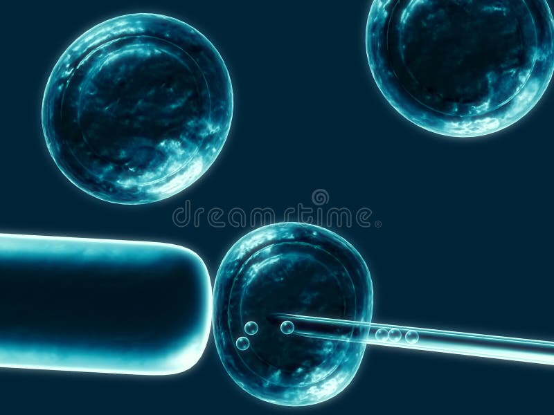
Free with trial 3d rendered illustration of cells and a pipette. Biotech illustration illustrations Gene manipulation. 3d rendered illustration of cells and a pipette
Free with trial DNA plant concept, can refer to alternative medicine, crop gene modification. Biotech illustration vectors DNA plant concept
Free with trial Human or animal cell. cross section. structure of a Eukaryotic cell. Vector diagram. Biotech illustration vectors Animal cell cut-away. Human or animal cell. cross section. structure of a Eukaryotic cell. Vector diagram
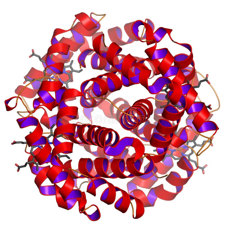
Free with trial Hemoglobin or haemoglobin (frequently abbreviated as Hb) is what makes our blood colored red. It transports oxygen from the lungs to the cells and CO2 back from the cells to the lung. Here, the globular shape of the protein (this is why it is called hemoGLOBIN) is visible. The heme groups (containing iron that can bind oxygen or CO2) are shown as ball-and-stick model. Rendered in great detail on white background. Fog simulates depth. Modifications such as other colorings, less fog, or perspecive views are available on request. Biotech illustration illustrations Globular protein. Hemoglobin or haemoglobin (frequently abbreviated as Hb) is what makes our blood colored red. It transports oxygen from the lungs to the cells and CO2 back from the cells to the lung. Here, the globular shape of the protein (this is why it is called hemoGLOBIN) is visible. The heme groups (containing iron that can bind oxygen or CO2) are shown as ball-and-stick model. Rendered in great detail on white background. Fog simulates depth. Modifications such as other colorings, less fog, or perspecive views are available on request.
Free with trial DNA strands on abstract medical background. Biotech illustration illustrations DNA strands
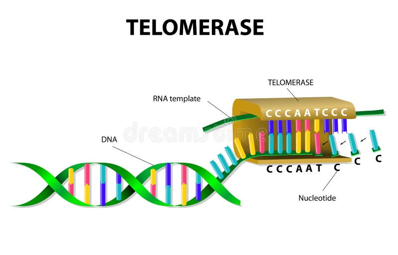
Free with trial Telomerase is an enzyme that lengthens telomeres by adding on repeating sequences of DNA. Telomerase binds to the ends of the telomere via an RNA template that is used for the attachment of a new strand of DNA. Biotech illustration vectors Telomerase elongates telomere. Telomerase is an enzyme that lengthens telomeres by adding on repeating sequences of DNA. Telomerase binds to the ends of the telomere via an RNA template that is used for the attachment of a new strand of DNA.
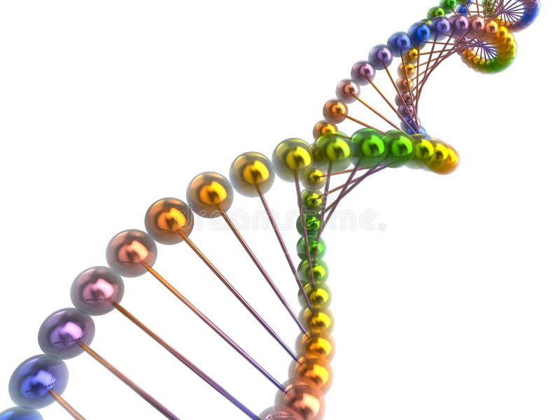
Free with trial DNA strand modern design, bright and colorful. Biotech illustration illustrations DNA strand bright and colorful. DNA strand modern design, bright and colorful
Free with trial Enzyme that produces fluorine compounds (Fluorinase). The protein consists of six identical chains, here colored from blue (N-terminus) to red (C-terminus), rainbow coloring. It is shown in cartoon representation: alpha-helices are represented by spirals, beta-sheets by arrows. The ball-and-stick molecules are the active sites. Detailed rendering. Biotech illustration illustrations Protein on white background. Enzyme that produces fluorine compounds (Fluorinase). The protein consists of six identical chains, here colored from blue (N-terminus) to red (C-terminus), rainbow coloring. It is shown in cartoon representation: alpha-helices are represented by spirals, beta-sheets by arrows. The ball-and-stick molecules are the active sites. Detailed rendering.
Free with trial Dna genetic code 3d concept abstract. Biotech illustration illustrations Dna code. Dna genetic code 3d concept abstract
Free with trial 3d rendered illustration of a double helix. Biotech illustration illustrations Dna model. 3d rendered illustration of a double helix
Free with trial DNA structure model on white surface. Biotech illustration illustrations DNA strand. DNA structure model on white surface
Free with trial DNA strand isolated on white background. Biotech illustration illustrations DNA strand
Free with trial Dna genetic code 3d concept abstract spiral. Biotech illustration illustrations Dna spiral. Dna genetic code 3d concept abstract spiral
Free with trial 3d rendered illustration of sperm. Biotech illustration illustrations Human sperm. 3d rendered illustration of sperm
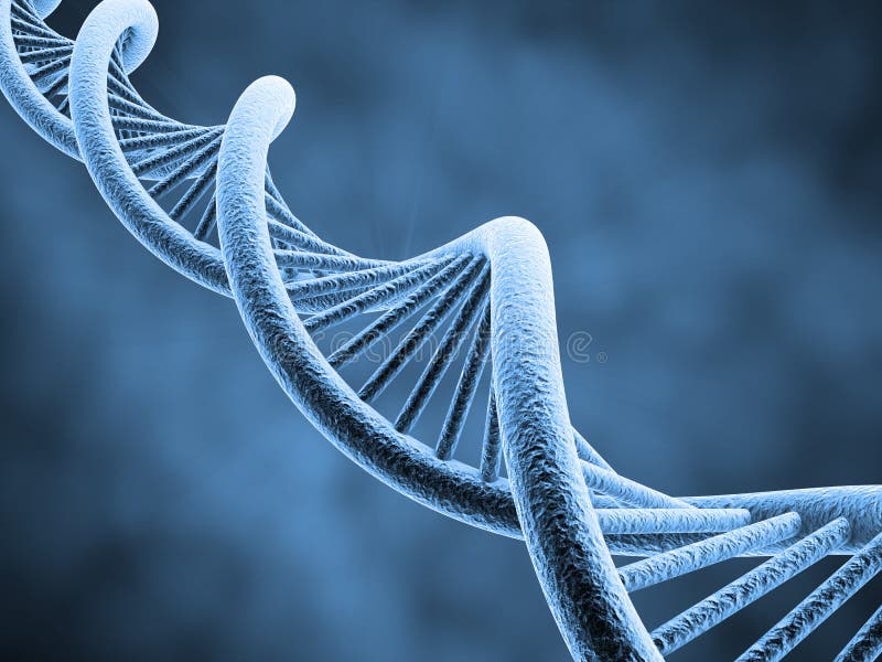
Free with trial High resolution render of DNA double helix. Biotech illustration illustrations DNA strings. High resolution render of DNA double helix
Free with trial 3d stem cell high resolution 3d render. Biotech illustration illustrations 3d stem cell
Free with trial 3D render of DNA strands on abstract background. Biotech illustration illustrations DNA abstract. 3D render of DNA strands on abstract background
Free with trial Abstract blur background with DNA strands. Biotech illustration illustrations DNA abstract. Abstract blur background with DNA strands
Free with trial 3D render of DNA strands on abstract background. Biotech illustration illustrations Abstract DNA. 3D render of DNA strands on abstract background
Free with trial 3D render of DNA strands on abstract background. Biotech illustration illustrations DNA abstract. 3D render of DNA strands on abstract background
Free with trial Research, Bio Technology and Science, Chemical laboratory infographic. Biotech illustration vectors Research, Bio Technology and Science infographic. Research, Bio Technology and Science, Chemical laboratory infographic
Free with trial Research, Bio Technology and Science, Chemical laboratory infographic. Biotech illustration vectors Research, Bio Technology and Science infographic. Research, Bio Technology and Science, Chemical laboratory infographic
Free with trial 3d model of a genetic code incorporated by the nature. Biotech illustration illustrations 3d genetic code. 3d model of a genetic code incorporated by the nature
Free with trial 3d model of a genetic code incorporated by the nature. Biotech illustration illustrations 3d genetic code. 3d model of a genetic code incorporated by the nature
Free with trial 3d rendered illustration of a cell manipulation. Biotech illustration illustrations Cell manipulation
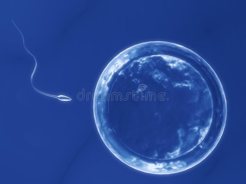
Free with trial 3d rendered illustration of a human egg and sperm. Biotech illustration illustrations Human egg with sperm. 3d rendered illustration of a human egg and sperm
Free with trial 3d rendered illustration of a human egg and sperm. Biotech illustration illustrations Human egg with sperm. 3d rendered illustration of a human egg and sperm
Free with trial DNA inside a pill - gene therapy concept. Biotech illustration illustrations Gene therapy concept
Free with trial DNA model isolated on white background. Biotech illustration illustrations DNA isolated on white. DNA model isolated on white background
Free with trial 3d artificial insemination 3d illustration. Biotech illustration illustrations Artificial insemination
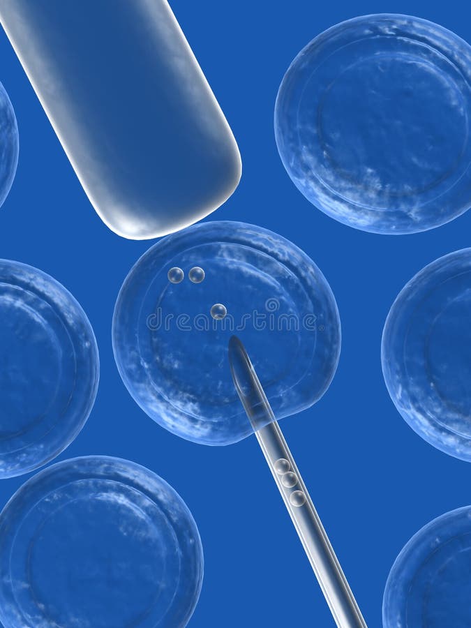
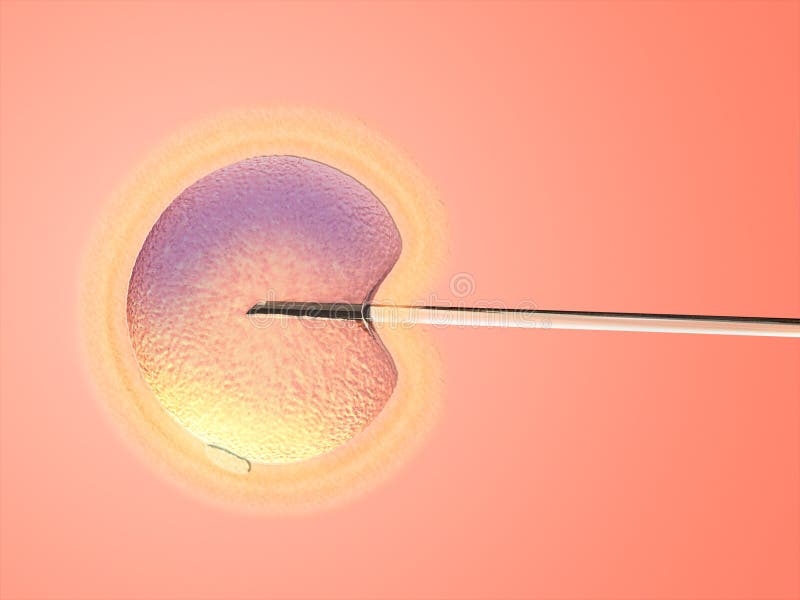
Free with trial Artificial insemination. Glass needle and female ovule on warm background. Digital illustration. Biotech illustration illustrations Artificial insemination
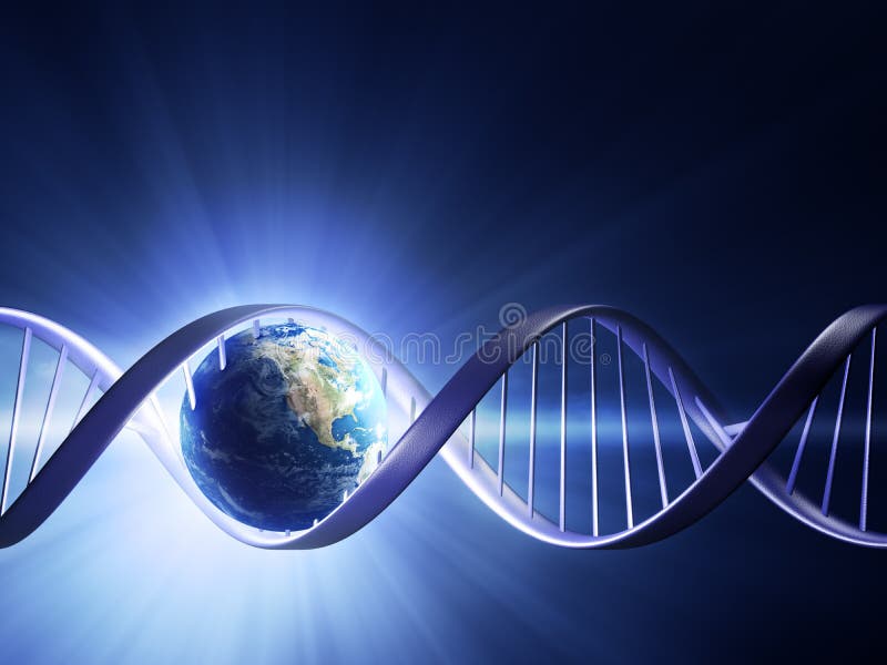
Free with trial Abstract render of earth inside a glowing DNA strand ( earth uv map from ). Biotech illustration illustrations Glowing earth DNA strand. Abstract render of earth inside a glowing DNA strand ( earth uv map from )
Free with trial DNA strand modern design, double helix. Biotech illustration illustrations DNA strand modern design
Free with trial Abstract render of earth inside a glowing DNA strand ( earth uv map from ). Biotech illustration illustrations Glowing earth DNA strand. Abstract render of earth inside a glowing DNA strand ( earth uv map from )
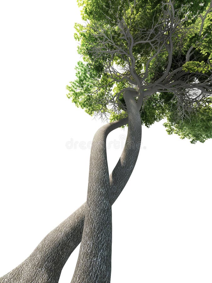
Free with trial Dna-modified tree. Transgenic concept. Biotech illustration illustrations Dna modified tree genetic. Dna-modified tree. Transgenic concept
Free with trial Graphic representative of medical technology and biotechnology industry. Biotech illustration illustrations Medical Technology Graphic. Graphic representative of medical technology and biotechnology industry.
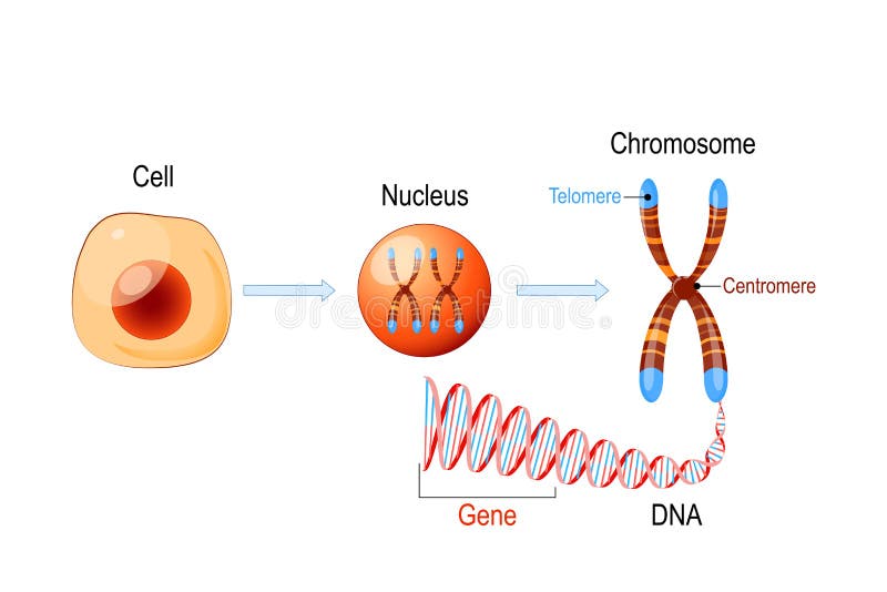
Free with trial Cell Structure. Nucleus with chromosomes, DNA molecule double helix, telomere and gene length of DNA that codes for a specific protein. Genome research. Biotech illustration vectors Cell Structure. Nucleus with chromosomes, DNA molecule, telomere and gene. Cell Structure. Nucleus with chromosomes, DNA molecule double helix, telomere and gene length of DNA that codes for a specific protein. Genome research
Free with trial Natural Alternative Herbal Medicine and Healthcare icon and element. Biotech illustration vectors Natural Alternative Herbal Medicine icon. Natural Alternative Herbal Medicine and Healthcare icon and element
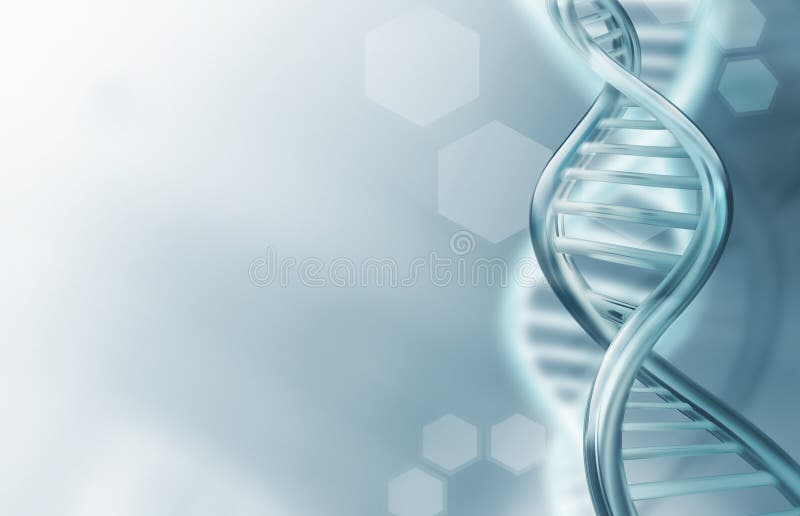
Free with trial Abstract science background with DNA strands. Biotech illustration illustrations DNA strands background. Abstract science background with DNA strands
Free with trial Natural Alternative Herbal Medicine and Healthcare icons and element set. Biotech illustration vectors Natural Alternative Herbal Medicine icons. Natural Alternative Herbal Medicine and Healthcare icons and element set
Free with trial Natural Alternative Herbal Medicine and Healthcare icons and element set. Biotech illustration vectors Natural Alternative Herbal Medicine icons. Natural Alternative Herbal Medicine and Healthcare icons and element set
Free with trial DNA, genetic symbol - abstract people icon. Biotech illustration vectors DNA, genetic symbol - people, man and woman icon. DNA, genetic symbol - abstract people icon
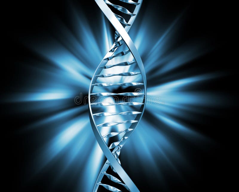
Free with trial 3D rendered double Helix / DNA. Biotech illustration illustrations DNA 1. 3D rendered double Helix / DNA.
Free with trial A Needle penetrating a stem cell and injecting manipulated DNA code. Biotech illustration illustrations Cloning - injecting DNA 1. A Needle penetrating a stem cell and injecting manipulated DNA code.
Free with trial 3D rendered double Helix / DNA. Biotech illustration illustrations Stylized DNA double Helix. 3D rendered double Helix / DNA.
Free with trial A Needle penetrating a stem cell and injecting manipulated DNA code. Biotech illustration illustrations Cloning - injecting DNA 2. A Needle penetrating a stem cell and injecting manipulated DNA code.
Free with trial Business icons and symbols of various industries / business sectors like consulting,tourism,hospitality,agriculture,renewable energy,real estate,consumer services,construction,financial services. Biotech illustration vectors Business icons and symbols of various industries / business sectors like consulting,tourism,hospitality,agriculture