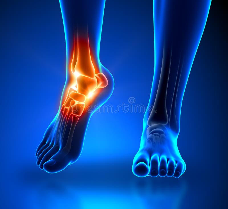
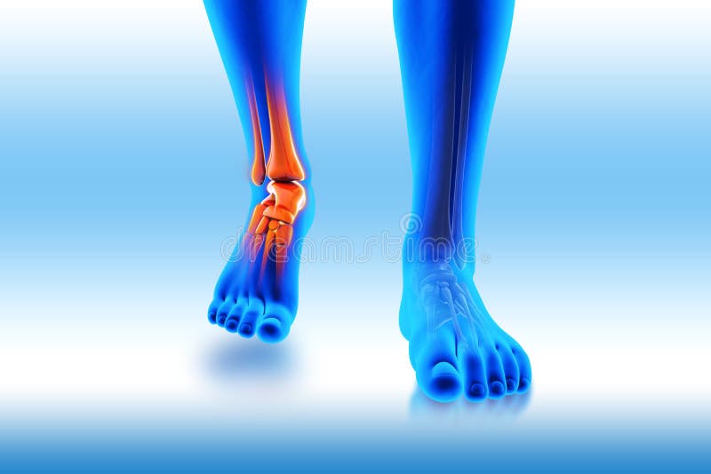
Free with trial Ankle pain - detail - pain concept. Bone joint damage illustrations Ankle pain - detail
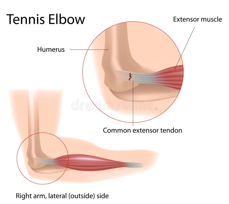
Free with trial A common condition of elbow pain caused by tear in the extensor tendon of the arm, eps8. Bone joint damage vectors Tennis elbow. A common condition of elbow pain caused by tear in the extensor tendon of the arm, eps8
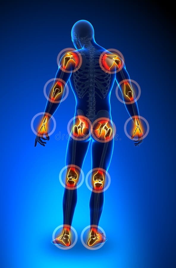
Free with trial Joints pain - full figure - All. Bone joint damage illustrations Joints pain - full figure
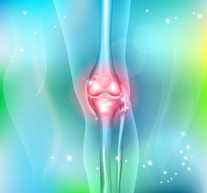
Free with trial Joint pain, joint anatomy beautiful abstract background. Bone joint damage vectors Joint pain
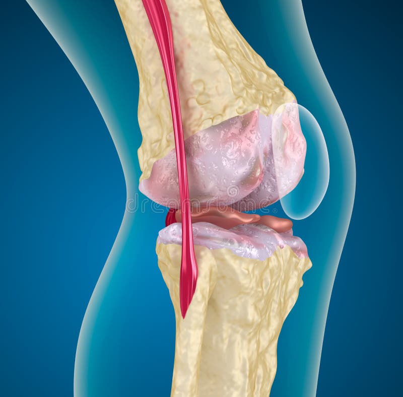
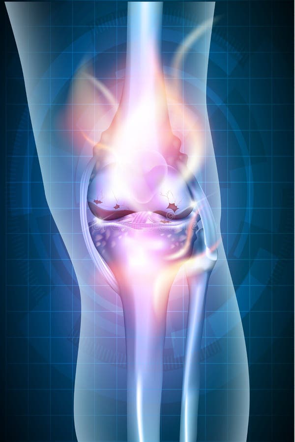
Free with trial Osteoporosis of the knee joint. Medical background. Bone joint damage illustrations Osteoporosis of the knee joint.
Free with trial Knee joint health care icon, abstract transparent shapes and wave at the background. Bone joint damage vectors Joint icon. Knee joint health care icon, abstract transparent shapes and wave at the background
Free with trial Joint treatment abstract background with beautiful glow and wave at the background. Bone joint damage vectors Joint background. Joint treatment abstract background with beautiful glow and wave at the background
Free with trial Knee joint repair concept icon, beautiful blue color and glow. Bone joint damage vectors Knee joint repair
Free with trial Tennis elbow medical fitness anatomy vector illustration diagram with arm bones, joint and muscles. Overuse of extensor muscles leading to pain. Bone joint damage vectors Tennis elbow medical fitness anatomy vector illustration diagram with arm bones, joint and muscles.
Free with trial Knee with Torn Meniscus Realistic Vector Medical Scheme with Damaged Knee Joint and Magnified Painful Meniscus Tear Anatomical Illustration. Musculoskeletal System and Joints Injuries, Meniscus Trauma. Bone joint damage vectors Injured Knee Joint with Torn Meniscus Vector Chart. Knee with Torn Meniscus Realistic Vector Medical Scheme with Damaged Knee Joint and Magnified Painful Meniscus Tear Anatomical Illustration. Musculoskeletal System and Joints Injuries, Meniscus Trauma
Free with trial Knee pain abstract background. Healthy joint and unhealthy painful joint with osteoarthritis. Bone joint damage vectors Knee pain abstract background
Free with trial Osteoarthritis, destruction of cartilage. Isolated on white. Bone joint damage vectors Osteoarthritis, unhealthy joint. Osteoarthritis, destruction of cartilage. Isolated on white
Free with trial The human rib cage is made up of 12 paired rib bones; each are symmetrically paired on a right and left side. Of all 24 ribs, the first seven pairs are often labeled as `true. ` These bones are connected to the costal cartilage, while the five other `false` sets are not. The ribcage also encloses the thoracic cavity and helps protect the heart and lungs from damage. There are 24 ribs in the human body, divided into two sets of 12 curved, flat bones. Each one is attached by cartilage at the back to the thoracic vertebrae. MEN and women have 12 pairs of ribs a few individuals have 13 or 11 pairs. The idea that men have fewer ribs than women is widespread but wrong, perhaps deriving from the biblical story of Eve being made from one of Adam`s ribs. Both men and women have 24 ribs, twelve on each side. Floating rib: One of the last two ribs. A rib is said to be `floating` if it does not attach to the sternum the breast bone or to another rib. There are usually 12 pairs of ribs in all. Each pair of ribs is attached to the building blocks of the spine the vertebrae in the back. The ribs partially enclose and protect the chest cavity, where many vital organs including the heart and the lungs are located. The rib cage is collectively made up of long, curved individual bones with joint-connections to the spinal vertebrae. Bone joint damage illustrations Ribs with Ligments anterior view. The human rib cage is made up of 12 paired rib bones; each are symmetrically paired on a right and left side. Of all 24 ribs, the first seven pairs are often labeled as `true.` These bones are connected to the costal cartilage, while the five other `false` sets are not. The ribcage also encloses the thoracic cavity and helps protect the heart and lungs from damage. There are 24 ribs in the human body, divided into two sets of 12 curved, flat bones. Each one is attached by cartilage at the back to the thoracic vertebrae. MEN and women have 12 pairs of ribs a few individuals have 13 or 11 pairs. The idea that men have fewer ribs than women is widespread but wrong, perhaps deriving from the biblical story of Eve being made from one of Adam`s ribs. Both men and women have 24 ribs, twelve on each side. Floating rib: One of the last two ribs. A rib is said to be `floating` if it does not attach to the sternum the breast bone or to another rib. There are usually 12 pairs of ribs in all. Each pair of ribs is attached to the building blocks of the spine the vertebrae in the back. The ribs partially enclose and protect the chest cavity, where many vital organs including the heart and the lungs are located. The rib cage is collectively made up of long, curved individual bones with joint-connections to the spinal vertebrae.
Free with trial Pain circles. Red painful target spot, targeting medication remedy circle and joint pain spots. Muscle pain, painful headaches or health healing sound wave isolated vector icons set. Bone joint damage vectors Pain circles. Red painful target spot, targeting medication remedy circle and joint pain spots isolated vector set. Pain circles. Red painful target spot, targeting medication remedy circle and joint pain spots. Muscle pain, painful headaches or health healing sound wave isolated vector icons set
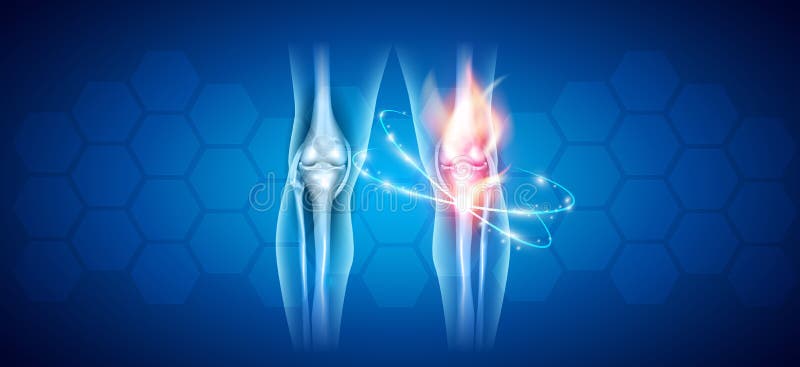
Free with trial Burning knee, painful knee and normal knee joint, abstract design. Human legs on a blue checkered background. Bone joint damage vectors Burning knee
Free with trial Vector illustration. Anatomy, front x-ray of an arthritic knee joint and a knee after unicompartmental or partial knee replacement. For advertising and medical publications. EPS 10. Bone joint damage vectors Meniscus _Arthritic knee and Partial knee replacement. Vector illustration. Anatomy, front x-ray of an arthritic knee joint and a knee after unicompartmental or partial knee replacement. For advertising and medical publications. EPS 10
Free with trial Arthroscopy procedure process explanation from medical view outline diagram. Labeled educational knee joint diagnosis and treatment with trimming instrument, scope and irrigation vector illustration. Bone joint damage vectors Arthroscopy procedure as knee diagnostics process explanation outline diagram. Arthroscopy procedure process explanation from medical view outline diagram. Labeled educational knee joint diagnosis and treatment with trimming instrument, scope and irrigation vector illustration.
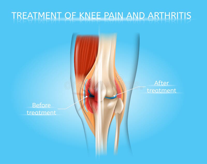
Free with trial Treatment of Knee Pain and Arthritis Realistic Vector Medical Poster or Scheme with Damaged by Disease and Healthy Human Knee Joint Before and After Treatment, Anatomical Cross Section Illustration. Bone joint damage vectors Knee Pain and Arthritis Treatment Vector Chart. Treatment of Knee Pain and Arthritis Realistic Vector Medical Poster or Scheme with Damaged by Disease and Healthy Human Knee Joint Before and After Treatment, Anatomical Cross Section Illustration
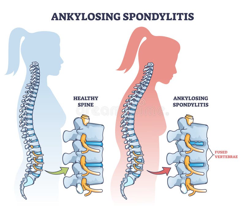
Free with trial Ankylosing Spondylitis Realistic Vector Medical Chart with Human Vertebral Column Joints Inflammation and Bones Fusion Anatomical Illustration. Spine joint bones arthritis symptoms orthopedic concept. Bone joint damage vectors Spine Joints Arthritis Symptoms Vector Infographic. Ankylosing Spondylitis Realistic Vector Medical Chart with Human Vertebral Column Joints Inflammation and Bones Fusion Anatomical Illustration. Spine joint bones arthritis symptoms orthopedic concept
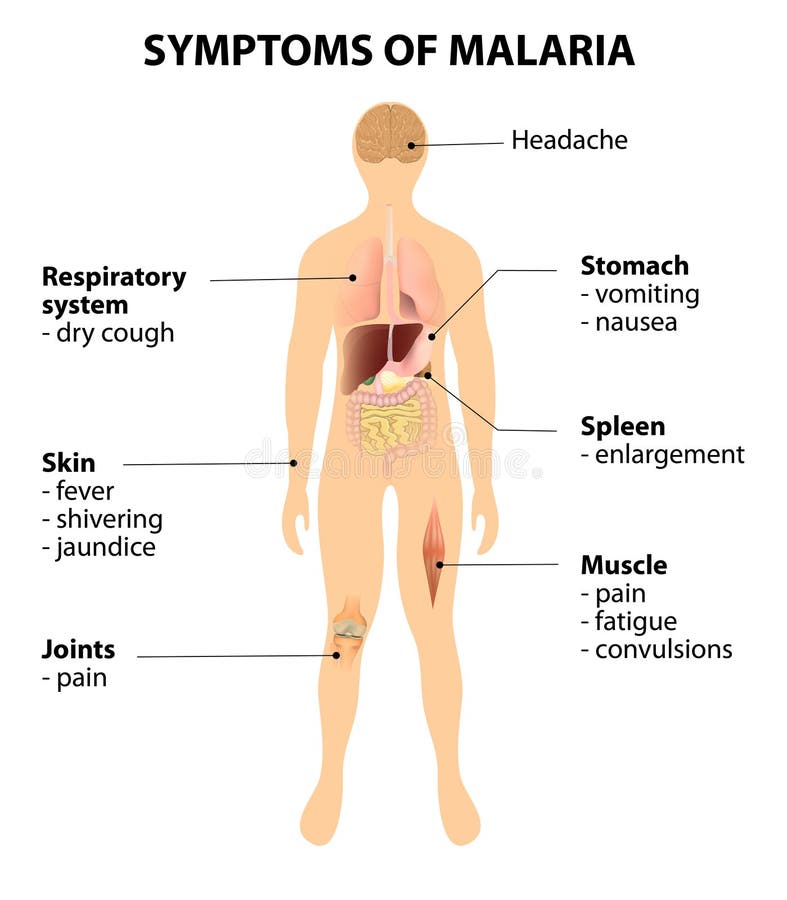
Free with trial Symptoms of malaria. How to Recognize the Signs of malaria. Disease and organs on silhouette man. Bone joint damage vectors Symptoms of malaria
Free with trial Vector diagram of forefoot pain, metatarsalgia symptom, tenderness in the balls of metatarsal bones of the foot. Used: Gradient, transparency, blend mode. Bone joint damage vectors Metatarsalgia
Free with trial Illustration of Plantar fasciitis. Bottom and side view of skeletal ankle. Used: gradient, transparency, Blend mode. Bone joint damage vectors Plantar fasciitis
Free with trial ILLUSTRATION OF A HEALTHY INTERVERTEBRAL DISC, THE DAMAGED INTERVERTEBRAL DISC, HERNIATION. Bone joint damage vectors DAMAGE TO THE SPINE. ILLUSTRATION OF A HEALTHY INTERVERTEBRAL DISC, THE DAMAGED INTERVERTEBRAL DISC, HERNIATION.
Free with trial Mechanism of formation of an ankle sprain and Grades of an ankle sprain (Eversion) illustration with external and skeletal (medial) view. Bone joint damage vectors Ankle sprain.Eversion. Mechanism of formation of an ankle sprain and Grades of an ankle sprain (Eversion) illustration with external and skeletal (medial) view
Free with trial Injuries in the foot: plantar fasciitis, heel spur and sesamoidit. Used: gradient, transparency, blend mode. Bone joint damage vectors Plantar fasciitis_heel spur_sesamoidit. Injuries in the foot: plantar fasciitis, heel spur and sesamoidit. Used: gradient, transparency, blend mode.
Free with trial Illustration of Mechanism of formation of a High ankle sprain (Syndesmotic Sprain) and Grades of high ankle sprain with external and skeletal (lateral and front) view of an ankle. Bone joint damage vectors High ankle sprain
Free with trial Mechanism of formation of an ankle sprain and Grades of an ankle sprain (Inversion) illustration with external and skeletal (lateral) view. Bone joint damage vectors Ankle sprain.Inversion. Mechanism of formation of an ankle sprain and Grades of an ankle sprain (Inversion) illustration with external and skeletal (lateral) view
Free with trial Vector illustration of healthy human foot and a medial ankle injury. Tarsal tunnel syndrome. EPS 10. Bone joint damage vectors Medial ankle injury_Tarsal tunnel syndrome. Vector illustration of healthy human foot and a medial ankle injury. Tarsal tunnel syndrome. EPS 10.
Free with trial Plantar fasciitis. disorder of the connective tissue which supports the arch of the foot. vector diagram for medical, educational and scientific use. Bone joint damage vectors Plantar fasciitis. disorder of the connective tissue which supports the arch of the foot
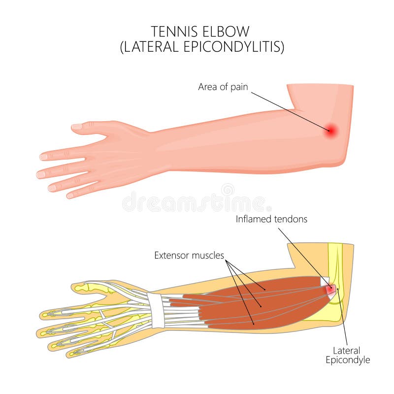
Free with trial Illustration of Lateral Epicondylitis or tennis elbow. Used: Gradient, transparency, blend mode. Bone joint damage vectors Lateral Epicondylitis or tennis elbow
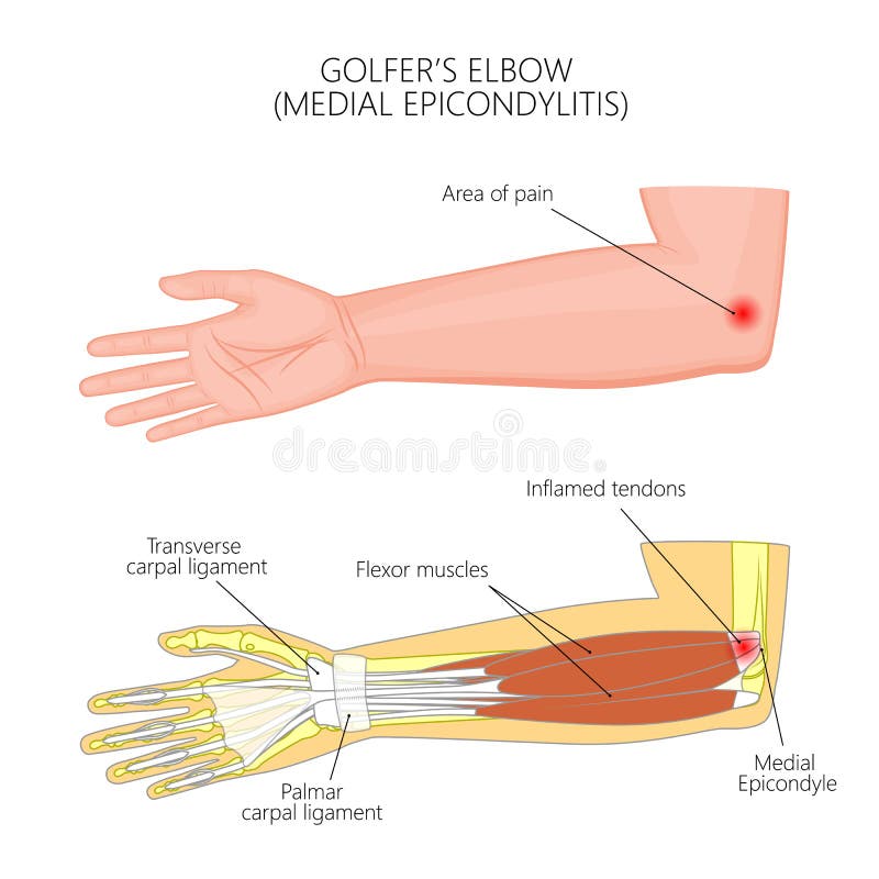
Free with trial Illustration of Medial Epicondylitis or golfers elbow. Used: Gradient, transparency, blend mode. Bone joint damage vectors Medial Epicondylitis or golfer
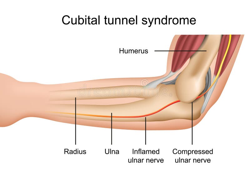
Free with trial Cubital tunnel syndrome, inflamed ulnar nerve medical vector illustration on white background eps 10 infographic. Bone joint damage vectors Cubital tunnel syndrome, inflamed ulnar nerve medical vector illustration on white background
Free with trial Rheumatoid arthritis. Healthy and damage joint. Close-up of bone, cartilage, and Cells in a joint capsule: synoviocytes, osteoclast, neutrophil, T lymphocyte, B-cell, macrophage, mast, plasma, and dendritic cell. Bone joint damage vectors Rheumatoid arthritis. Healthy and damage joint
Free with trial Damaged joint and normal joint design on an abstract grey glowing background. Bone joint damage vectors Joint
Free with trial Burning leg knee joint on a abstract blue checkered background. Bone joint damage illustrations Burning leg knee joint
Free with trial Human leg knee joint anatomy on a beautiful light blue background. Bone joint damage vectors Human leg and knee joint. Human leg knee joint anatomy on a beautiful light blue background
Free with trial Vector illustration of bursitis of the elbow joint. Bone joint damage vectors Bursitis of the elbow joint
Free with trial Joint problems and treatment abstract scientific background. Bone joint damage vectors Joint problems and treatment
Free with trial Knee joint pain abstract blue triangle background. Bone joint damage vectors Knee joint pain abstract triangle background
Free with trial Knee joint abstract treatment procedure illustration. Doctor with screwdriver and gears in the joint. Bone joint damage vectors Knee joint abstract treatment
Free with trial Ankylosing spondylitis as inflammatory spine bone disease outline diagram. Labeled educational anatomical comparison with healthy and damaged vertebrae vector illustration. Fused skeletal back parts. Bone joint damage vectors Ankylosing spondylitis as inflammatory spine bone disease outline diagram
Free with trial Osgood schlatter disease condition with leg and knee joint pain outline diagram. Labeled educational medical scheme with patella tendon inflammation and tibial tuberosity anatomy vector illustration. Bone joint damage vectors Osgood schlatter disease condition with knee joint pain outline diagram. Osgood schlatter disease condition with leg and knee joint pain outline diagram. Labeled educational medical scheme with patella tendon inflammation and tibial tuberosity anatomy vector illustration.
Free with trial Rotator cuff tear as shoulder muscle trauma or arm injury outline diagram. Labeled educational upper body anatomy with medical tendon torn away from bone vector illustration. Painful joint condition. Bone joint damage vectors Rotator cuff tear as shoulder muscle trauma or arm injury outline diagram
Free with trial Knee injuries with medical bone, ligament or muscle trauma outline diagram. Labeled educational inflammation disease collection vector illustration. ACL injury, meniscus, runners or tendinitis example. Bone joint damage vectors Knee injuries with medical bone, ligament and muscle trauma outline diagram. Knee injuries with medical bone, ligament or muscle trauma outline diagram. Labeled educational inflammation disease collection vector illustration. ACL injury, meniscus, runners or tendinitis example
Free with trial Osteoarthritis Vector Medical Poster with Magnification of Thinned Cartilage, Bone Ends Rub Together in Damaged Human Knee Joint Realistic Illustration. Musculoskeletal System Disease, Joints Injuries. Bone joint damage vectors Osteoarthritis Realistic Vector Medical Scheme. Osteoarthritis Vector Medical Poster with Magnification of Thinned Cartilage, Bone Ends Rub Together in Damaged Human Knee Joint Realistic Illustration. Musculoskeletal System Disease, Joints Injuries
Free with trial Vector illustration. Anatomy, front x-ray of an arthritic knee joint and a knee after total knee replacement. For advertising and medical publications. EPS 10. Bone joint damage vectors Meniscus _Arthritic knee and Total knee replacement. Vector illustration. Anatomy, front x-ray of an arthritic knee joint and a knee after total knee replacement. For advertising and medical publications. EPS 10
Free with trial Vector illustration of back pain. damage to the spine. Bone joint damage vectors Back pain. damage to the spine
Free with trial Normal leg knee joint at the blue background and unhealthy joint at the red background. Bone joint damage vectors Knee joints healthy and unhealthy. Normal leg knee joint at the blue background and unhealthy joint at the red background
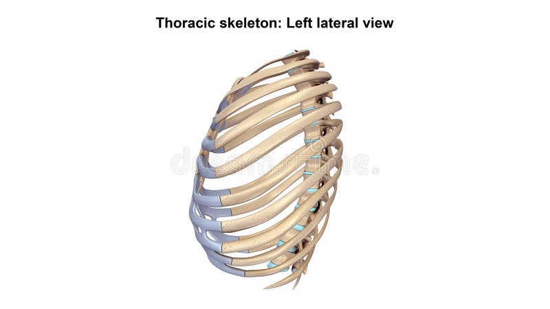
Free with trial Thoracic Skeleton The human rib cage is made up of 12 paired rib bones; each are symmetrically paired on a right and left side. Of all 24 ribs, the first seven pairs are often labeled as `true. ` These bones are connected to the costal cartilage, while the five other `false` sets are not. The ribcage also encloses the thoracic cavity and helps protect the heart and lungs from damage. There are 24 ribs in the human body, divided into two sets of 12 curved, flat bones. Each one is attached by cartilage at the back to the thoracic vertebrae. MEN and women have 12 pairs of ribs a few individuals have 13 or 11 pairs. The idea that men have fewer ribs than women is widespread but wrong, perhaps deriving from the biblical story of Eve being made from one of Adam`s ribs. Both men and women have 24 ribs, twelve on each side. Floating rib: One of the last two ribs. A rib is said to be `floating` if it does not attach to the sternum the breast bone or to another rib. There are usually 12 pairs of ribs in all. Each pair of ribs is attached to the building blocks of the spine the vertebrae in the back. The ribs partially enclose and protect the chest cavity, where many vital organs including the heart and the lungs are located. The rib cage is collectively made up of long, curved individual bones with joint-connections to the spinal vertebrae. Bone joint damage illustrations Thoracic Skeleton Lateral view. Thoracic Skeleton The human rib cage is made up of 12 paired rib bones; each are symmetrically paired on a right and left side. Of all 24 ribs, the first seven pairs are often labeled as `true.` These bones are connected to the costal cartilage, while the five other `false` sets are not. The ribcage also encloses the thoracic cavity and helps protect the heart and lungs from damage. There are 24 ribs in the human body, divided into two sets of 12 curved, flat bones. Each one is attached by cartilage at the back to the thoracic vertebrae. MEN and women have 12 pairs of ribs a few individuals have 13 or 11 pairs. The idea that men have fewer ribs than women is widespread but wrong, perhaps deriving from the biblical story of Eve being made from one of Adam`s ribs. Both men and women have 24 ribs, twelve on each side. Floating rib: One of the last two ribs. A rib is said to be `floating` if it does not attach to the sternum the breast bone or to another rib. There are usually 12 pairs of ribs in all. Each pair of ribs is attached to the building blocks of the spine the vertebrae in the back. The ribs partially enclose and protect the chest cavity, where many vital organs including the heart and the lungs are located. The rib cage is collectively made up of long, curved individual bones with joint-connections to the spinal vertebrae.
Free with trial Vector illustration of a healthy knee joint and an unhealthy knee with a patellar tendon rupture problem. Anatomy of the human knee, side view of the bent knee. EPS 10. Bone joint damage vectors Knee problem_Patellar tendon rupture. Vector illustration of a healthy knee joint and an unhealthy knee with a patellar tendon rupture problem. Anatomy of the human knee, side view of the bent knee. EPS 10.
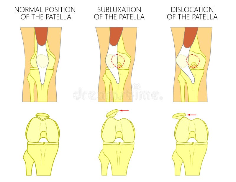
Free with trial Vector illustration of a healthy human knee joint and unhealthy knees with problem. Subluxation and dislocation of the patella or kneecap. Anatomy of human knee joint, front view of straight and bent knee. Bone joint damage vectors Knee joint problem_Dislocation of the patella. Vector illustration of a healthy human knee joint and unhealthy knees with problem. Subluxation and dislocation of the patella or kneecap. Anatomy of human knee joint, front view of straight and bent knee.
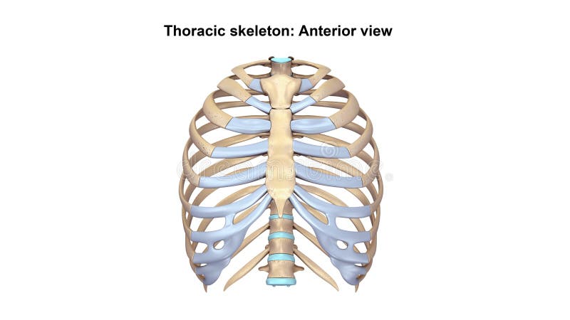
Free with trial Thoracic Skeleton The human rib cage is made up of 12 paired rib bones; each are symmetrically paired on a right and left side. Of all 24 ribs, the first seven pairs are often labeled as `true. ` These bones are connected to the costal cartilage, while the five other `false` sets are not. The ribcage also encloses the thoracic cavity and helps protect the heart and lungs from damage. There are 24 ribs in the human body, divided into two sets of 12 curved, flat bones. Each one is attached by cartilage at the back to the thoracic vertebrae. MEN and women have 12 pairs of ribs a few individuals have 13 or 11 pairs. The idea that men have fewer ribs than women is widespread but wrong, perhaps deriving from the biblical story of Eve being made from one of Adam`s ribs. Both men and women have 24 ribs, twelve on each side. Floating rib: One of the last two ribs. A rib is said to be `floating` if it does not attach to the sternum the breast bone or to another rib. There are usually 12 pairs of ribs in all. Each pair of ribs is attached to the building blocks of the spine the vertebrae in the back. The ribs partially enclose and protect the chest cavity, where many vital organs including the heart and the lungs are located. The rib cage is collectively made up of long, curved individual bones with joint-connections to the spinal vertebrae. Bone joint damage illustrations Thoracic Skeleton Anterior view. Thoracic Skeleton The human rib cage is made up of 12 paired rib bones; each are symmetrically paired on a right and left side. Of all 24 ribs, the first seven pairs are often labeled as `true.` These bones are connected to the costal cartilage, while the five other `false` sets are not. The ribcage also encloses the thoracic cavity and helps protect the heart and lungs from damage. There are 24 ribs in the human body, divided into two sets of 12 curved, flat bones. Each one is attached by cartilage at the back to the thoracic vertebrae. MEN and women have 12 pairs of ribs a few individuals have 13 or 11 pairs. The idea that men have fewer ribs than women is widespread but wrong, perhaps deriving from the biblical story of Eve being made from one of Adam`s ribs. Both men and women have 24 ribs, twelve on each side. Floating rib: One of the last two ribs. A rib is said to be `floating` if it does not attach to the sternum the breast bone or to another rib. There are usually 12 pairs of ribs in all. Each pair of ribs is attached to the building blocks of the spine the vertebrae in the back. The ribs partially enclose and protect the chest cavity, where many vital organs including the heart and the lungs are located. The rib cage is collectively made up of long, curved individual bones with joint-connections to the spinal vertebrae.
Free with trial Illustration diagram of normal knee joint and a knee with chondromalacia patella. Flexed bent knee. Front view. Bone joint damage vectors Chondromalacia patella. Flattened femoral groove. Illustration diagram of normal knee joint and a knee with chondromalacia patella. Flexed bent knee. Front view.
Free with trial ACL injury or trauma as tear or sprain of anterior cruciate outline concept. Labeled educational leg or knee pain anatomical explanation with bone and ligament medical structure vector illustration. Bone joint damage vectors ACL injury or trauma as tear or sprain of anterior cruciate outline concept
Free with trial Pain circles. Red painful target spot, targeting medication remedy circle and joint pain spots. Muscle pain, painful headaches or health healing sound wave isolated vector icons set. Bone joint damage vectors Pain circles. Red painful target spot, targeting medication remedy circle and joint pain spots. Muscle pain, painful
Free with trial Pain localization mark, set of abstract symbols of pain. Red circles for marking human pain. Headache, hurt body part marker, muscular joint. Vector illusration EPS 10. Bone joint damage vectors Pain localization mark, set of abstract symbols of pain. Red circles for marking human pain. Headache, hurt body part
Free with trial 3D image ankle pain - hurt trauma on a blue background. Bone joint damage illustrations Ankle pain - hurt trauma
Free with trial Healthy knee and knee with osteoarthritis on a white background. Bone joint damage vectors Healthy knee and knee with osteoarthritis
Free with trial Tennis elbow or lateral epicondylitis. Vector illustration for medical use. Bone joint damage vectors Tennis elbow or lateral epicondylitis.
Free with trial Human leg, knee anatomy, abstract grey mesh background. Bone joint damage vectors Human leg, knee anatomy
Free with trial Pain circles. Red painful target spot, targeting medication remedy circle and joint pain spots. Muscle pain, painful headaches or health healing sound wave isolated vector icons set. Bone joint damage vectors Pain circles. Red painful target spot
Free with trial Illustration diagram of normal leg with a healthy knee and a leg with over pronation of the foot arch and chondromalacia patella. Used: gradient, blend mode, transparency. Bone joint damage vectors Knee Pain and chondromalacia patella. Illustration diagram of normal leg with a healthy knee and a leg with over pronation of the foot arch and chondromalacia patella. Used: gradient, blend mode, transparency.
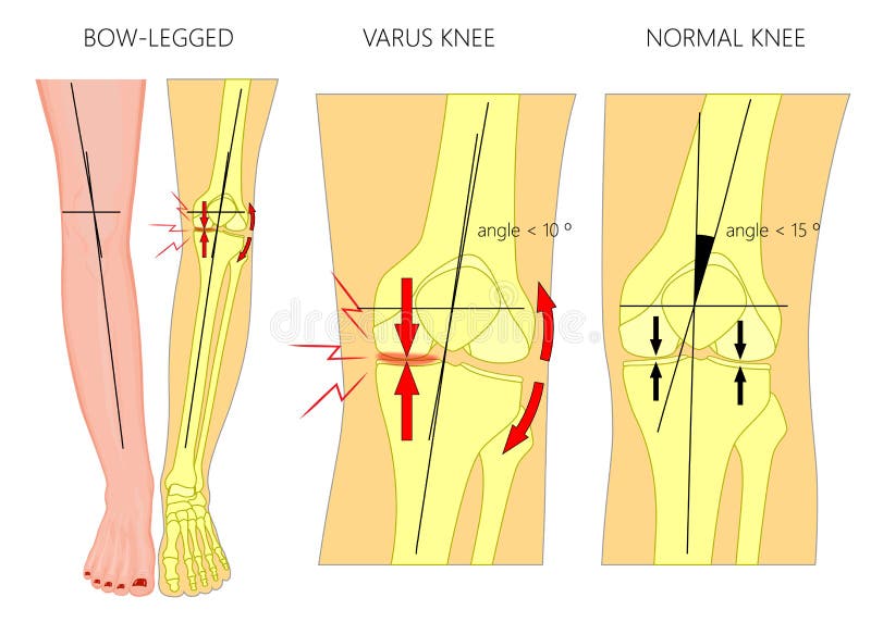
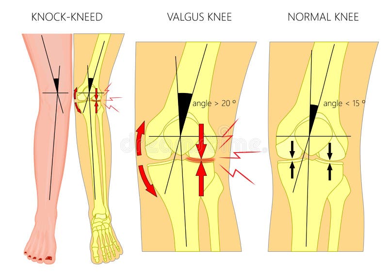
Free with trial Vector illustration diagram. Shapes of human legs. Normal and curved legs. Knock knees. Bowed legs. Genu valgum and genu varum. For advertising, medical publications. EPS 10. Bone joint damage vectors Shapes of the legs.Normal and curved legs.Knock knees.Bowed leg. Vector illustration diagram. Shapes of human legs. Normal and curved legs. Knock knees. Bowed legs. Genu valgum and genu varum. For advertising, medical publications. EPS 10.
Free with trial Vector illustration diagram. Shapes of human legs. Normal and curved legs. Knock knees. Bowed legs. Genu valgum and genu varum. For advertising, medical publications. EPS 10. Bone joint damage vectors Shapes of the legs.Normal and curved legs.Knock knees.Bowed leg. Vector illustration diagram. Shapes of human legs. Normal and curved legs. Knock knees. Bowed legs. Genu valgum and genu varum. For advertising, medical publications. EPS 10.
Free with trial Vector illustration diagram. Shapes of human legs. Normal and curved legs. Knock knees. Genu valgum and genu varum. For advertising, medical publications. EPS 10. Bone joint damage vectors Shapes of the legs.Normal and curved legs.Knock knees.Bowed leg. Vector illustration diagram. Shapes of human legs. Normal and curved legs. Knock knees. Genu valgum and genu varum. For advertising, medical publications. EPS 10.
Free with trial Spinal Cord Injury Vector Medical Scheme with Magnification of Fractured Vertebral Body and Damaged with Injury Spinal Cord Anatomical Realistic Illustration. Dangerous Back or Spine Trauma Concept. Bone joint damage vectors Human Spinal Cord Injury Anatomical Vector Scheme. Spinal Cord Injury Vector Medical Scheme with Magnification of Fractured Vertebral Body and Damaged with Injury Spinal Cord Anatomical Realistic Illustration. Dangerous Back or Spine Trauma Concept
Free with trial Rheumatoid Arthritis of Cervical Spine Vector Medical Scheme with Inflamed and Damaged Vertebral Column Synovial Joints Realistic Illustration. Human Musculoskeletal System Painful Diseases Concept. Bone joint damage vectors Rheumatoid Arthritis of Cervical Spine Vector
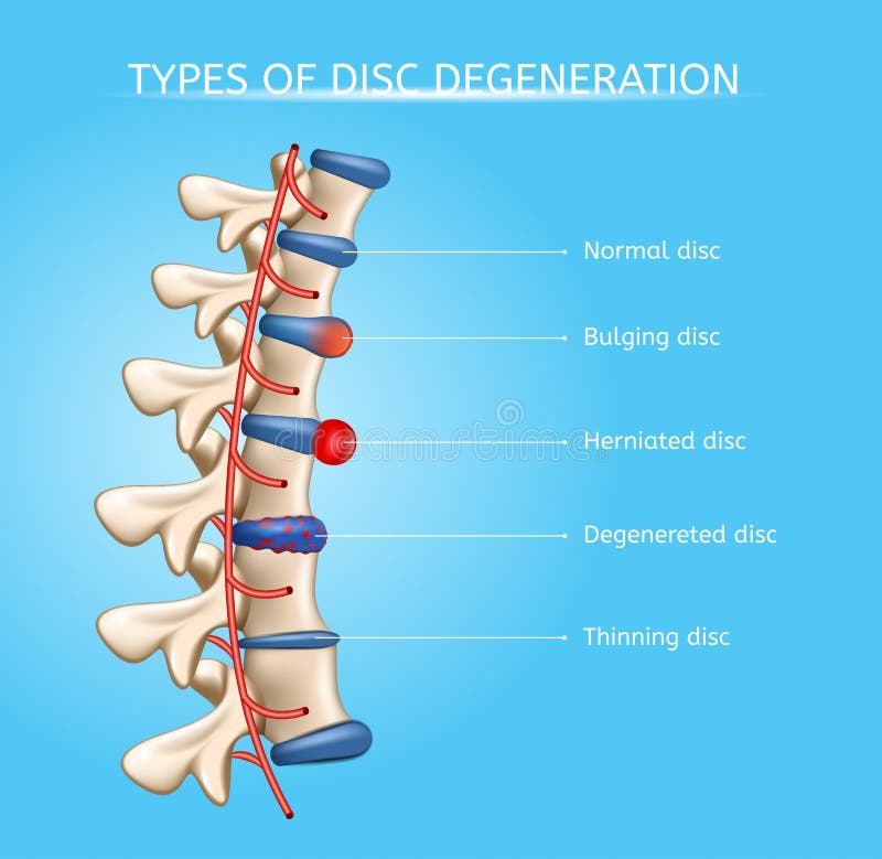
Free with trial Types of Spinal Disc Degeneration Vector Medical Scheme with Normal, Bulging, Herniated, Degenerated and Thinning Discs on Human Vertebral Column Illustration. Spinal Disc Diseases Orthopedic Concept. Bone joint damage vectors Intervertebral Disc Degeneration Types Vector. Types of Spinal Disc Degeneration Vector Medical Scheme with Normal, Bulging, Herniated, Degenerated and Thinning Discs on Human Vertebral Column Illustration. Spinal Disc Diseases Orthopedic Concept
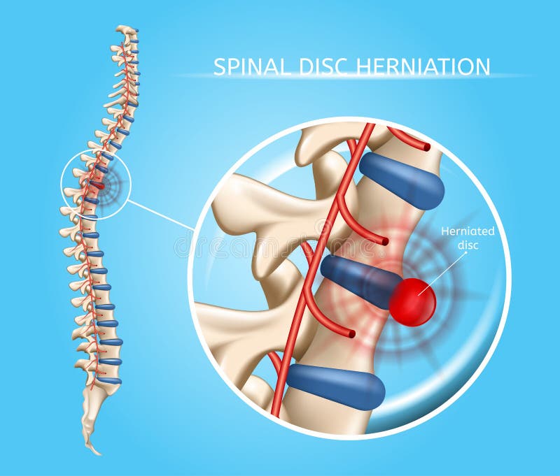
Free with trial Spinal Disk Herniation Vector Medical Scheme with Vertebral Column and Herniated Disc Anatomical Illustration on Blue Background. Chronic Spinal and Neck Pain Causes, Spine Joints Diseases Concept. Bone joint damage vectors Spinal Disk Herniation Vector Medical Scheme
Free with trial Set of icons of the joints and their treatment Cartilage damage, arthritis, osteoarthritis, restoration of cartilage pain relief icons. Flat icons in a round frames. Bone joint damage vectors Set of icons. Joints and their treatment. Flat icons in rounded frames. Set of icons of the joints and their treatment Cartilage damage, arthritis, osteoarthritis, restoration of cartilage pain relief icons. Flat icons in a round frames.
Free with trial Medical infographic scheme of osteoarthritis joint damage in human body, flat vector illustration isolated on white background. Osteoarthritis and arthritis disease. Bone joint damage vectors Medical scheme of osteoarthritis joint damage, flat vector illustration isolated. Medical infographic scheme of osteoarthritis joint damage in human body, flat vector illustration isolated on white background. Osteoarthritis and arthritis disease.
Free with trial Bone pain glyph icon, body and painful, joint ache sign, vector graphics, a solid pattern on a white background, eps 10. Bone joint damage vectors Bone pain glyph icon, body and painful, joint ache sign, vector graphics, a solid pattern on a white background.
Free with trial Bone pain line icon, body and painful, joint ache sign, vector graphics, a linear pattern on a white background, eps 10. Bone joint damage vectors Bone pain line icon, body and painful, joint ache sign, vector graphics, a linear pattern on a white background.
Free with trial Handwriting text Osteoarthritis. Concept meaning Degeneration of joint cartilage and the underlying bone. Bone joint damage illustrations Handwriting text Osteoarthritis. Concept meaning Degeneration of joint cartilage and the underlying bone
Free with trial Word writing text Osteoarthritis. Business concept for Degeneration of joint cartilage and the underlying bone. Bone joint damage illustrations Word writing text Osteoarthritis. Business concept for Degeneration of joint cartilage and the underlying bone
Free with trial Word writing text Osteoarthritis. Business concept for Degeneration of joint cartilage and the underlying bone. Bone joint damage illustrations Word writing text Osteoarthritis. Business concept for Degeneration of joint cartilage and the underlying bone
Free with trial Shown is a knee with erosion of the cartilage causing meniscus damage. There is also hypertrphyof the knee bone and lining. Bone joint damage illustrations Knee - Meniscus Damage and Hypertrophy. Shown is a knee with erosion of the cartilage causing meniscus damage. There is also hypertrphyof the knee bone and lining.
Free with trial Detailed 3D illustration of a human knee joint affected by osteoarthritis, highlighting cartilage erosion, bone spurs, and inflammation within the joint capsule. The image visually represents the pain and damage associated with this degenerative condition. Bone joint damage illustrations Knee Osteoarthritis Illustration Showing Joint Degeneration and Inflammation. Detailed 3D illustration of a human knee joint affected by osteoarthritis. Detailed 3D illustration of a human knee joint affected by osteoarthritis, highlighting cartilage erosion, bone spurs, and inflammation within the joint capsule. The image visually represents the pain and damage associated with this degenerative condition
Free with trial Hip labral tear anatomical explanation with medical labrum bone damage outline diagram. Labeled educational skeletal anatomy with pelvis, femur and torn labrum condition example vector illustration. Bone joint damage vectors Hip labral tear explanation as medical labrum bone damage outline diagram. Hip labral tear anatomical explanation with medical labrum bone damage outline diagram. Labeled educational skeletal anatomy with pelvis, femur and torn labrum condition example vector illustration.