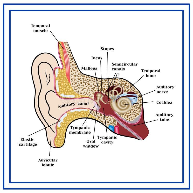
Free with trial The structure of the human ear. Bone structure vectors The structure of the human ear
Free with trial Sartorius muscle description with medical bones structure outline diagram. Labeled educational and anatomical scheme with skeletal physiology and muscular location in human leg vector illustration. Bone structure vectors Sartorius muscle description with medical bones structure outline diagram
Free with trial Rotator cuff anatomical structure and location explanation outline diagram. Labeled educational body part description with shoulder bones and muscle posterior or anterior view vector illustration. Bone structure vectors Rotator cuff anatomical structure and location explanation outline diagram
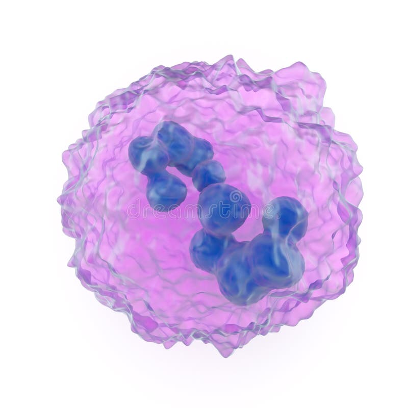
Free with trial Illustration showing the structure of a megakaryocyte, a large nucleated cell in the bone marrow which is the precursor for the production of platelets or thrombocytes. Bone structure illustrations Megakaryocyte
Free with trial Dog nail structure anatomy / fingernail vector / pink. Bone structure vectors Dog nail structure anatomy / fingernail vector
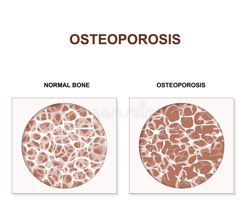
Free with trial Human bone disease- osteoporosis. Human bone structure. Bone structure vectors Human bone disease- osteoporosis
Free with trial Spine structure. Human skeleton, medicine. 3d vector icon. Bone structure vectors Spine structure. Human skeleton, medicine. 3d vector
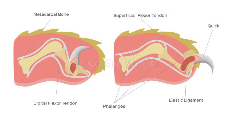
Free with trial Cat nail structure anatomy / vector on white. Bone structure vectors Cat nail structure anatomy / vector
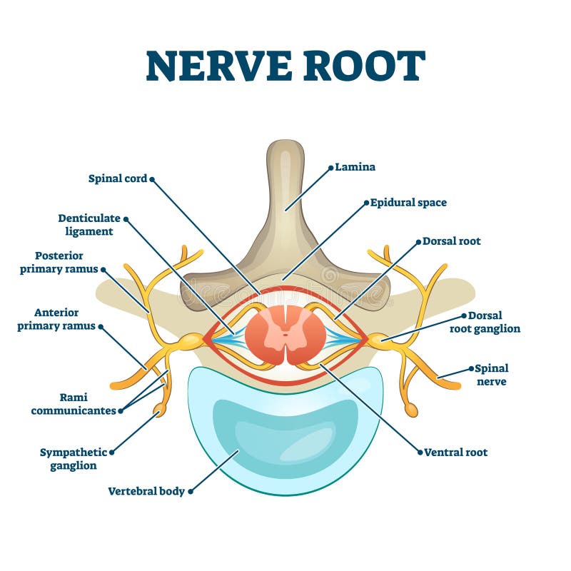
Free with trial Nerve root anatomical structure labeled cross section, vector illustration educational diagram. Medical information with root scheme. Human spine health guide as informative poster. Graphical example. Bone structure vectors Nerve root anatomical structure labeled cross section
Free with trial Osteosarcoma. Osteogenic sarcoma is a cancerous tumor in a bone. malignant neoplasm. Two human bones: healthy femur and bone with osteosarcoma. Vector illustration for biological, science, medical use. Bone structure vectors Two human bones: healthy femur and bone with osteosarcoma
Free with trial Deltoid muscle and skeletal shoulder anatomical structure outline diagram. Labeled educational bone description with anterior, lateral and posterior view vector illustration. Ball and socket joint. Bone structure vectors Deltoid muscle and skeletal shoulder anatomical structure outline diagram
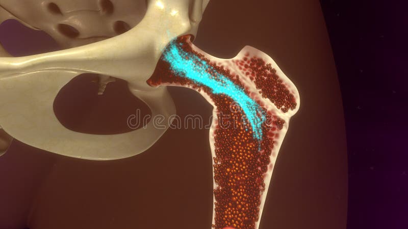
Free with trial Bone marrow is the spongy tissue inside some of the bones in the body, including the hip and thigh bones. Bone structure illustrations Bone marrow
Free with trial Cartilage elastic tissue location in body and leg structure outline diagram. Labeled educational description with healthy side view of orthopedic components vector illustration. Medical joint scheme. Bone structure vectors Cartilage elastic tissue location in body and leg structure outline diagram
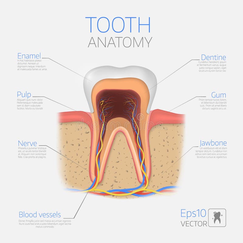
Free with trial Types of teeth and external and internal structure of tooth. Bone structure vectors Types and structure of teeth. Types of teeth and external and internal structure of tooth
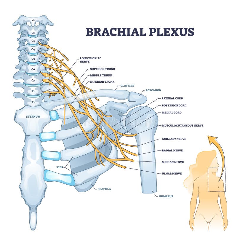
Free with trial Brachial plexus network of nerves in the shoulder structure outline concept. Labeled educational skeletal scheme with neurology graphic vector illustration. Human nervous system corde or trunk example. Bone structure vectors Brachial plexus network of nerves in the shoulder structure outline concept
Free with trial Rib cage anatomy, labeled vector illustration diagram. Medical human chest skeletal bone structure model. Numbered ribs, sternum, cartilage parts and clavicular articulation. Health care education. Bone structure vectors Rib cage anatomy, labeled vector illustration diagram
Free with trial Bone remodeling. Close-up of a Osteoclast resorbs bone, and Osteoblasts synthesized bone tissue. Vector illustration. Bone structure vectors Bone remodeling. Osteoclast resorbs bone, and Osteoblasts synthesized bone tissue. Bone remodeling. Close-up of a Osteoclast resorbs bone, and Osteoblasts synthesized bone tissue. Vector illustration
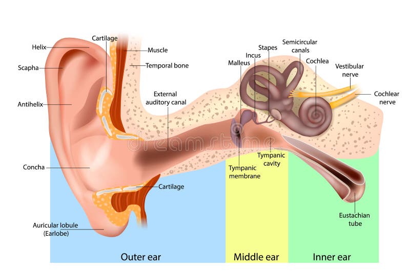
Free with trial Human Ear Anatomy. Ear structure anatomical diagram. The human ear consists of the Outer, Middle and Inner ear. Bone structure vectors Human Ear Anatomy. Ear structure diagram. The human ear consists of the Outer, Middle and Inner ear.
Free with trial Diaphragm muscle structure with transparent ribcage bones outline diagram. Labeled educational scheme with muscular system for central tendon, dome and openings for esophagus vector illustration. Bone structure vectors Diaphragm muscle structure with transparent ribcage bones outline diagram
Free with trial Osteoporosis stages. close-up of human bones with different density. bone disease. Vector illustration. Bone structure vectors Osteoporosis stages. aging process with bone. Osteoporosis stages. close-up of human bones with different density. bone disease. Vector illustration
Free with trial Bone remodeling process resorption, reversal, formation, and mineralization. Osteoblast, osteoclast, and osteocyte. Vector illustration of human bone cell types. Medical diagram. Bone structure vectors Bone remodeling process. Osteoblast, osteoclast, and osteocyte. Bone remodeling process resorption, reversal, formation, and mineralization. Osteoblast, osteoclast, and osteocyte. Vector illustration of human bone cell types. Medical diagram
Free with trial Obturator internus muscle with externus location near pelvis bone outline diagram. Labeled educational scheme with human hip muscular system and skeletal structure description vector illustration. Bone structure vectors Obturator internus muscle with externus location near pelvis outline diagram. Obturator internus muscle with externus location near pelvis bone outline diagram. Labeled educational scheme with human hip muscular system and skeletal structure description vector illustration.
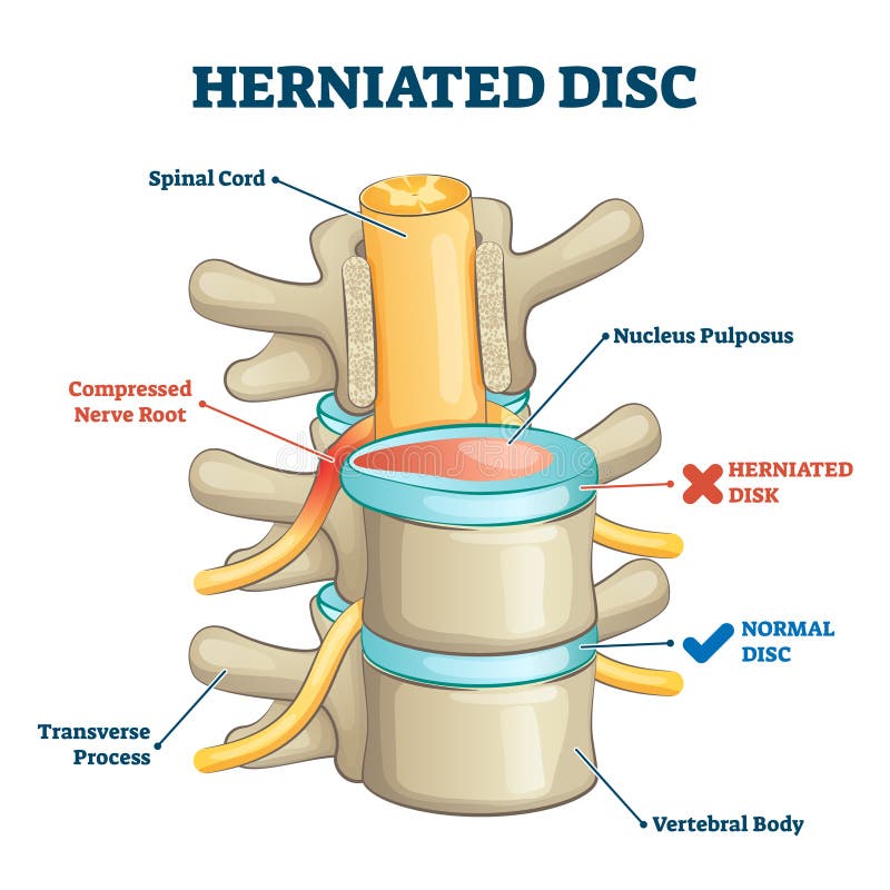
Free with trial Herniated disc injury 3D side view on spinal bone skeleton vector illustration. Medical condition with back trauma pain and nerve root compression by nucleus pulposus. Problematic example comparison. Bone structure vectors Herniated disc injury 3D side view on spine bone skeleton vector illustration. Herniated disc injury 3D side view on spinal bone skeleton vector illustration. Medical condition with back trauma pain and nerve root compression by nucleus pulposus. Problematic example comparison.
Free with trial Palmaris longus skeletal and muscular body structure for human arm outline diagram. Labeled educational scheme with anatomical and medical hand inner parts physical description vector illustration. Bone structure vectors Palmaris longus skeletal and muscular structure for human arm outline diagram. Palmaris longus skeletal and muscular body structure for human arm outline diagram. Labeled educational scheme with anatomical and medical hand inner parts physical description vector illustration.
Free with trial Humerus bone labeled vector illustration diagram. Long bone type in the upper arm. Skeleton anatomy scheme with greater tubercle, deltoid tuberosity, medial epicondyle, trochlea and other parts. Bone structure vectors Humerus bone labeled vector illustration diagram
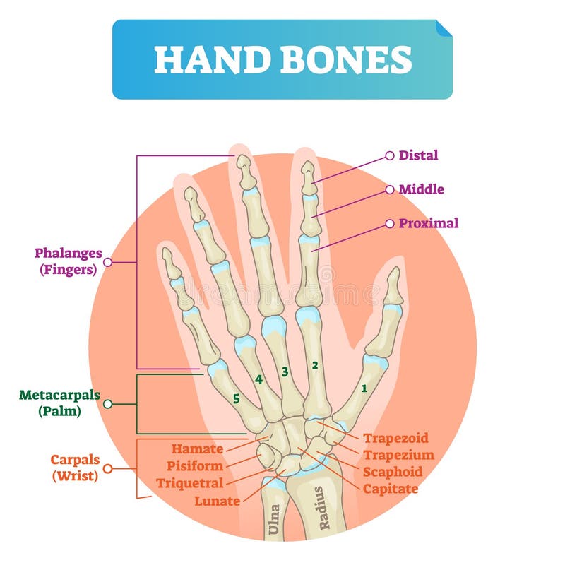
Free with trial Hand bones vector illustration. Labeled educational human arm structure with phalanges, metacarpals, hamate, pisiform, triquetral and lunate. Palm finger parts scheme. Bone structure vectors Hand bones vector illustration. Labeled educational arm structure. Hand bones vector illustration. Labeled educational human arm structure with phalanges, metacarpals, hamate, pisiform, triquetral and lunate. Palm finger parts scheme.
Free with trial Anatomy of healthy teeth and dental implant in jaw bone. Bone structure illustrations Anatomy of healthy teeth and dental implant in jaw bone - 3d rendering. Anatomy of healthy teeth and dental implant in jaw bone.
Free with trial Hip Joint Anatomy Infographic Diagram. for medical science education. vector drawing. type ball and socket synovial joint. cartoon illustration. structure chart. parts femur bone capsule scheme. Bone structure vectors Hip Joint Anatomy Infographic Diagram
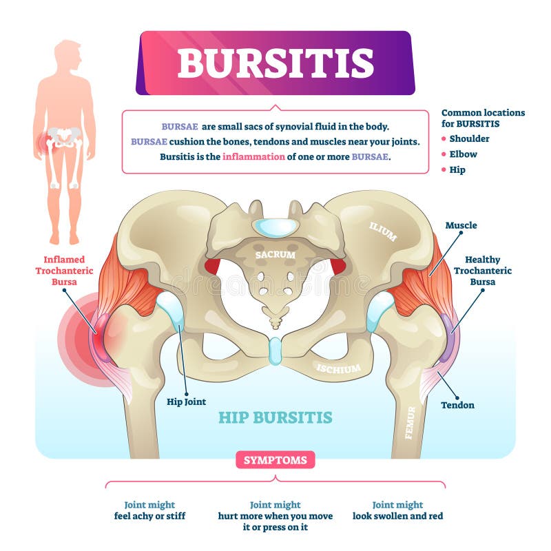
Free with trial Bursitis vector illustration. Labeled bursae synovial inflammation scheme. Bone and tendon illness and disease diagnosis. Educational chronic problem symptoms, causes and anatomical structure diagram. Bone structure vectors Bursitis vector illustration. Labeled bursae synovial inflammation scheme.
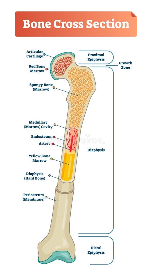
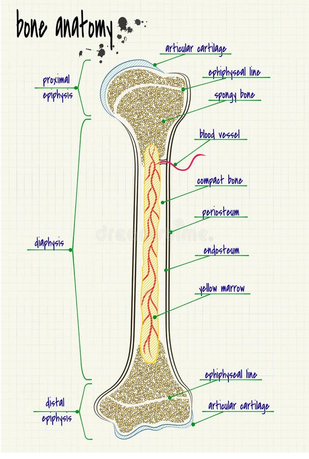
Free with trial Vector illustration scheme of bone cross section. Diagram with articular cartilage, marrow, spongy bone, medullary cavity, endosteum, diaphysis, and periosteum. Explaned distal and proximal epiphysis. Bone structure vectors Vector illustration scheme of bone cross section. Diagram with articular cartilage, marrow, medullary cavity and periosteum. Vector illustration scheme of bone cross section. Diagram with articular cartilage, marrow, spongy bone, medullary cavity, endosteum, diaphysis, and periosteum. Explaned distal and proximal epiphysis.
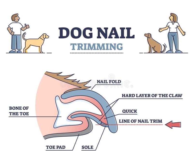
Free with trial Dog nail trimming with anatomical claw side view structure outline diagram. Animal fingernail care and hygiene with veterinary pedicure vector illustration. Labeled educational medical pet clipping. Bone structure vectors Dog nail trimming with anatomical claw side view structure outline diagram
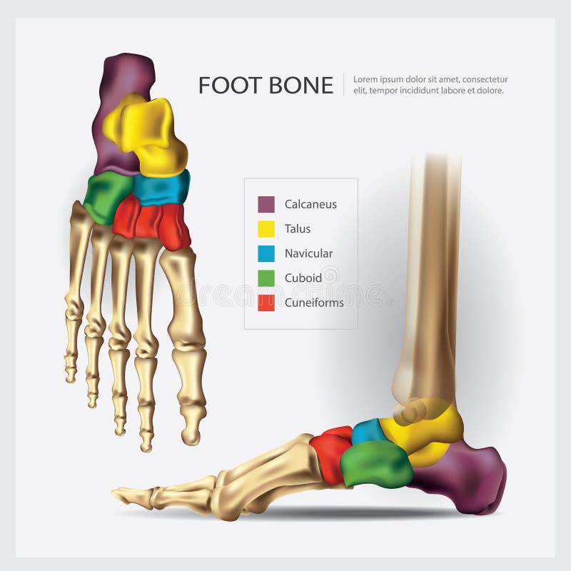
Free with trial Human Anatomy Foot Bone Vector Illustration. Bone structure vectors Human Anatomy Foot Bone
Free with trial Carpal bones with hand palm skeletal structure and anatomy outline diagram. Labeled educational scheme with medical left hand model and isolated hamate, trapezoid or pisiform bone vector illustration. Bone structure vectors Carpal bones with hand palm skeletal structure and anatomy outline diagram
Free with trial Peroneus brevis leg muscle with longus and tertius muscular part location outline diagram. Labeled educational foot skeletal structure from later view vector illustration. Tibia and metatarsal bone. Bone structure vectors Peroneus brevis leg muscle with longus and tertius location outline diagram. Peroneus brevis leg muscle with longus and tertius muscular part location outline diagram. Labeled educational foot skeletal structure from later view vector illustration. Tibia and metatarsal bone.
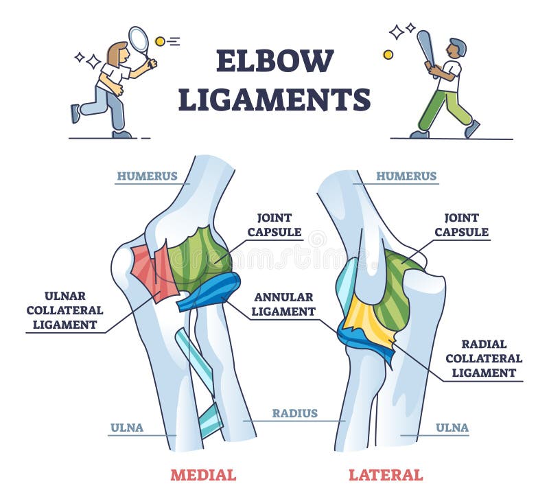
Free with trial Elbow ligaments with medical medial or lateral xray structure outline diagram. Labeled educational anatomical joint capsule, radius and ulna parts as inner medicine explanation vector illustration. Bone structure vectors Elbow ligaments with medical medial or lateral xray structure outline diagram
Free with trial Horse skeleton with animal skeletal system and bone anatomy outline diagram. Labeled educational scheme with biological detailed explanation vector illustration. Stallion zoology with internal parts. Bone structure vectors Horse skeleton with animal skeletal system and bone anatomy outline diagram. Labeled educational scheme with biological detailed explanation vector
Free with trial Four stages of osteoporosis. Landscape poster. Osteoporotic bone and healthy bone structure in comparison. Vector illustration useful for medical, educational or scientific graphic design. Bone structure vectors Osteoporosis Bones Poster. Four stages of osteoporosis. Landscape poster. Osteoporotic bone and healthy bone structure in comparison. Vector illustration useful for medical, educational or scientific graphic design.
Free with trial Vector illustration of the structure of the human spine. Bone structure vectors Illustration of the structure of the human spine
Free with trial Vector illustration of the structure of the human spine. Bone structure vectors Illustration of the structure of the human spine
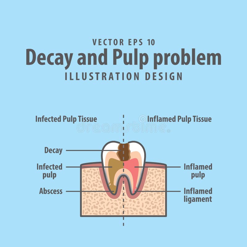
Free with trial Decay and Pulp problem cross-section structure inside tooth diagram and chart illustration vector on blue background. Dental concept. Bone structure vectors Decay and Pulp problem cross-section structure inside tooth
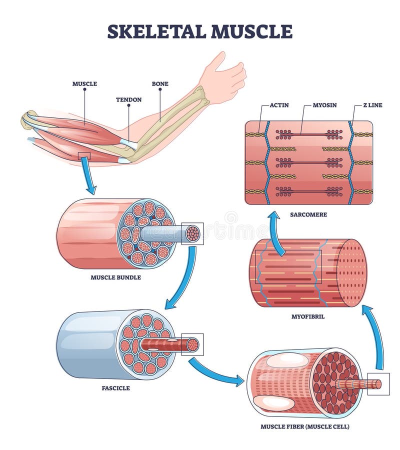
Free with trial Skeletal muscle structure layers with anatomical arm closeups outline diagram. Labeled educational descriptions with bundle, fascicle, fibers, myofibril and sarcomere sections vector illustration. Bone structure vectors Skeletal muscle structure layers with anatomical closeups outline diagram. Skeletal muscle structure layers with anatomical arm closeups outline diagram. Labeled educational descriptions with bundle, fascicle, fibers, myofibril and sarcomere sections vector illustration.
Free with trial Human spine structure vector illustration. Backbone and vertebral column anatomy with section names. Scoliosis concept and symbol of spinal surgery. Side lateral view isolated. Medical banner n. Bone structure vectors Human spine structure anatomy. Human spine structure vector illustration. Backbone and vertebral column anatomy with section names. Scoliosis concept and symbol of spinal surgery. Side lateral view isolated. Medical banner n
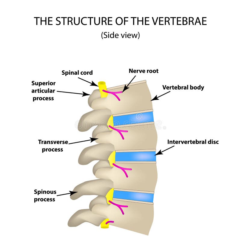
Free with trial The anatomical structure of the spine. Side view. The intervertebral discs. Infographics. Vector illustration on isolated background. Bone structure vectors The structure of the spine. Side view. The intervertebral discs. Infographics. Vector illustration on isolated background
Free with trial The anatomical structure of the intervertebral disc. Top view. Spine. Infographics. Vector illustration on isolated background. Bone structure vectors The anatomical structure of the intervertebral disc. Top view. Spine. Infographics. Vector illustration on isolated background
Free with trial Anatomical training poster. Fingernail Anatomy. Cross-section of the finger. Structure of human nail, vector. Bone structure vectors Anatomical training poster. Fingernail Anatomy. Cross-section of the finger. Structure of human nail
Free with trial Vector tooth structure. Cross Section Anatomy with all parts. Medical infogtaphic template. Bone structure vectors Vector tooth structure. Cross Section Anatomy with all parts.
Free with trial Tooth in a cut. Medical diagram of the structure of the inside cross-section of the tooth. Vector infographic concept isolated on white background. Bone structure vectors Tooth in a cut. Medical diagram of the structure of the inside cross-section of the tooth. Vector infographic concept
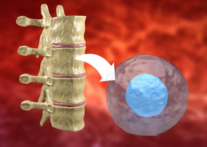
Free with trial Therapy with stem cells taken from the bone marrow to treat diseases of the human body. 3D rendering. Bone structure illustrations Therapy with stem cells taken from the bone marrow to treat diseases of the human body
Free with trial Shining bone with lytic lesion. Bone structure vectors Osteoporosis. Shining bone with lytic lesion
Free with trial Herniated disk concept and spine pain diagnostic as a human spinal system symbol as medical health problem and anatomy symbol with the skeletal bone structure and intervertebral discs closeup. Bone structure illustrations Herniated Disk
Free with trial Human Spine concept as medical health care anatomy symbol with the skeletal spinal bone structure closeup on a dark blue background as blank copy space. Bone structure illustrations Human Spine Concept
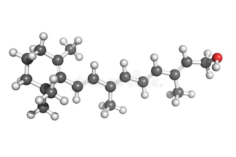
Free with trial Ball and stick model of retinol (vitamin A). Atoms are coloured according to convention (carbon-grey, hydrogen-white, oxygen-red). Vitamin A is essential for vision, skin health, bone growth and teeth mineralization. Bone structure illustrations Vitamin A structure. Ball and stick model of retinol (vitamin A). Atoms are coloured according to convention (carbon-grey, hydrogen-white, oxygen-red). Vitamin A is essential for vision, skin health, bone growth and teeth mineralization.
Free with trial 3d rendering of the female structure of muscle in gymnastic pose as illustration. Bone structure illustrations Structure of muscle of a sporty woman. 3d rendering of the female structure of muscle in gymnastic pose as illustration
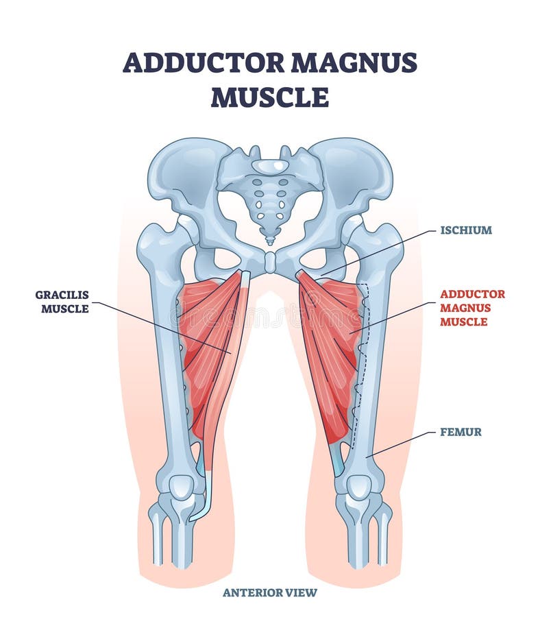
Free with trial Adductor magnus muscle with ischium and femur skeleton outline diagram. Labeled educational gracilis muscular system from anterior view vector illustration. Human body hips and legs inner structure. Bone structure vectors Adductor magnus muscle with ischium and femur skeleton outline diagram
Free with trial Vector illustration anatomy of human legs and diagram of human bones isolated on white background. For advertising and medical publications. EPS 10. Bone structure vectors Bone fracture_Human leg anatomy and skeleton. Vector illustration anatomy of human legs and diagram of human bones isolated on white background. For advertising and medical publications. EPS 10.
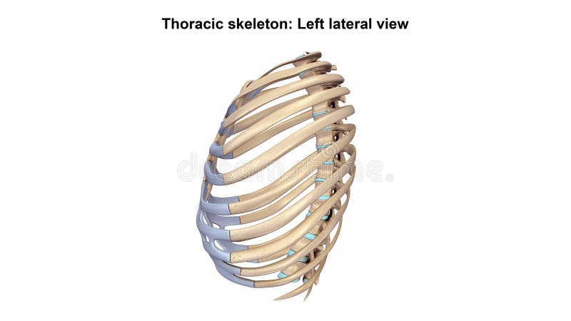
Free with trial Thoracic Skeleton The human rib cage is made up of 12 paired rib bones; each are symmetrically paired on a right and left side. Of all 24 ribs, the first seven pairs are often labeled as `true. ` These bones are connected to the costal cartilage, while the five other `false` sets are not. The ribcage also encloses the thoracic cavity and helps protect the heart and lungs from damage. There are 24 ribs in the human body, divided into two sets of 12 curved, flat bones. Each one is attached by cartilage at the back to the thoracic vertebrae. MEN and women have 12 pairs of ribs a few individuals have 13 or 11 pairs. The idea that men have fewer ribs than women is widespread but wrong, perhaps deriving from the biblical story of Eve being made from one of Adam`s ribs. Both men and women have 24 ribs, twelve on each side. Floating rib: One of the last two ribs. A rib is said to be `floating` if it does not attach to the sternum the breast bone or to another rib. There are usually 12 pairs of ribs in all. Each pair of ribs is attached to the building blocks of the spine the vertebrae in the back. The ribs partially enclose and protect the chest cavity, where many vital organs including the heart and the lungs are located. The rib cage is collectively made up of long, curved individual bones with joint-connections to the spinal vertebrae. Bone structure illustrations Thoracic Skeleton Lateral view. Thoracic Skeleton The human rib cage is made up of 12 paired rib bones; each are symmetrically paired on a right and left side. Of all 24 ribs, the first seven pairs are often labeled as `true.` These bones are connected to the costal cartilage, while the five other `false` sets are not. The ribcage also encloses the thoracic cavity and helps protect the heart and lungs from damage. There are 24 ribs in the human body, divided into two sets of 12 curved, flat bones. Each one is attached by cartilage at the back to the thoracic vertebrae. MEN and women have 12 pairs of ribs a few individuals have 13 or 11 pairs. The idea that men have fewer ribs than women is widespread but wrong, perhaps deriving from the biblical story of Eve being made from one of Adam`s ribs. Both men and women have 24 ribs, twelve on each side. Floating rib: One of the last two ribs. A rib is said to be `floating` if it does not attach to the sternum the breast bone or to another rib. There are usually 12 pairs of ribs in all. Each pair of ribs is attached to the building blocks of the spine the vertebrae in the back. The ribs partially enclose and protect the chest cavity, where many vital organs including the heart and the lungs are located. The rib cage is collectively made up of long, curved individual bones with joint-connections to the spinal vertebrae.
Free with trial Mesenchymal stem cells are multipotent stromal cells. differentiate into osteoblast bone, chondrocyte cartilage, myocytes muscle. Bone structure vectors Mesenchymal stem cells are multipotent stromal cells
Free with trial Erythropoietin. Glycoprotein cytokine secreted by the kidney in response to cellular hypoxia that stimulates red blood cell production erythropoiesis in the bone marrow. Vector illustration. Bone structure illustrations Erythropoietin and erythropoiesis. Erythropoietin. Glycoprotein cytokine secreted by the kidney in response to cellular hypoxia that stimulates red blood cell production erythropoiesis in the bone marrow. Vector illustration
Free with trial Medical illustration showing tooth decay affecting enamel and dentin with visible pulp and nerve structure. Suitable for dental education and healthcare materials. Bone structure illustrations Dental Anatomy Illustration with Tooth Decay and Pulp Structure. Medical illustration showing tooth decay affecting enamel and dentin with visible pulp and nerve structure. Suitable for dental education and healthcare materials.
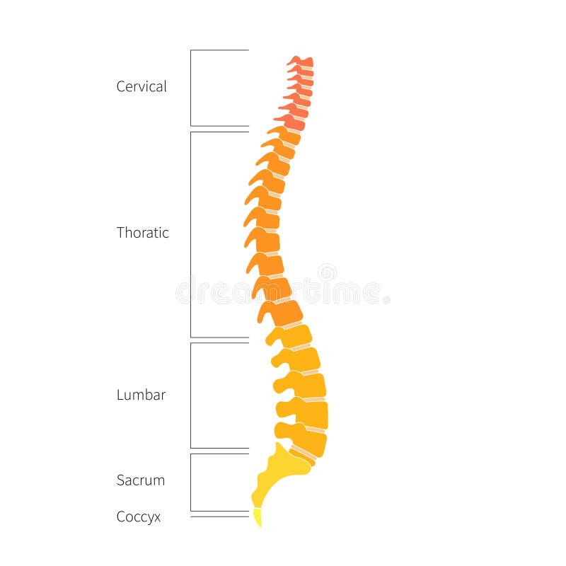
Free with trial Vertebral Column spine structure of human body. View with all vertebrae groups. cervical, thoracic, lumbar, sacrum and coccyx. Bone structure vectors Vertebral Column spine structure. Vertebral Column spine structure of human body. View with all vertebrae groups. cervical, thoracic, lumbar, sacrum and coccyx.
Free with trial Estrogen action. Oestrogen, or estradiol is the primary female sex hormone. Woman silhouette with highlighted internal organs liver, uterus, ovary, brain, adrenal gland, bone, muscle and heart. vector illustration for medical, educational and science use. Bone structure vectors Estrogen action. Woman silhouette with highlighted internal organs. Estrogen action. Oestrogen, or estradiol is the primary female sex hormone. Woman silhouette with highlighted internal organs liver, uterus, ovary, brain, adrenal gland, bone, muscle and heart. vector illustration for medical, educational and science use
Free with trial Hormones produced by the parathyroid gland. Parathyroid hormone PTH. parathormone. parathyrin is regulates the serum calcium in bone, kidney, and intestine. Bone structure vectors Hormones produced by the parathyroid gland. Parathyroid hormone PTH
Free with trial Anatomy of dog paws with forelimb and hindlimb bones vector illustration. Educational labeled skeleton comparison with zoological inside structure scheme. Animal legs inner closeup examination model. Bone structure vectors Anatomy of dog paws with forelimb and hindlimb bones vector illustration
Free with trial Osteoporosis is a disease characterized by low bone mass and deterioration of bone tissue, leading to fragility and fractures. Bone structure vectors Osteoporosis
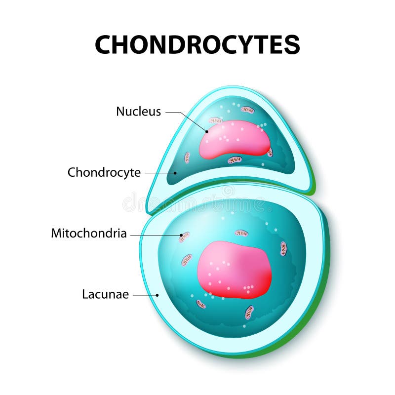
Free with trial Chondrocytes is cells found in healthy cartilage. Chondrocyte lie in lacunae spaces around cells. Bone structure vectors Structure of the chondrocytes. Chondrocytes is cells found in healthy cartilage. Chondrocyte lie in lacunae spaces around cells.
Free with trial Herniated disc injury as labeled spinal pain explanation vector illustration. Medical condition with back bone trauma and nerve root compression by nucleus pulposus. Problematic example comparison. Bone structure vectors Herniated disc injury as labeled spinal pain explanation vector illustration
Free with trial Vector illustration of tissues of the internal environment: bone and cartilage. Bone structure vectors Tissue of the internal environment. Vector illustration of tissues of the internal environment: bone and cartilage
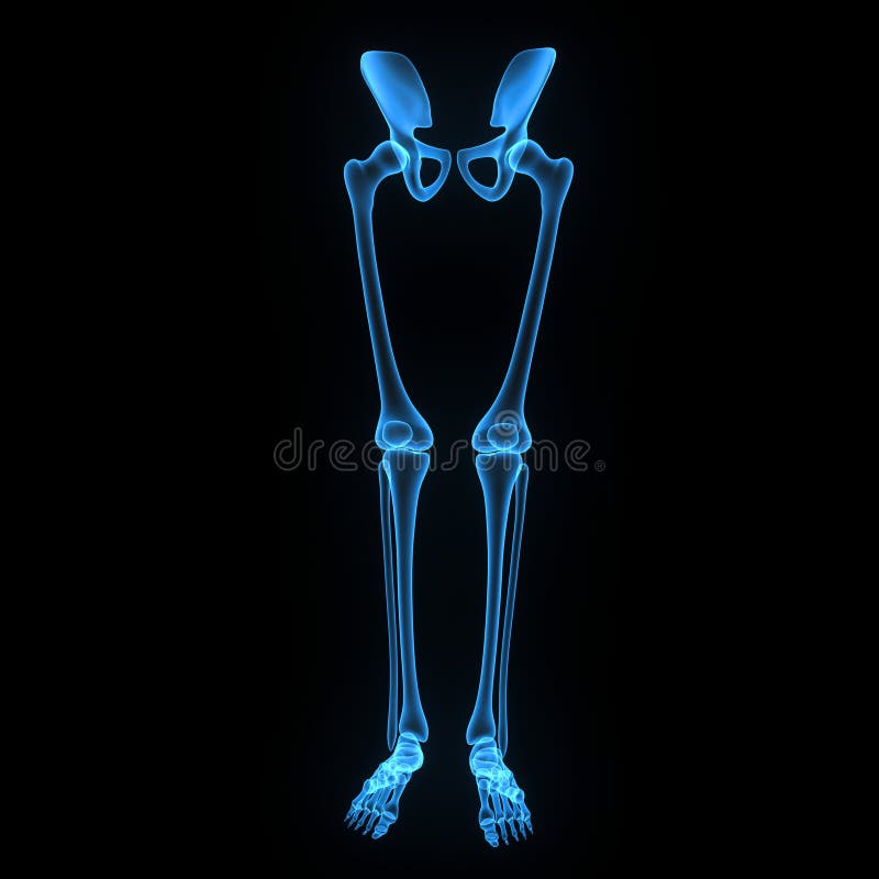
Free with trial The hip joint is one of the most important joints in the human body. It allows us to walk, run, and jump. It bears our body’s weight and the force of the strong muscles of the hip and leg. Yet the hip joint is also one of our most flexible joints and allows a greater range of motion than all other joints in the body except for the shoulder. The hip joint is a ball-and-socket synovial joint formed between the os coxa (hip bone) and the femur. A round, cup-shaped structure on the os coax, known as the acetabulum, forms the socket for the hip joint. The rounded head of the femur forms the ball of the joint. The tibia, sometimes known as the shin bone, is the larger and stronger of the two lower leg bones. It forms the knee joint with the femur and the ankle joint with the fibula and tarsus. The fibula is the long, thin and lateral bone of the lower leg. It runs parallel to the tibia, or shin bone, and plays a significant role in stabilizing the ankle and supporting the muscles of the lower leg. The bones of the ankle and foot form the most distal region of the lower limb in the appendicular skeleton. These bones are responsible for the propulsion, balance, and support of the body’s weight through many diverse activities, such as standing, walking, running, and jumping. Bone structure illustrations Skeleton: Hip, Femur, Tibia, Fibula, Ankle and Foot bones. The hip joint is one of the most important joints in the human body. It allows us to walk, run, and jump. It bears our body’s weight and the force of the strong muscles of the hip and leg. Yet the hip joint is also one of our most flexible joints and allows a greater range of motion than all other joints in the body except for the shoulder. The hip joint is a ball-and-socket synovial joint formed between the os coxa (hip bone) and the femur. A round, cup-shaped structure on the os coax, known as the acetabulum, forms the socket for the hip joint. The rounded head of the femur forms the ball of the joint. The tibia, sometimes known as the shin bone, is the larger and stronger of the two lower leg bones. It forms the knee joint with the femur and the ankle joint with the fibula and tarsus. The fibula is the long, thin and lateral bone of the lower leg. It runs parallel to the tibia, or shin bone, and plays a significant role in stabilizing the ankle and supporting the muscles of the lower leg. The bones of the ankle and foot form the most distal region of the lower limb in the appendicular skeleton. These bones are responsible for the propulsion, balance, and support of the body’s weight through many diverse activities, such as standing, walking, running, and jumping.
Free with trial Human Health Infographic Elements Objects. Heart, brain, liver, lungs, stomach, intestine, skin hair structure, red blood cells, fat and immune cells, body parts, bones etc. Bone structure vectors Human Health Infographic Elements Objects.
Free with trial Parathyroid hormone and Calcium metabolism. parathormone or parathyrin PTH that regulates serum calcium through its effects on bone, kidney, and the intestine. vector illustration. Bone structure vectors Parathyroid hormone and Calcium metabolism
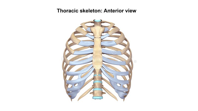
Free with trial Thoracic Skeleton The human rib cage is made up of 12 paired rib bones; each are symmetrically paired on a right and left side. Of all 24 ribs, the first seven pairs are often labeled as `true. ` These bones are connected to the costal cartilage, while the five other `false` sets are not. The ribcage also encloses the thoracic cavity and helps protect the heart and lungs from damage. There are 24 ribs in the human body, divided into two sets of 12 curved, flat bones. Each one is attached by cartilage at the back to the thoracic vertebrae. MEN and women have 12 pairs of ribs a few individuals have 13 or 11 pairs. The idea that men have fewer ribs than women is widespread but wrong, perhaps deriving from the biblical story of Eve being made from one of Adam`s ribs. Both men and women have 24 ribs, twelve on each side. Floating rib: One of the last two ribs. A rib is said to be `floating` if it does not attach to the sternum the breast bone or to another rib. There are usually 12 pairs of ribs in all. Each pair of ribs is attached to the building blocks of the spine the vertebrae in the back. The ribs partially enclose and protect the chest cavity, where many vital organs including the heart and the lungs are located. The rib cage is collectively made up of long, curved individual bones with joint-connections to the spinal vertebrae. Bone structure illustrations Thoracic Skeleton Anterior view. Thoracic Skeleton The human rib cage is made up of 12 paired rib bones; each are symmetrically paired on a right and left side. Of all 24 ribs, the first seven pairs are often labeled as `true.` These bones are connected to the costal cartilage, while the five other `false` sets are not. The ribcage also encloses the thoracic cavity and helps protect the heart and lungs from damage. There are 24 ribs in the human body, divided into two sets of 12 curved, flat bones. Each one is attached by cartilage at the back to the thoracic vertebrae. MEN and women have 12 pairs of ribs a few individuals have 13 or 11 pairs. The idea that men have fewer ribs than women is widespread but wrong, perhaps deriving from the biblical story of Eve being made from one of Adam`s ribs. Both men and women have 24 ribs, twelve on each side. Floating rib: One of the last two ribs. A rib is said to be `floating` if it does not attach to the sternum the breast bone or to another rib. There are usually 12 pairs of ribs in all. Each pair of ribs is attached to the building blocks of the spine the vertebrae in the back. The ribs partially enclose and protect the chest cavity, where many vital organs including the heart and the lungs are located. The rib cage is collectively made up of long, curved individual bones with joint-connections to the spinal vertebrae.
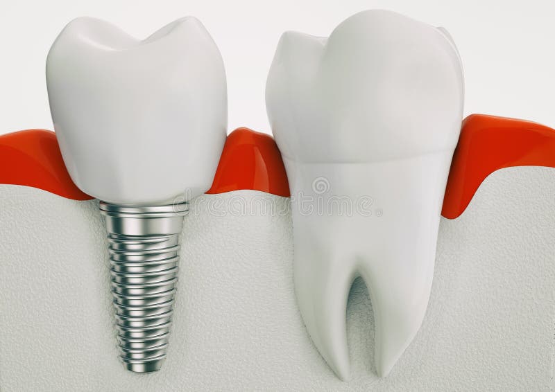
Free with trial Dental implant, and Normal tooth on the white background. tooth anatomy. illustration showing closeup cut away of the bone of lower jaw, and part of Dental implant Crown, Abutment, Implant. Bone structure vectors Dental implant, and Normal tooth
Free with trial Vector illustration of the structure of the cervical vertebrae. Bone structure vectors Structure of the cervical vertebrae
Free with trial Perichondrium as hyaline, fibrous and elastic cartilage membrane outline diagram. Labeled educational tissue layer structure with cartilaginous ground substance, halo and collagen vector illustration. Bone structure vectors Perichondrium as hyaline and elastic cartilage membrane outline diagram. Perichondrium as hyaline, fibrous and elastic cartilage membrane outline diagram. Labeled educational tissue layer structure with cartilaginous ground substance, halo and collagen vector illustration.
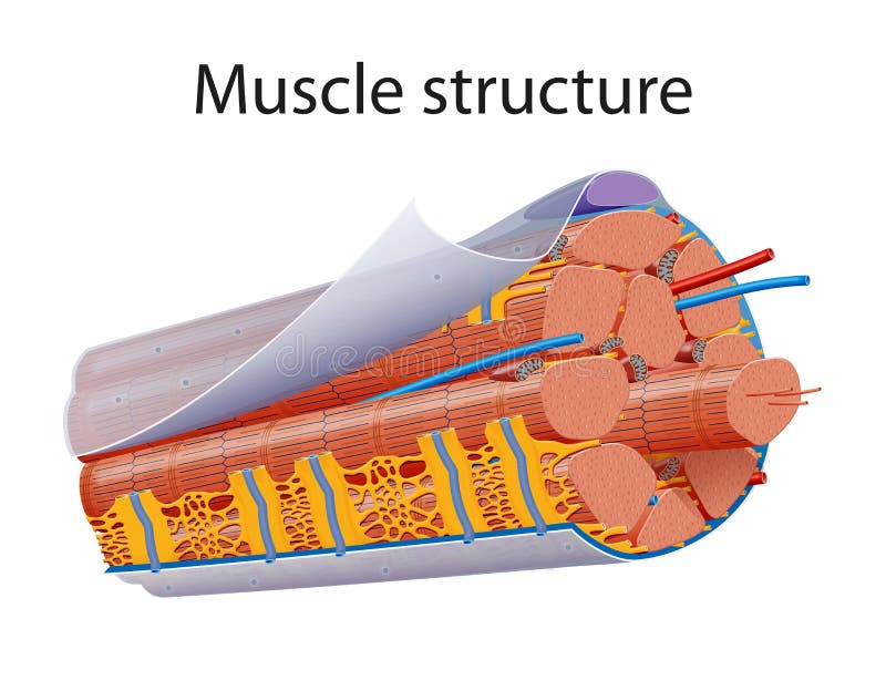
Free with trial Skeletal muscles are organs of the vertebrate muscular system that are mostly attached by tendons to bones of the skeleton. The muscle cells of skeletal muscles are much longer than in the other types of muscle tissue, and are often known as muscle fibers. Bone structure illustrations Illustration of Structure Skeletal Muscle with sarcomere. Skeletal muscles are organs of the vertebrate muscular system that are mostly attached by tendons to bones of the skeleton. The muscle cells of skeletal muscles are much longer than in the other types of muscle tissue, and are often known as muscle fibers
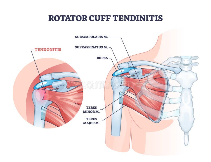
Free with trial Rotator cuff tendinitis as shoulder muscular inflammation outline diagram. Labeled educational anatomical structure and medical description with body muscle injury and pain cause vector illustration. Bone structure vectors Rotator cuff tendinitis as shoulder muscular inflammation outline diagram
Free with trial Vertebral Column Anatomy Infographic Diagram for medical science education. vector spine of human body. drawing all vertebra classification. structure part of skeletal system. anterior posterior lateral view. Bone structure vectors Vertebral Column Anatomy Infographic Diagram
Free with trial Brachioradialis muscle medical location with anatomical bones outline diagram. Labeled educational scheme with hands inner structure and muscular description vector illustration. Body and arm parts. Bone structure vectors Brachioradialis muscle medical location with anatomical bones outline diagram
Free with trial Different human cell types icon set. Stock vector illustration of bone, nerve, epithelial, muscle, blood, stem, sperm and oocyte in a circle. Medicine and biology collection. Bone structure vectors Different human cell types