Free with trial Anatomy of a euglena. Euglena freshwater protozoan. It is composed of chlorophyll and has a rudimentary eye. Vector diagram. Cell flagellate vectors Structure of a euglena. Anatomy of a euglena. Euglena freshwater protozoan. It is composed of chlorophyll and has a rudimentary eye. Vector diagram
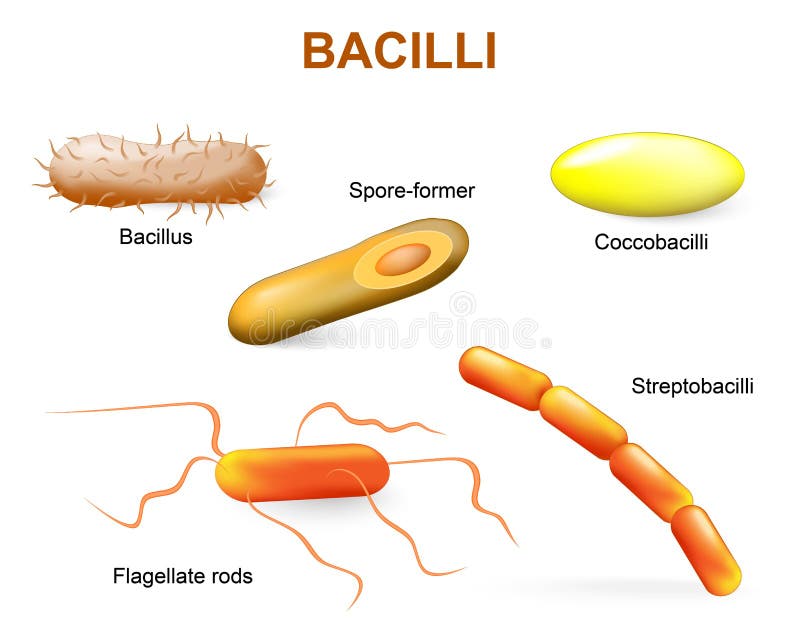
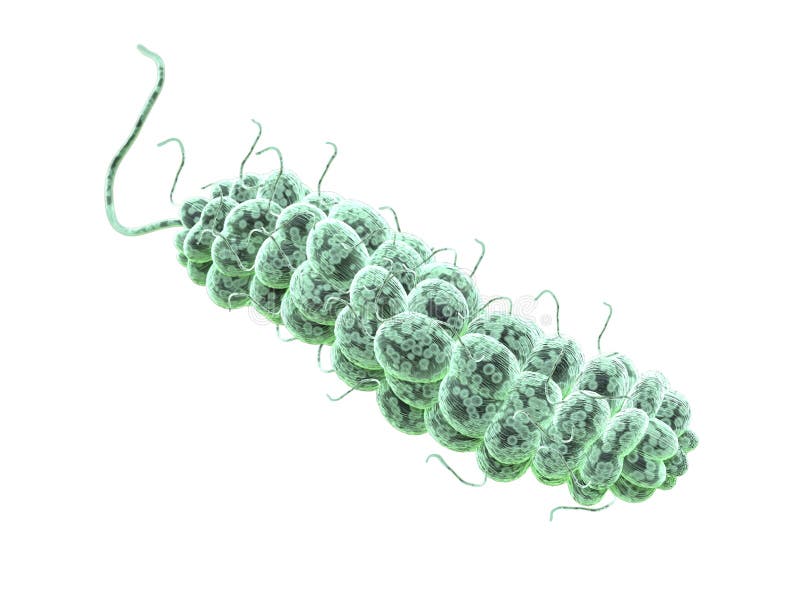
Free with trial Bacillii. Common bacteria infecting human. Cell flagellate vectors Types of bacteria. bacilli. Bacillii. Common bacteria infecting human.
Free with trial Euglena is a genus of unicellular flagellate protists. It is the best known and most widely studied member of the phylum Euglenophyta, a diverse group containing some 44 genera and at least 800 species. Cell flagellate illustrations Euglena
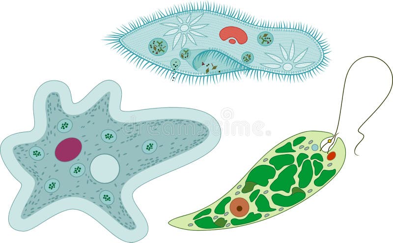
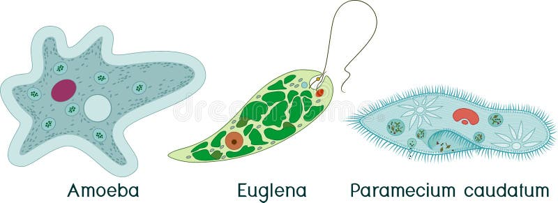
Free with trial Set of unicellular organisms protozoa: Paramecium caudatum, Amoeba proteus and Euglena viridis isolated on white background. Cell flagellate vectors Set of unicellular organisms protozoa: Paramecium caudatum, Amoeba proteus and Euglena viridis
Free with trial Cross section of a Chlamydomonas. Structure of the algae cell. Vector diagram for educational, biological, and science use. Cell flagellate vectors Cross section of a Chlamydomonas. Structure of the algae cell
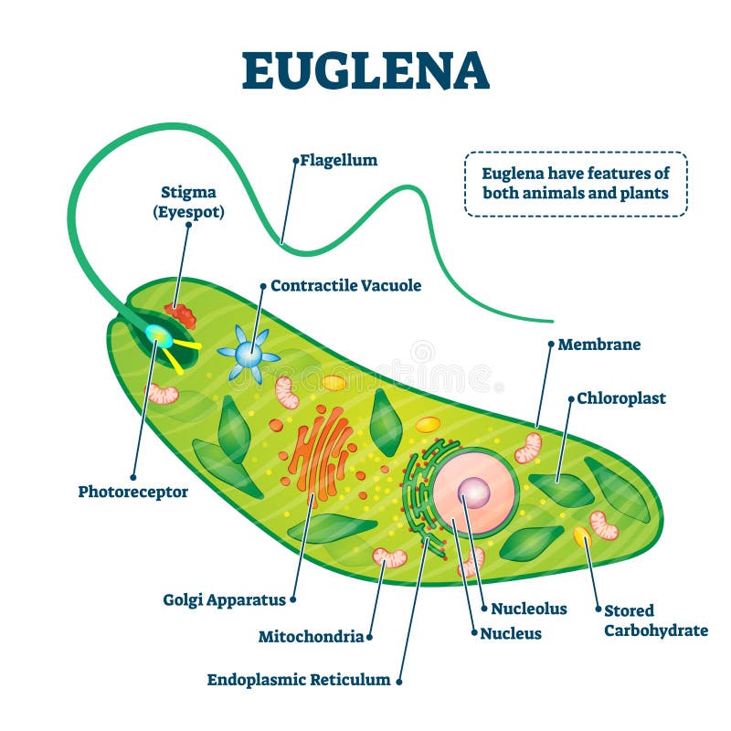
Free with trial Euglena vector illustration. Labeled microorganism structure and description. Biological inner parts scheme for genus of single cell flagellate eukaryotes. Diagram with flagellum and stigma location. Cell flagellate vectors Euglena vector illustration. Labeled microorganism structure or description. Euglena vector illustration. Labeled microorganism structure and description. Biological inner parts scheme for genus of single cell flagellate eukaryotes. Diagram with flagellum and stigma location.
Free with trial Set of unicellular organisms protozoa: Paramecium caudatum, Amoeba proteus and Euglena viridis isolated on white background. Cell flagellate vectors Set of unicellular organisms protozoa: Paramecium caudatum, Amoeba proteus and Euglena viridis
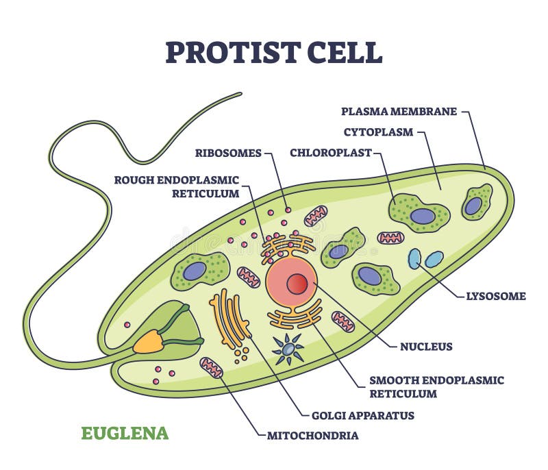
Free with trial Protist cell anatomy with euglena microorganism structure outline diagram. Labeled educational scheme with green organism parts description vector illustration. Eukaryotic biological inner structure. Cell flagellate vectors Protist cell anatomy with euglena microorganism structure outline diagram
Free with trial Structure of Chlamydomonas cell isolated on white background. Cell flagellate vectors Structure of Chlamydomonas cell
Free with trial Large number of E coli Bacteria. This is shape of Bacteria. it is flagellate rods type category. E coli Bacteria. Cell flagellate illustrations E coli Bacteria. flagellate rods type category. Large number of E coli Bacteria. This is shape of Bacteria. it is flagellate rods type category. E coli Bacteria
Free with trial 3D illustration of salmonella typhi Bacteria. This is shape of Bacteria. E coli Bacteria. flagellate rods. Cell flagellate illustrations 3D illustration of salmonella typhi Bacteria. flagellate rods. 3D illustration of salmonella typhi Bacteria. This is shape of Bacteria. E coli Bacteria. flagellate rods.
Free with trial 3D illustration of salmonella typhi Bacteria. This is shape of Bacteria. E coli Bacteria. flagellate rods. Cell flagellate illustrations 3D illustration of salmonella typhi Bacteria. flagellate rods. 3D illustration of salmonella typhi Bacteria. This is shape of Bacteria. E coli Bacteria. flagellate rods
Free with trial Large number of E coli Bacteria. This is shape of Bacteria. it is flagellate rods type category. E coli Bacteria. Cell flagellate illustrations E coli Bacteria. flagellate rods type category. Large number of E coli Bacteria. This is shape of Bacteria. it is flagellate rods type category. E coli Bacteria.
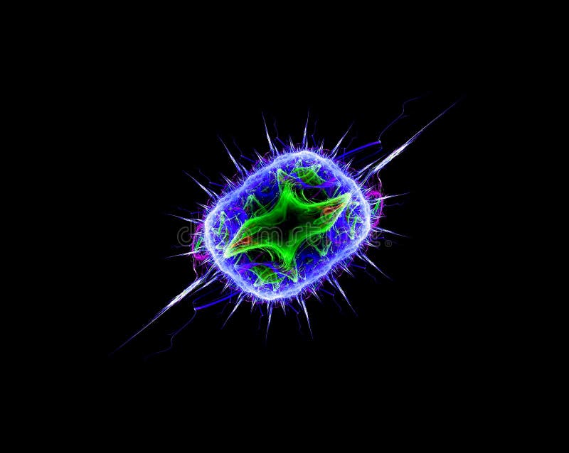
Free with trial Computer generated illustration, 3D fractal-flagellate bacteria-like, at microscopic view. Cell flagellate illustrations Fluorescent Bacteria. Computer generated illustration, 3D fractal-flagellate bacteria-like, at microscopic view.
Free with trial Volvox is a polyphyletic genus in the volvocine green algae clade. Each mature Volvox colony is composed of up to thousands of cells from two differentiated cell types: numerous flagellate somatic cells and a smaller number of germ cells lacking in soma that are embedded in the surface of a hollow sphere or coenobium containing an extracellular matrix[ made of glycoproteins. Cell flagellate illustrations Volvox aureus - Wimperkugel - green alga. Volvox is a polyphyletic genus in the volvocine green algae clade. Each mature Volvox colony is composed of up to thousands of cells from two differentiated cell types: numerous flagellate somatic cells and a smaller number of germ cells lacking in soma that are embedded in the surface of a hollow sphere or coenobium containing an extracellular matrix[ made of glycoproteins
Free with trial Euglena is a genus of single-celled flagellate protists. It is the best known and most widely studied member of the class Euglenoidea, a diverse group containing some 54 genera and at least 800 species. Cell flagellate illustrations Euglena
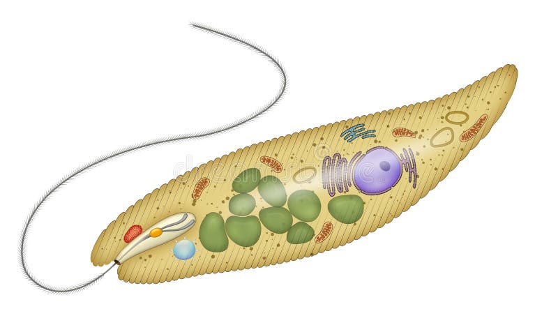
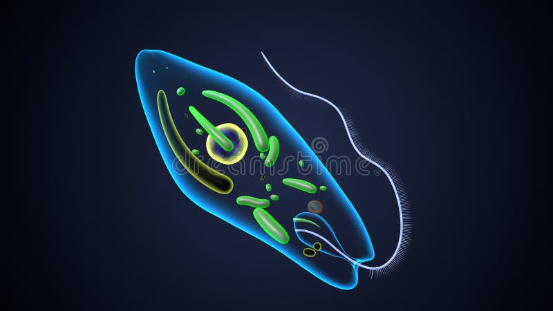
Free with trial Euglena is a genus of single cell flagellate eukaryotes. It is the best known and most widely studied member of the class Euglenoidea, a diverse group containing some 54 genera and at least 800 species. Species of Euglena are found in fresh water and salt water. Cell flagellate illustrations Anatomy of a euglena. Euglena is a genus of single cell flagellate eukaryotes. It is the best known and most widely studied member of the class Euglenoidea, a diverse group containing some 54 genera and at least 800 species. Species of Euglena are found in fresh water and salt water
Free with trial Vector illustrationof unicellular organisms. Amoeba proteus Paramecium caudatum and Euglena viridis. Protozoa. Cell flagellate vectors Vector illustrationof unicellular organisms. Amoeba proteus Paramecium caudatum and Euglena viridis. Protozoa
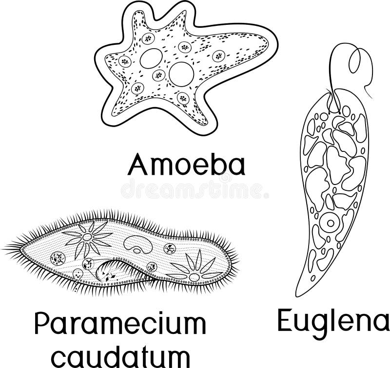
Free with trial Coloring page. Set of unicellular organisms protozoa: Paramecium caudatum, Amoeba proteus and Euglena viridis isolated on white background. Cell flagellate vectors Coloring page. Set of unicellular organisms protozoa: Paramecium caudatum, Amoeba proteus and Euglena viridis
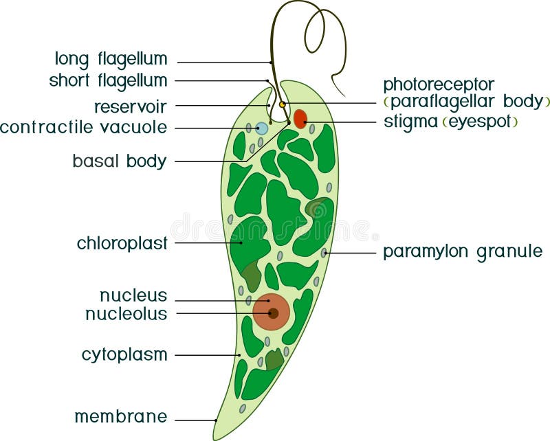
Free with trial Diagram of Euglena. Structure of Euglena viridis with titles. Cell flagellate vectors Diagram of Euglena. Structure of Euglena viridis with different organelles. Diagram of Euglena. Structure of Euglena viridis with titles
Free with trial Coloring page with structure of Euglena viridis with titles isolated on white background. Cell flagellate vectors Coloring page with structure of Euglena viridis with titles
Free with trial Diagram of Euglena. Structure of Euglena viridis with titles isolated on white background. Cell flagellate vectors Diagram of Euglena. Structure of Euglena viridis with titles
Free with trial Set of unicellular organisms protozoa: Paramecium caudatum, Amoeba proteus, Chlamydomonas and Euglena viridis on white background. Cell flagellate vectors Set of unicellular organisms protozoa: Paramecium caudatum, Amoeba proteus, Chlamydomonas and Euglena viridis
Free with trial Chlamydomonas is a genus of green algae belonging to the division Chlorophyta. These single-celled organisms are known for their flagellated cells and their ability to perform photosynthesis. Chlamydomonas is commonly found in freshwater environments such as ponds, lakes, and rivers, although some species can also thrive in soil or marine habitats. Cell flagellate vectors Chlamydomonas
Free with trial Set of microscopic unicellular organisms: protozoa Paramecium caudatum, Amoeba proteus, Chlamydomonas, Euglena viridis and green algae Chlorella, Spirogyra isolated on white background. Cell flagellate vectors Set of microscopic unicellular organisms: protozoa Paramecium caudatum, Amoeba proteus, Chlamydomonas
Free with trial Coloring page. Set of unicellular organisms protozoa: Paramecium caudatum, Amoeba proteus, Chlamydomonas and Euglena viridis on white background. Cell flagellate vectors Coloring page. Set of unicellular organisms protozoa: Paramecium caudatum, Amoeba proteus, Chlamydomonas and Euglena viridis
Free with trial Set of microscopic unicellular organisms: protozoa Paramecium caudatum, Amoeba proteus, Chlamydomonas, Euglena viridis, green algae Chlorella, Spirogyra and bacteria isolated on white background. Cell flagellate vectors Set of microscopic unicellular organisms: protozoa Paramecium caudatum, Amoeba proteus, Chlamydomonas
Free with trial Protozoa Paramecium caudatum, Amoeba proteus, Chlamydomonas, Euglena viridis, green algae Chlorella, Spirogyra under magnifying glass isolated on white background. Cell flagellate vectors Protozoa Paramecium caudatum, Amoeba proteus, Chlamydomonas, Euglena viridis, green algae Chlorella, Spirogyra
Free with trial Protozoa, protista and amoeba microorganism cells, vector micro organism. Ameba and protist unicellular cells in lab microscope, protozoan eukaryotic organism types. Cell flagellate vectors Protozoa, protista and amoeba microorganism cells
Free with trial Green Algae Chlorella proteus science icon with nucleus, vacuole, contractile. Biology education laboratory cartoon protozoa organism. Bold bright unicellular microorganism. Vector illustration isolated on white. Cell flagellate vectors Green Algae Chlorella proteus science icon with nucleus, vacuole, contractile. Biology education laboratory cartoon
Free with trial Paramecium Caudatum proteus science icon with nucleus, vacuole, contractile. Biology education laboratory cartoon protozoa organism. Bold bright unicellular microorganism. Vector illustration isolated on white. Cell flagellate vectors Paramecium Caudatum proteus science icon with nucleus, vacuole, contractile. Biology education laboratory cartoon
Free with trial Euglena structure anatomy diagram poster chart, medical illustration vector. Science biology study. Single cell flagellate eukaryotes, chloroplast, nucleolus, mitochondrion, flagellum, photoreceptor. Cell flagellate vectors Euglena structure anatomy diagram poster chart, medical illustration vector. Science biology study. Single cell flagellate
Free with trial Euglena structure anatomy diagram poster chart, medical illustration vector. Science biology study. Single cell flagellate eukaryotes, chloroplast, nucleolus, mitochondrion, flagellum, photoreceptor. Cell flagellate vectors Euglena structure anatomy diagram poster chart, medical illustration vector. Science biology study. Single cell flagellate
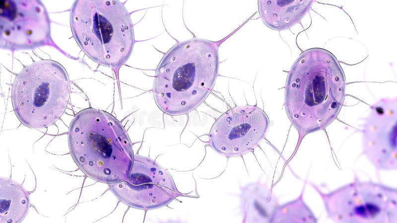
Free with trial A detailed microscopic view of isolated flagellate protozoa on a white background, showcasing their intricate cellular structures. Cell flagellate illustrations Microscopic View of Flagellate Protozoa Isolated on White Background. A detailed microscopic view of isolated flagellate protozoa on a white background, showcasing their intricate cellular structures.
Free with trial This captivating view unveils flagellate protozoa, showcasing their unique structure and movement. Cell flagellate illustrations Exploring the Intricate World of Flagellate Protozoa Revealed Under Microscopic Examination. This captivating view unveils flagellate protozoa, showcasing their unique structure and movement.
Free with trial Chlamydomonas structure anatomy diagram poster chart, medical illustration vector. Science biology study. Green algae unicellular flagellate, basal, chloroplast, pyrenoid, vacuole, eyespot. Cell flagellate vectors Chlamydomonas structure anatomy diagram poster chart, medical illustration vector. Science biology study. Green algae unicellular
Free with trial Conceptual image of flagellate bacterium. Cell flagellate illustrations Conceptual image of flagellate bacterium.
Free with trial Conceptual image of flagellate bacterium. Cell flagellate illustrations Conceptual image of flagellate bacterium.
Free with trial Conceptual image of flagellate bacterium. Cell flagellate illustrations Conceptual image of flagellate bacterium.
Free with trial Detailed, vibrant 3D rendering of diverse microscopic bacteria and virus organisms, including rod and flagellate shapes, in various colors (blue, pink, orange, green) on a stark black background. Cell flagellate illustrations Detailed, vibrant 3D rendering of diverse microscopic bacteria and virus organisms, including rod and flagellate shapes, in
Free with trial Magnified view showcases cercozoa, a diverse group of amoeboid and flagellate protozoa, demonstrating their varied shapes and textures. Cell flagellate illustrations Exploring the Intricate World of Cercozoa and Their Role in Microbial Ecosystems. Magnified view showcases cercozoa, a diverse group of amoeboid and flagellate protozoa, demonstrating their varied shapes and textures.
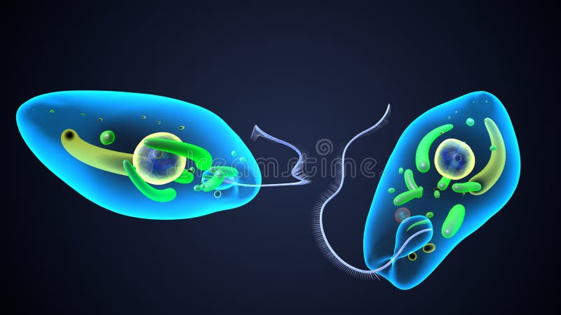
Free with trial Euglena is a genus of single cell flagellate eukaryotes. It is the best known and most widely studied member of the class euglenoidea, a diverse group containing some 54 genera and at least 200 species. Cell flagellate illustrations Euglena reproduction anatomy. 3d render. Euglena is a genus of single cell flagellate eukaryotes. It is the best known and most widely studied member of the class euglenoidea, a diverse group containing some 54 genera and at least 200 species
Free with trial Euglena is a genus of single cell flagellate eukaryotes. It is the best known and most widely studied member of the class euglenoidea, a diverse group containing some 54 genera and at least 200 species. Cell flagellate illustrations Euglena reproduction anatomy. 3d render. Euglena is a genus of single cell flagellate eukaryotes. It is the best known and most widely studied member of the class euglenoidea, a diverse group containing some 54 genera and at least 200 species
Free with trial Euglena is a genus of single cell flagellate eukaryotes. It is the best known and most widely studied member of the class euglenoidea, a diverse group containing some 54 genera and at least 200 species. Cell flagellate illustrations Euglena reproduction anatomy. 3d render. Euglena is a genus of single cell flagellate eukaryotes. It is the best known and most widely studied member of the class euglenoidea, a diverse group containing some 54 genera and at least 200 species
Free with trial Euglena is a genus of single cell flagellate eukaryotes. It is the best known and most widely studied member of the class euglenoidea, a diverse group containing some 54 genera and at least 200 species. Cell flagellate illustrations Euglena reproduction anatomy. 3d render. Euglena is a genus of single cell flagellate eukaryotes. It is the best known and most widely studied member of the class euglenoidea, a diverse group containing some 54 genera and at least 200 species
Free with trial Euglena is a genus of single cell flagellate eukaryotes. It is the best known and most widely studied member of the class euglenoidea, a diverse group containing some 54 genera and at least 200 species. Cell flagellate illustrations Euglena reproduction anatomy. 3d render. Euglena is a genus of single cell flagellate eukaryotes. It is the best known and most widely studied member of the class euglenoidea, a diverse group containing some 54 genera and at least 200 species
Free with trial Euglena is a genus of single cell flagellate eukaryotes. It is the best known and most widely studied member of the class euglenoidea, a diverse group containing some 54 genera and at least 200 species. Cell flagellate illustrations Euglena reproduction anatomy. 3d render. Euglena is a genus of single cell flagellate eukaryotes. It is the best known and most widely studied member of the class euglenoidea, a diverse group containing some 54 genera and at least 200 species
Free with trial Euglena is a genus of single cell flagellate eukaryotes. It is the best known and most widely studied member of the class euglenoidea, a diverse group containing some 54 genera and at least 200 species. Cell flagellate illustrations Euglena reproduction anatomy. 3d render. Euglena is a genus of single cell flagellate eukaryotes. It is the best known and most widely studied member of the class euglenoidea, a diverse group containing some 54 genera and at least 200 species
Free with trial Euglena is a genus of single cell flagellate eukaryotes. It is the best known and most widely studied member of the class euglenoidea, a diverse group containing some 54 genera and at least 200 species. Cell flagellate illustrations Euglena reproduction anatomy. 3d render. Euglena is a genus of single cell flagellate eukaryotes. It is the best known and most widely studied member of the class euglenoidea, a diverse group containing some 54 genera and at least 200 species
Free with trial Euglena is a genus of single cell flagellate eukaryotes. It is the best known and most widely studied member of the class euglenoidea, a diverse group containing some 54 genera and at least 200 species. Cell flagellate illustrations Euglena reproduction anatomy. 3d render. Euglena is a genus of single cell flagellate eukaryotes. It is the best known and most widely studied member of the class euglenoidea, a diverse group containing some 54 genera and at least 200 species
Free with trial Euglena is a genus of single cell flagellate eukaryotes. It is the best known and most widely studied member of the class euglenoidea, a diverse group containing some 54 genera and at least 200 species. Cell flagellate illustrations Euglena reproduction anatomy. 3d render. Euglena is a genus of single cell flagellate eukaryotes. It is the best known and most widely studied member of the class euglenoidea, a diverse group containing some 54 genera and at least 200 species
Free with trial Euglena is a genus of single cell flagellate eukaryotes. It is the best known and most widely studied member of the class euglenoidea, a diverse group containing some 54 genera and at least 200 species. Cell flagellate illustrations Euglena reproduction anatomy. 3d render. Euglena is a genus of single cell flagellate eukaryotes. It is the best known and most widely studied member of the class euglenoidea, a diverse group containing some 54 genera and at least 200 species
Free with trial Euglena is a genus of single cell flagellate eukaryotes. It is the best known and most widely studied member of the class euglenoidea, a diverse group containing some 54 genera and at least 200 species. Cell flagellate illustrations Euglena reproduction anatomy. 3d render. Euglena is a genus of single cell flagellate eukaryotes. It is the best known and most widely studied member of the class euglenoidea, a diverse group containing some 54 genera and at least 200 species
Free with trial Euglena is a genus of single cell flagellate eukaryotes. It is the best known and most widely studied member of the class euglenoidea, a diverse group containing some 54 genera and at least 200 species. Cell flagellate illustrations Euglena reproduction anatomy. 3d render. Euglena is a genus of single cell flagellate eukaryotes. It is the best known and most widely studied member of the class euglenoidea, a diverse group containing some 54 genera and at least 200 species
Free with trial Euglena is a genus of single cell flagellate eukaryotes. It is the best known and most widely studied member of the class euglenoidea, a diverse group containing some 54 genera and at least 200 species. Cell flagellate illustrations Euglena reproduction anatomy. 3d render. Euglena is a genus of single cell flagellate eukaryotes. It is the best known and most widely studied member of the class euglenoidea, a diverse group containing some 54 genera and at least 200 species
Free with trial Euglena is a genus of single cell flagellate eukaryotes. It is the best known and most widely studied member of the class euglenoidea, a diverse group containing some 54 genera and at least 200 species. Cell flagellate illustrations Euglena reproduction anatomy. 3d render. Euglena is a genus of single cell flagellate eukaryotes. It is the best known and most widely studied member of the class euglenoidea, a diverse group containing some 54 genera and at least 200 species
Free with trial Euglena is a genus of single cell flagellate eukaryotes. It is the best known and most widely studied member of the class euglenoidea, a diverse group containing some 54 genera and at least 200 species. Cell flagellate illustrations Euglena reproduction anatomy. 3d render. Euglena is a genus of single cell flagellate eukaryotes. It is the best known and most widely studied member of the class euglenoidea, a diverse group containing some 54 genera and at least 200 species
Free with trial Euglena is a genus of single cell flagellate eukaryotes. It is the best known and most widely studied member of the class euglenoidea, a diverse group containing some 54 genera and at least 200 species. Cell flagellate illustrations Euglena reproduction anatomy. 3d render. Euglena is a genus of single cell flagellate eukaryotes. It is the best known and most widely studied member of the class euglenoidea, a diverse group containing some 54 genera and at least 200 species
Free with trial Euglena is a genus of single cell flagellate eukaryotes. It is the best known and most widely studied member of the class euglenoidea, a diverse group containing some 54 genera and at least 200 species. Cell flagellate illustrations Euglena reproduction anatomy. 3d render. Euglena is a genus of single cell flagellate eukaryotes. It is the best known and most widely studied member of the class euglenoidea, a diverse group containing some 54 genera and at least 200 species
Free with trial Euglena is a genus of single cell flagellate eukaryotes. It is the best known and most widely studied member of the class euglenoidea, a diverse group containing some 54 genera and at least 200 species. Cell flagellate illustrations Euglena reproduction anatomy. 3d render. Euglena is a genus of single cell flagellate eukaryotes. It is the best known and most widely studied member of the class euglenoidea, a diverse group containing some 54 genera and at least 200 species
Free with trial Euglena is a genus of single cell flagellate eukaryotes. It is the best known and most widely studied member of the class euglenoidea, a diverse group containing some 54 genera and at least 200 species. Cell flagellate illustrations Euglena reproduction anatomy. 3d render. Euglena is a genus of single cell flagellate eukaryotes. It is the best known and most widely studied member of the class euglenoidea, a diverse group containing some 54 genera and at least 200 species
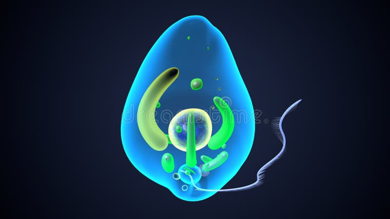
Free with trial Detailed rendering of a euglena cell showing flagellum and other structures against a dark backdrop Great for science and educational materials. Cell flagellate illustrations Examining Microscopic Parasite Euglena with Visible Structures and Organelles. Detailed rendering of a euglena cell showing flagellum and other structures against a dark backdrop Great for science and educational materials
Free with trial Delve into the intriguing world of cercozoa, where amoeboid and flagellate protozoa exhibit their distinct forms. Observing these microbes reveals the beauty of life at a microscopic level. Cell flagellate illustrations Fascinating Microscopic View of Cercozoa Showcasing Unique Shapes and Vibrant Colors. Delve into the intriguing world of cercozoa, where amoeboid and flagellate protozoa exhibit their distinct forms. Observing these microbes reveals the beauty of life at a microscopic level.
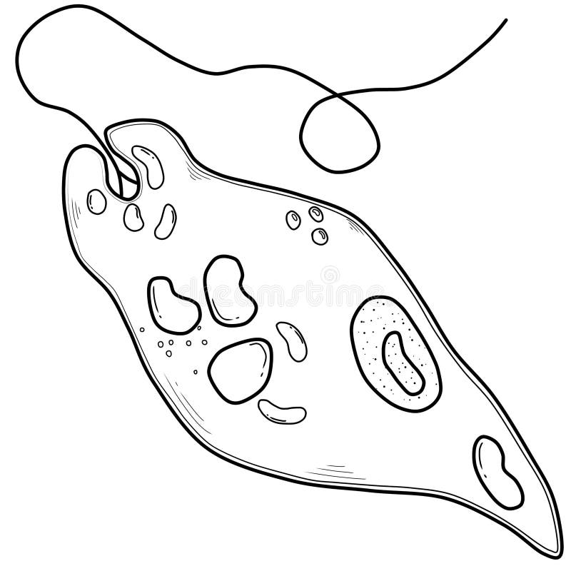
Free with trial Black and White Sketch of Euglena for use about biology article. Cell flagellate illustrations Black and White Sketch of Euglena
Free with trial This image showcases a detailed 3D rendering of a microscopic organism, possibly a type of bacteria or protist, generated by AI. The visualization highlights its intricate structure, including numerous flagella and internal organelles. The surrounding environment suggests an aquatic setting. Cell flagellate illustrations AI-Generated Microscopic Organism. This image showcases a detailed 3D rendering of a microscopic organism, possibly a type of bacteria or protist, generated by AI. The visualization highlights its intricate structure, including numerous flagella and internal organelles. The surrounding environment suggests an aquatic setting.
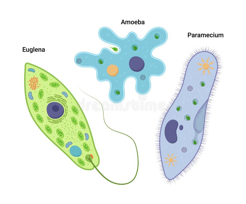
Free with trial A vibrant 3D illustration showcasing three distinct types of protozoa: a Paramecium with its characteristic slipper shape and cilia, a Euglena with its flagellum and red eyespot, and an Amoeba with its pseudopods. These single-celled organisms are depicted in a fluid, light blue environment with small bubbles, highlighting the complexity and diversity of microscopic life. Ideal for educational materials, scientific research, and biological concepts. Cell flagellate illustrations Microscopic World: Diverse Protozoa in Aquatic Environment. A vibrant 3D illustration showcasing three distinct types of protozoa: a Paramecium with its characteristic slipper shape and cilia, a Euglena with its flagellum and red eyespot, and an Amoeba with its pseudopods. These single-celled organisms are depicted in a fluid, light blue environment with small bubbles, highlighting the complexity and diversity of microscopic life. Ideal for educational materials, scientific research, and biological concepts.
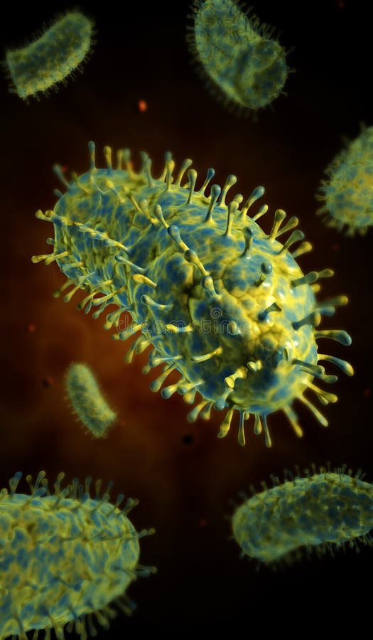
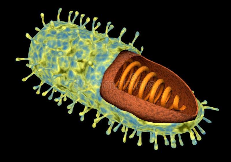
Free with trial Conceptual image of rabies virus. Cell flagellate illustrations Conceptual image of rabies virus.
Free with trial Conceptual image of rabies virus. Cell flagellate illustrations Conceptual image of rabies virus.
Free with trial Conceptual image of rabies virus. Cell flagellate illustrations Conceptual image of rabies virus.
Free with trial Conceptual medical illustration of Euglena. Cell flagellate illustrations Conceptual image of Euglena. Conceptual medical illustration of Euglena
Free with trial Conceptual medical illustration of Euglena. Cell flagellate illustrations Conceptual image of Euglena. Conceptual medical illustration of Euglena
Free with trial Conceptual medical illustration of Euglena. Cell flagellate illustrations Conceptual image of Euglena. Conceptual medical illustration of Euglena
Free with trial A vibrant blue dinoflagellate displays bright orange internal structures. Detailed, colorful rendering of a dinoflagellate with vibrant orange and blue coloring. Cell flagellate illustrations A vibrant blue dinoflagellate displays bright orange internal structures. Detailed, colorful rendering of a dinoflagellate with vibrant orange and blue coloring
Free with trial Biomedical illustration of the Giardia parasite inside the human intestines. Cell flagellate illustrations Biomedical illustration of the Giardia parasite inside the human intestines.
Free with trial Conceptual medical illustration of Trypanosoma. Cell flagellate illustrations Conceptual image of Trypanosoma. Conceptual medical illustration of Trypanosoma
Free with trial Conceptual medical illustration of Trypanosoma. Cell flagellate illustrations Conceptual image of Trypanosoma. Conceptual medical illustration of Trypanosoma
Free with trial Conceptual medical illustration of Trypanosoma. Cell flagellate illustrations Conceptual image of Trypanosoma. Conceptual medical illustration of Trypanosoma
Free with trial Conceptual medical illustration of Trypanosoma. Cell flagellate illustrations Conceptual image of Trypanosoma. Conceptual medical illustration of Trypanosoma
Free with trial Microscopic view of bacteria or virus cells dividing and multiplying rapidly in a petri dish. Cell flagellate illustrations Microscopic view of bacteria or virus cells dividing and multiplying
Free with trial Conceptual image of influenza causing flu virus. Cell flagellate illustrations Conceptual image of influenza causing flu virus.