Free with trial Vector illustration of a colon cut. Cell muscle vectors Small intestines cut. Vector illustration of a colon cut
Free with trial The stomach wall consists of 4 layers of tissue. From deep external to superficial internal these are the serosa, muscularis externa, submucosa and mucosa. This layered arrangement follows the same general structure in all regions of the stomach, and throughout the entire gastrointestinal tract. Cell muscle illustrations Stomach wall layers and gastric glands detailed anatomy. The stomach wall consists of 4 layers of tissue. From deep external to superficial internal these are the serosa, muscularis externa, submucosa and mucosa. This layered arrangement follows the same general structure in all regions of the stomach, and throughout the entire gastrointestinal tract.
Free with trial Muscle is a soft tissue found in most animals. Muscle cells contain protein filaments of actin and myosin that slide past one another, producing a contraction that changes both the length and the shape of the cell. Muscles function to produce force and motion. Cell muscle illustrations Movement of muscles. Muscle is a soft tissue found in most animals. Muscle cells contain protein filaments of actin and myosin that slide past one another, producing a contraction that changes both the length and the shape of the cell. Muscles function to produce force and motion.
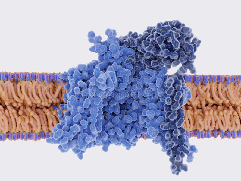
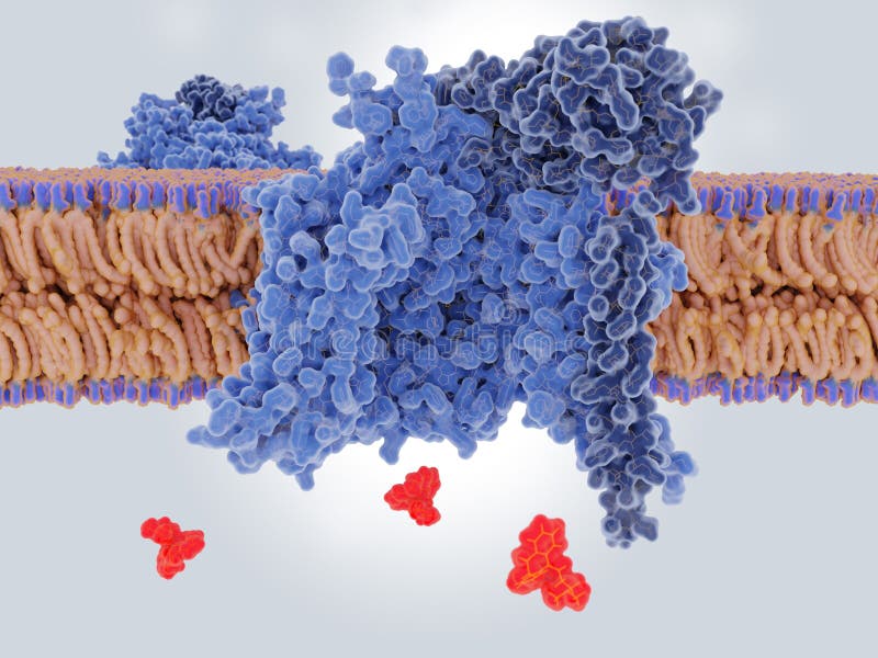
Free with trial Na+ channels are composed of a large alpha subunit and one or more regulatory beta subunits pink. They generate and propagate action potentials in neurons and muscle. Source: PDB entry 6AGF. Cell muscle illustrations The voltage-gated sodium channel in the open conformation,, top view. Na+ channels are composed of a large alpha subunit and one or more regulatory beta subunits pink. They generate and propagate action potentials in neurons and muscle. Source: PDB entry 6AGF
Free with trial Na+ channels are composed of a large alpha subunit and one or more regulatory beta subunits dark blue. They generate and propagate action potentials in neurons and muscle. Source: PDB entry 6AGF. Cell muscle illustrations The voltage-gated sodium channel in the open conformation,side view. Na+ channels are composed of a large alpha subunit and one or more regulatory beta subunits dark blue. They generate and propagate action potentials in neurons and muscle. Source: PDB entry 6AGF
Free with trial Surface Mucous Cells in Stomach Wall Structure. Human Stomach Layers and Foveolar Cell Diagram. Gastric Mucosa and Stomach Epithelium with Surface Mucous Cells Illustration. Cell muscle vectors Surface Mucous Cells in Stomach Wall Structure. Human Stomach Layers and Foveolar Cell Diagram. Gastric Mucosa and
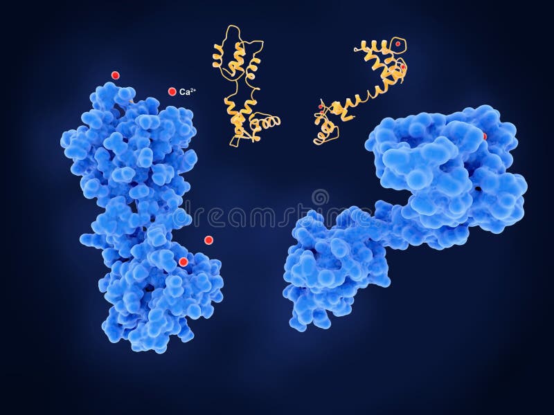
Free with trial Calmodulin is an intracelular target of calcium ions. Once activated by Ca2+ red, calmodulin interacts with various target proteins such as kinases and phosphatases.. It plays an important role in long-term memory. and muscle contraction. CaM has 4 calcium binding sites. PDB entry 3CLN, 1CFD. Cell muscle illustrations Calmodulin, calcium free left, inactive and activated right conformations. Calmodulin is an intracelular target of calcium ions. Once activated by Ca2+ red, calmodulin interacts with various target proteins such as kinases and phosphatases.. It plays an important role in long-term memory. and muscle contraction. CaM has 4 calcium binding sites. PDB entry 3CLN, 1CFD
Free with trial Microbes in the body. Immune protection against infection. Study of cell structure under the microscope. 3d illustration of infected cells. Cell muscle illustrations Microbes in the body. Immune protection against infection
Free with trial Vector illustration of a Neck pain from cell phone use. Cell muscle vectors Vector illustration of a Neck pain from cell phone use
Free with trial Clostridium tetani. Pathogenic bacterium causing infection Tetanus, also known as lockjaw muscle spasms. infection disease outbreak. red blood cells and bacteria with spores on red background. vector. Illustration easy editable for Your color. Cell muscle vectors Red blood cells and bacteria Clostridium tetani with spores on r. Clostridium tetani. Pathogenic bacterium causing infection Tetanus, also known as lockjaw muscle spasms. infection disease outbreak. red blood cells and bacteria with spores on red background. vector. Illustration easy editable for Your color
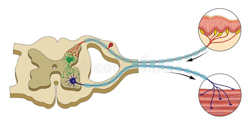
Free with trial A neuromuscular junction or myoneural junction is a chemical synapse between a motor neuron and a muscle fiber. It allows the motor neuron to transmit a signal to the muscle fiber, causing muscle contraction. Cell muscle illustrations Synapse of the nervous system. Neuromuscular. A neuromuscular junction or myoneural junction is a chemical synapse between a motor neuron and a muscle fiber. It allows the motor neuron to transmit a signal to the muscle fiber, causing muscle contraction.
Free with trial Vector illustration of anatomy of the muscular fibers for medical publications. Cell muscle vectors Anatomical illustration of muscle fibers for medical journals. Vector illustration of anatomy of the muscular fibers for medical publications
Free with trial Colony of microorganisms under the microscope. Germs and viruses in humans. Cell research. 3D illustration on the subject of microbiology. Cell muscle illustrations Colony of microorganisms under the microscope. Germs and viruses in humans
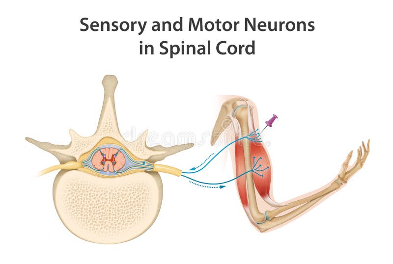
Free with trial Motor neurons of the spinal cord are part of the central nervous system and connect to muscles, glands and organs throughout the body. These neurons transmit impulses from the spinal cord to skeletal and smooth muscles, and so directly control all of our muscle movements. Cell muscle illustrations Sensory and Motor Neurons in Spinal Cord. Motor neurons of the spinal cord are part of the central nervous system and connect to muscles, glands and organs throughout the body. These neurons transmit impulses from the spinal cord to skeletal and smooth muscles, and so directly control all of our muscle movements.
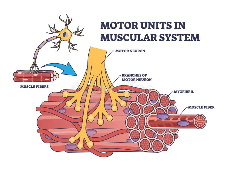
Free with trial Motor units in muscular system with fibers neuron anatomy outline diagram. Labeled educational medical scheme with myofibril and muscle fiber closeup vector illustration. Nerve functional contraction. Cell muscle vectors Motor units in muscular system with fibers neuron anatomy outline diagram
Free with trial Stem cells. These cells can become any tissue in the body. Explanation of stem cell application. Genetic research. Vector Illustration for medical, science, and educational use. Cell muscle vectors Stem cells, and human organs on a white background. Stem cells. These cells can become any tissue in the body. Explanation of stem cell application. Genetic research. Vector Illustration for medical, science, and educational use
Free with trial Different human tissue types set. Stock vector illustration of epithelial, muscle, stem, liver cells forming organs. Medicine and biology collection. Cell muscle vectors Different human tissue types
Free with trial Microorganisms under the microscope. Microbes and viruses in humans. Cell research. 3D illustration on the subject of microbiology. Cell muscle illustrations Microorganisms under the microscope. Microbes and viruses in humans
Free with trial Arteries carry blood away from the heart, and veins carry blood towards the heart. With the exception of pulmonary blood vessels, arteries carry oxygenated blood and veins carry deoxygenated blood. Arteries have thick walls with muscle tissue. Veins have thinner walls and use valves to keep your blood flowing. Cell muscle illustrations Structure of arteries, veins and capillaries. Arteries carry blood away from the heart, and veins carry blood towards the heart. With the exception of pulmonary blood vessels, arteries carry oxygenated blood and veins carry deoxygenated blood. Arteries have thick walls with muscle tissue. Veins have thinner walls and use valves to keep your blood flowing
Free with trial Amitriptyline is used as an antidepressant and antimigraine drug. It blocks sodium channels on neuronal membrans. Sodium channels generate and propagate action potentials in neurons and muscle. Source: PDB entry 6AGF. Cell muscle illustrations Antidepressive drug amitriptyline binding to and blocking a neuronal sodium channel. Amitriptyline is used as an antidepressant and antimigraine drug. It blocks sodium channels on neuronal membrans. Sodium channels generate and propagate action potentials in neurons and muscle. Source: PDB entry 6AGF
Free with trial Multiple sclerosis demyelination compared with medical healthy nerves outline diagram. Labeled educational scheme with anatomical and medical autoimmune disease muscle contraction vector illustration. Cell muscle vectors Multiple sclerosis demyelination compared with healthy nerves outline diagram. Multiple sclerosis demyelination compared with medical healthy nerves outline diagram. Labeled educational scheme with anatomical and medical autoimmune disease muscle contraction vector illustration
Free with trial ATP molecule converts into ADP, and ADP converts into AMT. Hydrolysis. Adenosine triphosphate stores energy for the cell. ATP breaks down into Adenosine diphosphate and a phosphate group. Adenosine monophosphate. Releasing energy for cellular processes. Detailed Vector illustration. Medical poster. Schematic diagram. Cell muscle vectors ATP molecule converts into ADP, and ADP converts into AMT. Hydrolysis
Free with trial Protein kinase C epsilon type, an enzyme which modulates cardiac cell metabolism through its actions at mitochondria. 3d rendering. Cell muscle illustrations Protein kinase C epsilon type, an enzyme which modulates cardiac
Free with trial Cute happy funny sperm cell. Vector flat line cartoon kawaii character illustration icon. Fertilization concept. Cell muscle vectors Cute happy funny sperm cell. Vector flat
Free with trial It has three main layers, the epidermis, the dermis and the subcutaneous layer. The epidermis is an elastic layer on the outside that is continually being regenerated. It includes the following: Keratinocytes - the main cells of the epidermis formed by cell division at its base. Cell muscle illustrations Diagram of human skin structure. It has three main layers, the epidermis, the dermis and the subcutaneous layer. The epidermis is an elastic layer on the outside that is continually being regenerated. It includes the following: Keratinocytes - the main cells of the epidermis formed by cell division at its base.
Free with trial Reflex is an involuntary, stereotyped response of an effector tissue that occurs when a receptor is stimulated. These reflexes are carried out by the sequential activation of a certain number of neurons that are interconnected. The last neuron usually innervates the effector tissue, which is usually a muscle. Cell muscle illustrations Spinal Reflex Arc Anatomical Scheme. Reflex is an involuntary, stereotyped response of an effector tissue that occurs when a receptor is stimulated. These reflexes are carried out by the sequential activation of a certain number of neurons that are interconnected. The last neuron usually innervates the effector tissue, which is usually a muscle
Free with trial Different human tissue types set. Stock vector illustration of epithelial, muscle, stem, liver cells forming organs. Medicine and biology collection. Cell muscle vectors Different human tissue types
Free with trial Female breast anatomy lactation composition with isolated profile views of breasts and milk duct cell spots vector illustration. Cell muscle vectors Female Lactation Anatomy Composition. Female breast anatomy lactation composition with isolated profile views of breasts and milk duct cell spots vector illustration
Free with trial Histamine. Immune response and Allergic reaction. Histamine-releasing cells produce of Histamine then Stomach secretes gastric acid, smooth muscle is reduced and in the bronchi there are difficulties with breathing, Goblet cells secrete mucus, and Skin becomes redness and itching. Vector poster. Isometric flat illustration. Cell muscle vectors Histamine. Immune response and Allergic reaction
Free with trial Arteries have thick walls with muscle tissue. Veins have thinner walls and use valves to keep your blood flowing. Artery vs Capillary. Arteries carry blood from your heart to your organs. Cell muscle illustrations Illustration of difference between arteries and veins. Arteries have thick walls with muscle tissue. Veins have thinner walls and use valves to keep your blood flowing. Artery vs Capillary. Arteries carry blood from your heart to your organs.
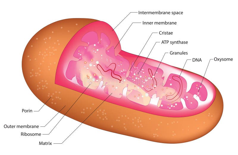
Free with trial Biological structure of mitochondria in the cell. Cell muscle vectors Mitochondrion. Biological structure of mitochondria in the cell
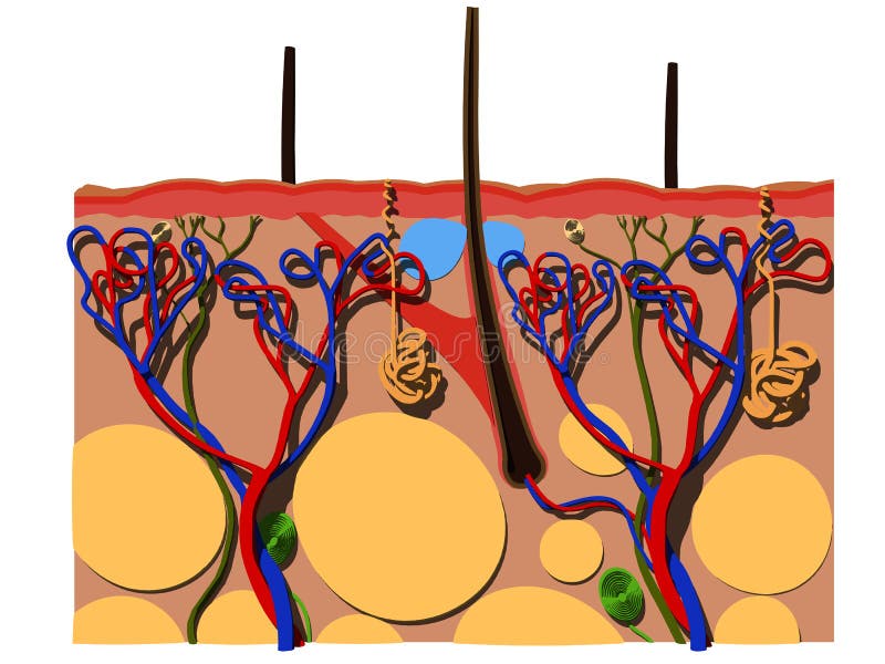
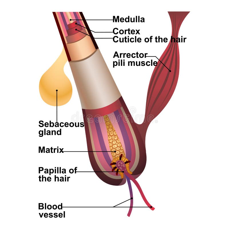
Free with trial Detailed explanation of hair structure and anatomy. Close-up of blood vessel papilla matrix arrector pili muscle cortex cuticle and medulla vector illustration. Isolated on white. Cell muscle vectors Detailed explanation of hair structure and anatomy
Free with trial Detailed explanation of hair structure and anatomy. Close-up of blood vessel papilla matrix arrector pili muscle cortex cuticle and medulla vector illustration. Isolated on white. Cell muscle vectors Detailed explanation of hair structure and anatomy
Free with trial Types of tissues. Medical infographics with anatomical structure and organ of human body. Nervous, epithelial, connective and muscle fiber part. Cartoon flat vector illustration isolated on background. Cell muscle vectors Types of tissues vector illustration. Types of tissues. Medical infographics with anatomical structure and organ of human body. Nervous, epithelial, connective and muscle fiber part. Cartoon flat vector illustration isolated on background
Free with trial Hand of a green orc holding a cell phone in a white background. This monster in clipping path is very useful for graphic design creations, 3d illustration. Cell muscle illustrations Hand of a green orc holding a cell phone in a white background
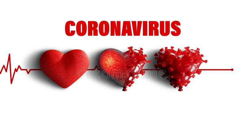
Free with trial 3D render - The course of heart disease after coronavirus infection. Covid-19 attacks the heart muscle causing its inflammation. This can lead to a heart attack. Copy space for text, white isolated background. Cell muscle illustrations 3D render - Corona Virus Heart damage process. Covid-19 epidemic. 3D render - The course of heart disease after coronavirus infection. Covid-19 attacks the heart muscle causing its inflammation. This can lead to a heart attack. Copy space for text, white isolated background.
Free with trial 3D render - The course of heart disease after coronavirus infection. Covid-19 attacks the heart muscle causing its inflammation. This can lead to a heart attack. Copy space for text, white isolated background. Cell muscle illustrations 3D render - The course of heart disease after coronavirus infection
Free with trial Lipoma fatty tumor medical poster. Not cancer, benign tumor under skin, fat lump in human body. Skin layers structure epidermis, dermis and hypodermis and muscle medical flat vector illustration. Cell muscle vectors Lipoma medical poster. Lipoma fatty tumor medical poster. Not cancer, benign tumor under skin, fat lump in human body. Skin layers structure epidermis, dermis and hypodermis and muscle medical flat vector illustration
Free with trial Cultured lab-grown meat infographics. Synthetic in vitro food concept. Biotechnological process with muscle stem cells, beef and tissue in laboratory. Color vector illustration. Cell muscle illustrations Cultured lab-grown meat infographics. Synthetic in vitro food concept. Biotechnological process with muscle stem cells
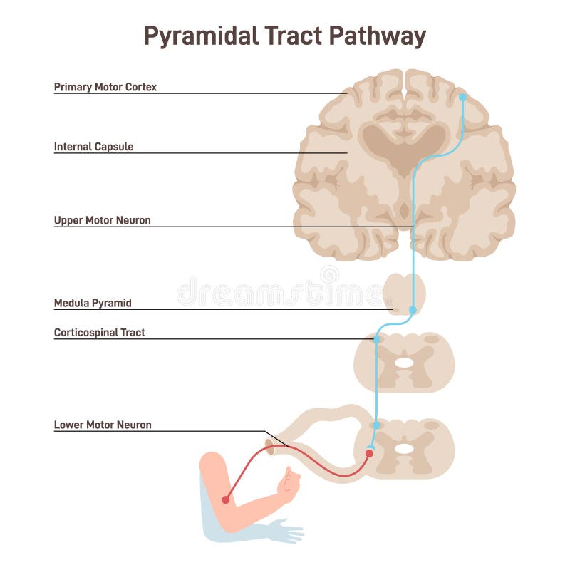
Free with trial Pyramidal tract pathway. Somatic nervous system, voluntary control of the body and face muscles. Upper motor neurons differences in body muscle control. Flat vector illustration. Cell muscle vectors Pyramidal tract pathway. Somatic nervous system, voluntary control
Free with trial 3D render - The course of heart disease after coronavirus infection. Covid-19 attacks the heart muscle causing its inflammation. This can lead to a heart attack. Copy space for text, white isolated background. Cell muscle illustrations 3D render - The course of heart disease after coronavirus infection
Free with trial 3D render - The course of heart disease after coronavirus infection. Covid-19 attacks the heart muscle causing its inflammation. This can lead to a heart attack. Copy space for text, white isolated background. Cell muscle illustrations 3D render - The course of heart disease after coronavirus infection
Free with trial 3D render – Concept of healthy and damaged heart by disease called myocarditis or inflammation of the heart muscle caused by Corona virus – Covid-19. Copy space for text, white isolated background. Cell muscle illustrations 3D render – Concept of healthy and damaged heart by disease called myocarditis
Free with trial Young man Positive business man showing new brand,latest smartphone. Man holding cell,mobile phone in hand and gesturing making thumbs up sign. Flat vector illustration isolated on white background. Cell muscle vectors Young man Positive business man showing new brand, latest smartphone. Young man Positive business man showing new brand,latest smartphone. Man holding cell,mobile phone in hand and gesturing making thumbs up sign. Flat vector illustration isolated on white background
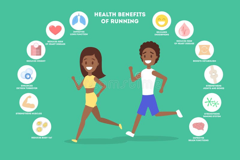
Free with trial Benefits of running or jogging infographic. Idea of healthy and active lifestyle. Immune improvement and muscle building. Weight loss. Isolated flat vector illustration. Cell muscle vectors Benefits of running or jogging infographic. Idea of healthy and active lifestyle
Free with trial Benefits of running or jogging infographic. Idea of healthy and active lifestyle. Immune improvement and muscle building. Weight loss. Isolated flat vector illustration. Cell muscle vectors Benefits of running or jogging infographic. Idea of healthy and active lifestyle
Free with trial Goblet cells are specialized epithelial cells found in the lining of various organs, particularly in the respiratory and digestive tracts. They are named for their goblet-like shape, with a wide base and a narrower opening at the top. Goblet cells are responsible for secreting mucins, which are glycoproteins that form mucous. Cell muscle vectors Goblet cell
Free with trial Lipoma fatty tumor medical poster. Not cancer, benign tumor under skin, fat lump in human body. Skin layers structure epidermis, dermis and hypodermis and muscle medical flat vector illustration. Cell muscle vectors Lipoma medical poster. Lipoma fatty tumor medical poster. Not cancer, benign tumor under skin, fat lump in human body. Skin layers structure epidermis, dermis and hypodermis and muscle medical flat vector illustration
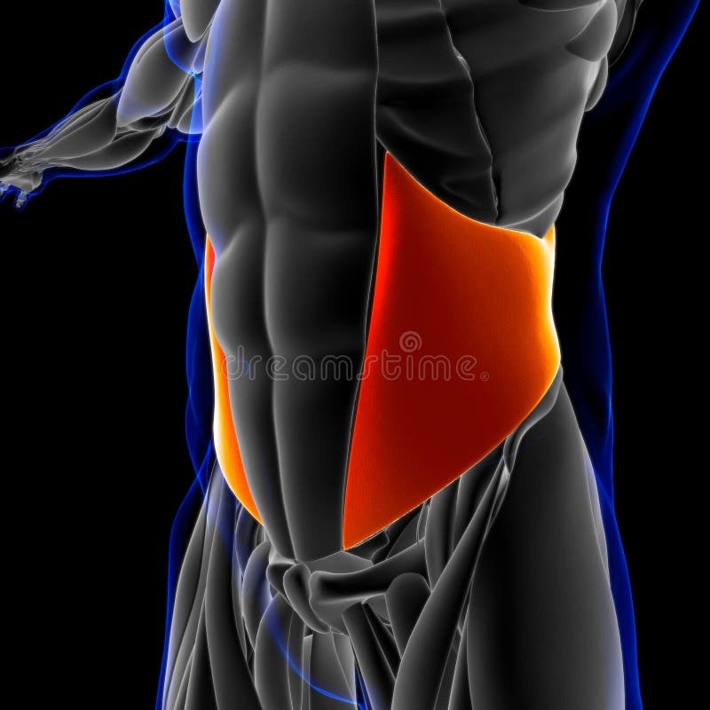
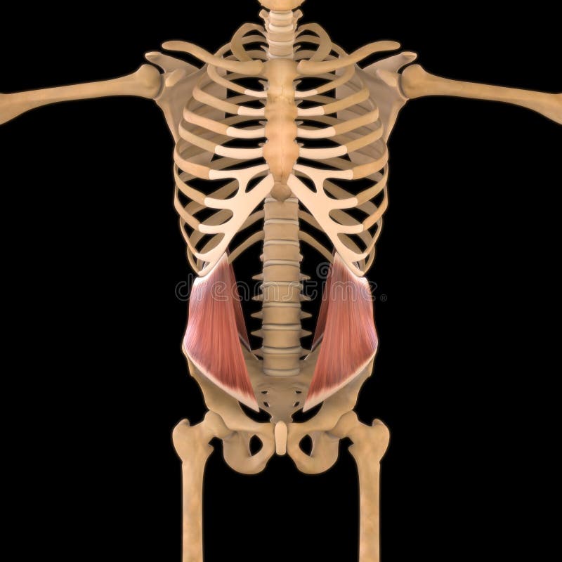
Free with trial 3D Illustration, Muscle is a soft tissue, Muscle cells contain proteins , producing a contraction that changes both the length and the shape of the cell. Muscles function to produce force and motion. Cell muscle illustrations Internal Oblique Anatomy For Medical Concept 3D Illustration. 3D Illustration, Muscle is a soft tissue, Muscle cells contain proteins , producing a contraction that changes both the length and the shape of the cell. Muscles function to produce force and motion
Free with trial Mesenchymal stem cells (MSCs) are a type of multipotent adult stem cell found in various tissues throughout the body, including bone marrow, adipose tissue (fat), umbilical cord tissue, and dental pulp. They have the capacity to differentiate into a variety of cell types, including osteoblasts (bone cells), chondrocytes (cartilage cells), and adipocytes (fat cells), among others. Cell muscle vectors Mesenchymal stem cells
Free with trial 3D Illustration, Muscle is a soft tissue, Muscle cells contain proteins , producing a contraction that changes both the length and the shape of the cell. Muscles function to produce force and motion. Cell muscle illustrations Internal Oblique Anatomy For Medical Concept 3D Illustration. 3D Illustration, Muscle is a soft tissue, Muscle cells contain proteins , producing a contraction that changes both the length and the shape of the cell. Muscles function to produce force and motion
Free with trial Powered by ATP, the pump moves sodium and potassium ions in opposite directions. Enzyme ATPase. Vector illustration. Cell muscle vectors Sodium potassium pump. K, Na pump. Active transport through cell membrane. Powered by ATP, the pump moves sodium and potassium ions in opposite directions. Enzyme ATPase. Vector illustration.
Free with trial This image is generated using AI tool. Cell muscle illustrations Fibroblast Cell Anatomy: Understanding the Structure and Composition. This image is generated using AI tool
Free with trial Human Internal Organ Anatomy Icons Set Vector. Stomach And Liver, Heart And Lung, Intestine And Gland, Muscle And Skin People Organ Line. Healthcare And Medicine Black Contour Illustrations. Cell muscle illustrations Human Internal Organ Anatomy Icons Set Vector . Human Internal Organ Anatomy Icons Set Vector. Stomach And Liver, Heart And Lung, Intestine And Gland, Muscle And Skin People Organ Line. Healthcare And Medicine Black Contour Illustrations .
Free with trial This microscopic image reveals the intricate details of glucose transporter proteins, crucial for cellular glucose uptake. These specialized proteins act as gatekeepers, facilitating the movement of glucose, a vital source of energy, across cell membranes. The image showcases the complex molecular structures of these transporters, highlighting their critical role in maintaining cellular energy. Cell muscle illustrations Cellular Glucose Uptake A Microscopic Exploration of Glucose Transporter Proteins. This microscopic image reveals the intricate details of glucose transporter proteins, crucial for cellular glucose uptake. These specialized proteins act as gatekeepers, facilitating the movement of glucose, a vital source of energy, across cell membranes. The image showcases the complex molecular structures of these transporters, highlighting their critical role in maintaining cellular energy
Free with trial Icon Blood Cell. related to Human Organ symbol. comic style. simple design editable. simple illustration. Cell muscle illustrations Icon Blood Cell. related to Human Organ symbol. comic style. simple design editable. simple illustration
Free with trial Blood clot formation, or hemostasis, is a rapid, multistep process that seals damaged blood vessels to stop bleeding. It involves direct vasoconstriction, the formation of a temporary platelet plug and the activation of clotting factors to form a stable fibrin mesh. Cell muscle illustrations Blood clot formation, or hemostasis. Is a rapid, multistep process that seals damaged blood vessels to stop bleeding. It involves direct vasoconstriction, the. Blood clot formation, or hemostasis, is a rapid, multistep process that seals damaged blood vessels to stop bleeding. It involves direct vasoconstriction, the formation of a temporary platelet plug and the activation of clotting factors to form a stable fibrin mesh
Free with trial Enteric nervous system overview shows gut-brain axis with myenteric plexus, submucosal plexus, and neurons regulating motility and secretion. Outline diagram. Cell muscle vectors Enteric nervous system overview shows gut-brain axis with myenteric
Free with trial Pattern designed using 3D modeled and rendered. Cell muscle illustrations Art pattern. Pattern designed using 3D modeled and rendered.
Free with trial Pattern designed using 3D modeled and rendered. Cell muscle illustrations Colorful structure. Pattern designed using 3D modeled and rendered.
Free with trial 3D modeled futuristic building and rendered. Cell muscle illustrations Futuristic Building
Free with trial Pattern designed using 3D modeling methods and rendered. Cell muscle illustrations Gum pattern. Pattern designed using 3D modeling methods and rendered.
Free with trial Gas exchange is the process by which oxygen from the lungs enters the blood and carbon dioxide from the blood enters the lungs. This exchange occurs in the lungs between the alveoli (tiny air sacs) and the capillaries (tiny blood vessels) that surround them. Cell muscle vectors Oxygen and Carbon Dioxide Exchange. Gas exchange is the process by which oxygen from the lungs enters the blood and carbon dioxide from the blood enters the lungs. This exchange occurs in the lungs between the alveoli (tiny air sacs) and the capillaries (tiny blood vessels) that surround them.
Free with trial 3D art illustration of anatomy of Front view of Human skin cutway diagram with name. Cell muscle illustrations Front view of Human skin cutway diagram
Free with trial Pattern designed using 3D modeled and rendered. Made and designed from scratch. Cell muscle illustrations 3D structure. Pattern designed using 3D modeled and rendered. Made and designed from scratch.
Free with trial Insulin function. INS. Fat accumulation, Ribosomal protein synthesis, Energy storage in liver, Blood sugar regulation, Glucose uptake. Hormone produced by beta cells of the pancreatic islets. Detailed vector poster. Cell muscle vectors Insulin role. Metabolic and Anabolic Functions of INS Pancreatic hormone. Insulin function. INS. Fat accumulation, Ribosomal protein synthesis, Energy storage in liver, Blood sugar regulation, Glucose uptake. Hormone produced by beta cells of the pancreatic islets. Detailed vector poster
Free with trial 3D art illustration of anatomy of Front view of Human skin cutway diagram. Cell muscle illustrations Front view of Human skin cutway diagram
Free with trial Epithelia are tissues consisting of closely apposed cells without intervening intercellular substances. Cell muscle illustrations G Unicellular Epithelium. Epithelia are tissues consisting of closely apposed cells without intervening intercellular substances.
Free with trial Digital illustration of Human hair structure anatomy in colour background. Cell muscle illustrations Human hair structure
Free with trial Motor neurons are large, multipolar lower motor neurons of the brainstem and spinal cord. Cell muscle illustrations Motor neurons
Free with trial Blood vessels consist of arteries, arterioles, capillaries, venules, and veins. Vessel networks deliver blood to all tissues in a directed and regulated manner. Arteries and veins are composed of three tissue layers. ... It consists of circularly arranged elastic fibers, connective tissue, and smooth muscle cells. Cell muscle illustrations Structure of the blood vessels. Blood vessels consist of arteries, arterioles, capillaries, venules, and veins. Vessel networks deliver blood to all tissues in a directed and regulated manner. Arteries and veins are composed of three tissue layers. ... It consists of circularly arranged elastic fibers, connective tissue, and smooth muscle cells.
Free with trial Fibrillin glycoprotein molecule on a white background. Cell muscle illustrations Fibrillin glycoprotein molecule
Free with trial Digital illustration of human skin anatomy. Cell muscle illustrations Human skin anatomy
Free with trial The body will use nutrients that provide energy when exercising, By based on factors from the blood system And primarily muscles, Waste such as perspiration and heat is released. Cell muscle vectors Changing energy into active muscles. The body will use nutrients that provide energy when exercising, By based on factors from the blood system And primarily muscles, Waste such as perspiration and heat is released
Free with trial A reflex arc is a neural pathway that controls a reflex. In vertebrates, most sensory neurons do not pass directly into the brain, but synapse in the spinal cord. This allows for faster reflex actions to occur by activating spinal motor neurons without the delay of routing signals through the brain. Cell muscle illustrations Spinal Reflex Arc illustration. Central nervous system. A reflex arc is a neural pathway that controls a reflex. In vertebrates, most sensory neurons do not pass directly into the brain, but synapse in the spinal cord. This allows for faster reflex actions to occur by activating spinal motor neurons without the delay of routing signals through the brain.
Free with trial 3d illustration of human skin anatomy. Cell muscle illustrations 3d human skin anatomy. 3d illustration of human skin anatomy
Free with trial Eight selfies infographic Illustrator. Cell muscle vectors Eight selfies infographic Illustrator