Free with trial Phases of epithelial repair. Drawing to show phases of tissue repair, specifically oral mucosal tissue. Cellularity vectors Phases of epithelial repair. Drawing to show phases of tissue repair, specifically oral mucosal tissue
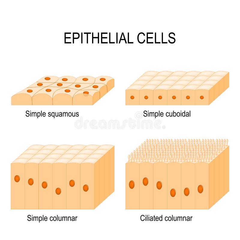
Free with trial Types of epithelial cells: cilliated columnar, simple columnar, simple cuboidal, and simple squamous cells. Cellularity vectors Types of epithelial cells. Cilliated columnar, simple columnar, simple cuboidal, and simple squamous cells
Free with trial Types of epithelial cells: cilliated columnar, simple columnar, simple cuboidal, and simple squamous cells. Cellularity vectors Types of epithelial cells. Cilliated columnar, simple columnar, simple cuboidal, and simple squamous cells
Free with trial Skin Cells and Structure Layers of epidermis. cornified stratum corneum, clear or translucent layer lucidum, granular stratum granulosum, spinous spinosum, basal or germinal layer stratum basale or germinativum. Cellularity vectors Skin Cells and Structure Layers of epidermis. Cornified stratum corneum, clear or translucent layer lucidum, granular stratum granulosum, spinous spinosum
Free with trial Simple columnar epithelium / Epithelial tissue. On white. Cellularity vectors Simple columnar epithelium / Epithelial tissue. On white
Free with trial Vaginal atrophy. Thinning, drying and inflammation of the vaginal walls that may occur when your body has less estrogen. Epithelial cells destruction. Flat vector illustration. Cellularity vectors Vaginal atrophy. Thinning, drying and inflammation of the vaginal walls. That may occur when your body has less estrogen. Epithelial cells destruction. Flat
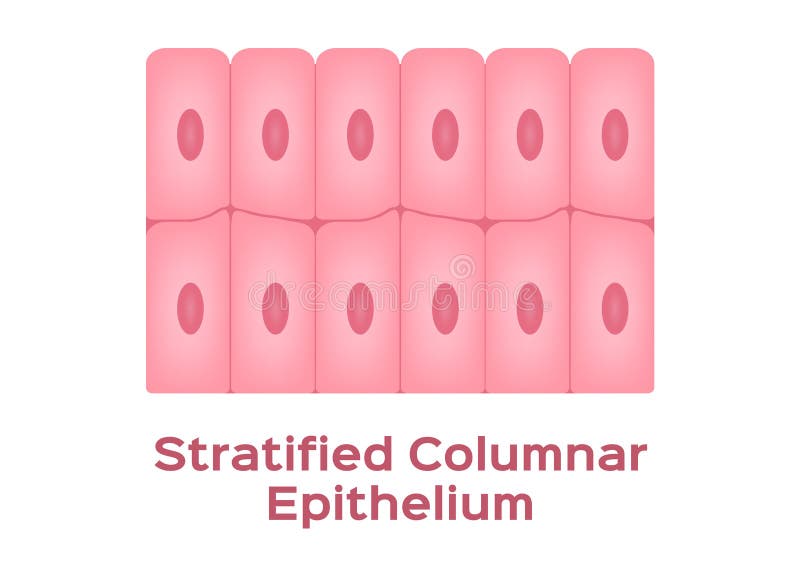
Free with trial Stratified columnar epithelium / Epithelial tissue. On white. Cellularity vectors Stratified columnar epithelium / Epithelial tissue. On white
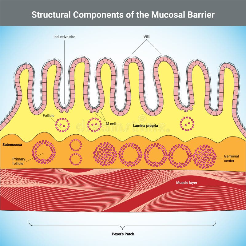
Free with trial Structural Components of the Mucosal Barrier medical illustration. Cellularity vectors Structural Components of the Mucosal Barrier medical illustration. Structural Components of the Mucosal Barrier vector medical illustration
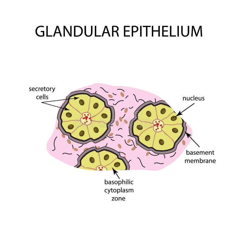
Free with trial The structure of the glandular epithelium. Infographics Vector illustration on background. Cellularity vectors The structure of the glandular epithelium. Infographics. Vector illustration on background. The structure of the glandular epithelium. Infographics Vector. The structure of the glandular epithelium. Infographics Vector illustration on background
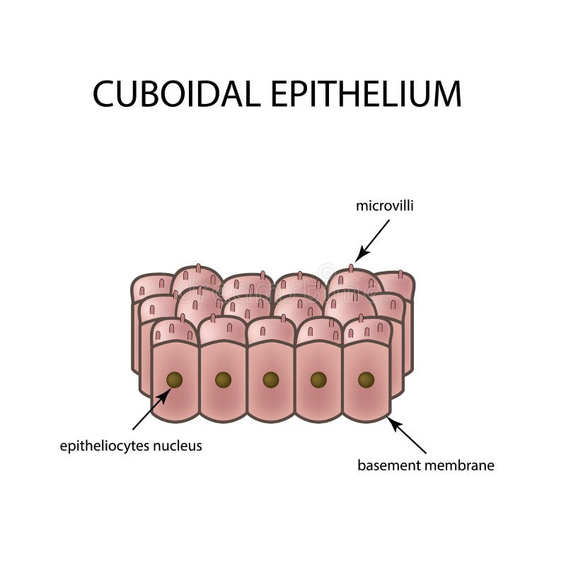
Free with trial The structure of cubic epithelium. Infographics Vector illustration on background. Cellularity vectors The structure of cubic epithelium. Infographics. Vector illustration on background. The structure of cubic epithelium. Infographics Vector illustration on. The structure of cubic epithelium. Infographics Vector illustration on background
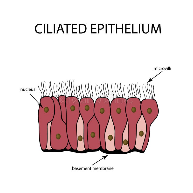
Free with trial The structure of the ciliated epithelium. Infographics. Vector illustration on background. Cellularity vectors The structure of the ciliated epithelium. Infographics. Vector illustration on background.
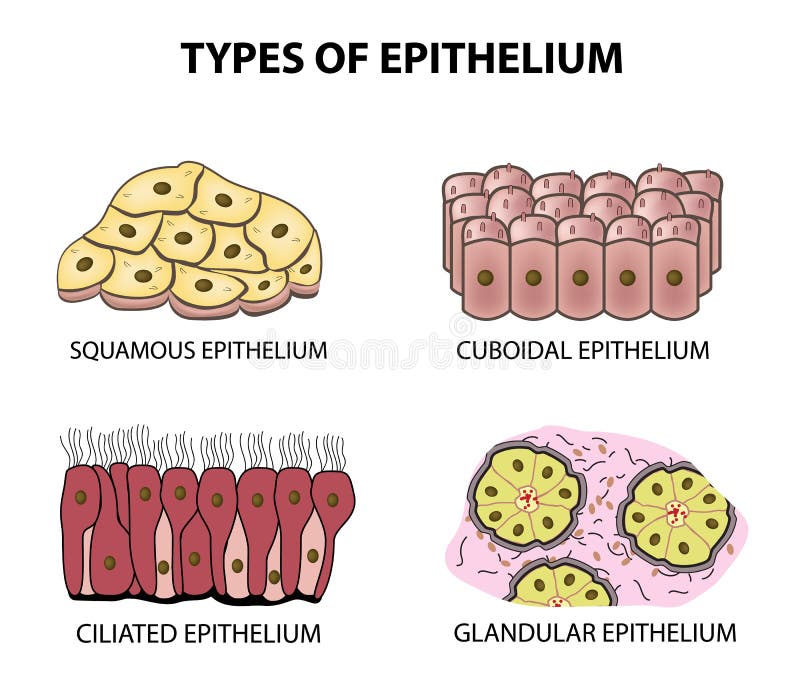
Free with trial Types of epithelium. squamous, cubic, ciliated, glandular. Set Infographics Vector illustration on background. Cellularity vectors Types of epithelium. squamous, cubic, ciliated, glandular. Set. Infographics. Vector illustration on background. Types of epithelium. squamous, cubic, ciliated. Types of epithelium. squamous, cubic, ciliated, glandular. Set Infographics Vector illustration on background
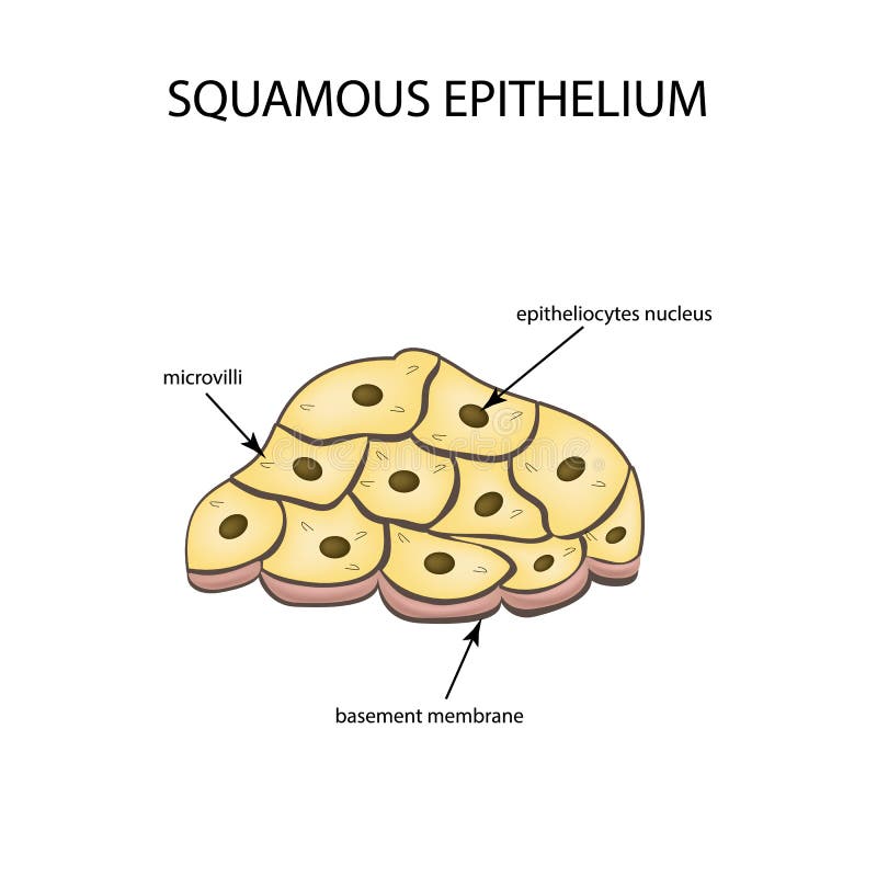
Free with trial The structure of the squamous epithelium. Infographics Vector illustration on background. Cellularity vectors The structure of the squamous epithelium. Infographics. Vector illustration on background. The structure of the squamous epithelium. Infographics Vector. The structure of the squamous epithelium. Infographics Vector illustration on background
Free with trial The image presents a detailed anatomical cross section illustration of a biological vascular structure, likely a blood vessel. Cellularity illustrations An illustrated anatomical cross section of a biological vessel structure. The image presents a detailed anatomical cross section illustration of a biological. The image presents a detailed anatomical cross section illustration of a biological vascular structure, likely a blood vessel
Free with trial Illustration of cancer cell division. Cellularity illustrations Illustration of cancer cell division. Illustration of cancer cell division
Free with trial High-resolution image of colorful pluripotent stem cells viewed under a microscope, showcasing their dynamic structures and vibrant colors, ideal for scientific presentations and educational materials. Cellularity illustrations Colorful Pluripotent Stem Cells Observed Under a Microscope. High-resolution image of colorful pluripotent stem cells viewed under a microscope, showcasing their. High-resolution image of colorful pluripotent stem cells viewed under a microscope, showcasing their dynamic structures and vibrant colors, ideal for scientific presentations and educational materials
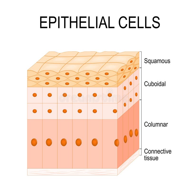
Free with trial Types of EpitheliumTypes of epithelium. Epithelial cells in a variety of configurations. Flat illustration. Cellularity vectors Types of epithelium. Epithelial cells in a variety of configurations. Types of EpitheliumTypes of epithelium. Epithelial cells in a variety of configurations. Types of EpitheliumTypes of epithelium. Epithelial cells in a variety of configurations. Flat illustration.
Free with trial 3d medical of bone marrow aspiration model showing the needle, syringe, and glass slide smear, with a zoomed inset of cellularity, myeloid-to-erythroid ratio, and cells. Cellularity illustrations 3d medical of bone marrow aspiration model showing the needle, syringe, and glass slide smear. With a zoomed inset of cellularity, myeloid-to-erythroid ratio
Free with trial An showing red blood cells erythrocytes traveling through a blood vessel. The cells are biconcave discs and appear in varying orientations and sizes emphasizing depth. The vessel wall is depicted as a tunnel like structure with a slightly transparent elastic surface possibly with smooth muscle and endothelial layers. The image highlights the cellularity and dynamic environment within the circulatory system absent of specific cellular detail but focusing on movement and flow. Cellularity illustrations Structure with a slightly transparent elastic surface possibly. An showing red blood cells erythrocytes traveling through a blood vessel. The cells are biconcave. An showing red blood cells erythrocytes traveling through a blood vessel. The cells are biconcave discs and appear in varying orientations and sizes emphasizing depth. The vessel wall is depicted as a tunnel like structure with a slightly transparent elastic surface possibly with smooth muscle and endothelial layers. The image highlights the cellularity and dynamic environment within the circulatory system absent of specific cellular detail but focusing on movement and flow.
Free with trial Histopathological slide revealing a dense proliferation of cells with visible vascular components in tumor tissue. Cellularity illustrations Microscopic view of tumor tissue with high cellularity and vascularity. Histopathological slide revealing a dense proliferation of cells with visible vascular. Histopathological slide revealing a dense proliferation of cells with visible vascular components in tumor tissue
Free with trial Educational graphic depicting bone marrow cellularity and chromosome analysis for myelodysplastic syndrome awareness during december. Cellularity illustrations Myelodysplastic syndrome awareness medical educational infographic with myelodysplast. Educational graphic depicting bone marrow cellularity and chromosome. Educational graphic depicting bone marrow cellularity and chromosome analysis for myelodysplastic syndrome awareness during december
Free with trial Cosmic Cellularity. Abstract depiction of cell-like structures in a cosmic nebula. Generative Ai. Cellularity illustrations Cosmic Cellularity. Abstract depiction of cell-like structures in a cosmic nebula. Generative Ai
Free with trial Cosmic Cellularity. Abstract depiction of cell-like structures in a cosmic nebula. Generative Ai. Cellularity illustrations Cosmic Cellularity. Abstract depiction of cell-like structures in a cosmic nebula. Generative Ai
Free with trial This image presents a stunning three dimensional microscopic representation of a eukaryotic cell detailing its complex internal structures and organelles. This intricate biological model clearly shows the nucleus the endoplasmic reticulum and mitochondria in high resolution Rendered by Ai allowing for an in depth study of cellular anatomy. The surrounding plasma membrane and the viscous cytoplasm. Cellularity illustrations Detailed 3D rendering of an animal cell structure. This image presents a stunning three dimensional microscopic representation of a eukaryotic cell detailing its. This image presents a stunning three dimensional microscopic representation of a eukaryotic cell detailing its complex internal structures and organelles. This intricate biological model clearly shows the nucleus the endoplasmic reticulum and mitochondria in high resolution Rendered by Ai allowing for an in depth study of cellular anatomy. The surrounding plasma membrane and the viscous cytoplasm
Free with trial A stunning macro view reveals a complex internal structure composed of multiple colored layers, each exhibiting a fractal-like cellular pattern. Rendered by Ai This intricate design resembles advanced composite materials or biological tissue architecture. The vibrant color gradient contrasts sharply with the dark, porous material surrounding the core layers. Cellularity illustrations Rainbow cross section of porous molecular material. A stunning macro view reveals a complex internal structure composed of multiple colored layers, each. A stunning macro view reveals a complex internal structure composed of multiple colored layers, each exhibiting a fractal-like cellular pattern. Rendered by Ai This intricate design resembles advanced composite materials or biological tissue architecture. The vibrant color gradient contrasts sharply with the dark, porous material surrounding the core layers
Free with trial This striking visualization captures a complex microscopic entity featuring a spiky blue membrane surrounding a central spherical component. The image blends scientific accuracy with stunning digital artistry. Rendered by Ai, this detailed illustration is ideal for educational materials or medical research abstracts. Its vibrant colors and high contrast detail highlight the mysterious nature of. Cellularity illustrations Futuristic spiky cell structure visualization. This striking visualization captures a complex microscopic entity featuring a spiky blue membrane surrounding a. This striking visualization captures a complex microscopic entity featuring a spiky blue membrane surrounding a central spherical component. The image blends scientific accuracy with stunning digital artistry. Rendered by Ai, this detailed illustration is ideal for educational materials or medical research abstracts. Its vibrant colors and high contrast detail highlight the mysterious nature of
Free with trial This striking false-color scanning electron micrograph reveals a biological filament perhaps an axon or hypha traversing a dense web of extracellular matrix. The intricate texture of the surrounding material contrasts sharply with the smooth, cyan structure. Rendered by Ai This detailed visualization provides insights into cellular networking and biological structure at the nanoscale. Cellularity illustrations Microscopic view of a glowing axon across tissue. This striking false-color scanning electron micrograph reveals a biological filament perhaps an axon or hypha. This striking false-color scanning electron micrograph reveals a biological filament perhaps an axon or hypha traversing a dense web of extracellular matrix. The intricate texture of the surrounding material contrasts sharply with the smooth, cyan structure. Rendered by Ai This detailed visualization provides insights into cellular networking and biological structure at the nanoscale
Free with trial A highly detailed microscopic visualization depicts a large complex cell extending fibrous structures into a dense biological matrix. This intricate visualization shows various spherical microbial and cellular components floating within the environment, capturing a moment of intense immunological activity. Rendered by Ai, the image uses vibrant greens and yellows to highlight the chaotic and. Cellularity illustrations Immune cell attack in a complex biological matrix. A highly detailed microscopic visualization depicts a large complex cell extending fibrous structures into a. A highly detailed microscopic visualization depicts a large complex cell extending fibrous structures into a dense biological matrix. This intricate visualization shows various spherical microbial and cellular components floating within the environment, capturing a moment of intense immunological activity. Rendered by Ai, the image uses vibrant greens and yellows to highlight the chaotic and
Free with trial This image displays a dramatic microscopic aggregation of spherical particles, suggesting cellular components or microbial clusters. The elements are interconnected by fine, web-like fibrous strands, illustrating complex biological assembly. Rendered by Ai, this visualization highlights critical structures relevant to biochemistry and materials science research. The monochrome blue palette. Cellularity illustrations Nanoscopic visualization of cellular interactions. This image displays a dramatic microscopic aggregation of spherical particles, suggesting cellular components. This image displays a dramatic microscopic aggregation of spherical particles, suggesting cellular components or microbial clusters. The elements are interconnected by fine, web-like fibrous strands, illustrating complex biological assembly. Rendered by Ai, this visualization highlights critical structures relevant to biochemistry and materials science research. The monochrome blue palette
Free with trial This stunning close-up depicts a complex biological or technological network featuring intricate gray matter and crystalline structures. Rendered by Ai, the image highlights the flow of energy across the matrix via bright orange tendrils that illuminate the surrounding environment. Transparent spherical objects interact dynamically within the dense filamentous connections showcasing molecular. Cellularity illustrations Microscopic cellular energy pathway visualization. This stunning close-up depicts a complex biological or technological network featuring intricate gray matter. This stunning close-up depicts a complex biological or technological network featuring intricate gray matter and crystalline structures. Rendered by Ai, the image highlights the flow of energy across the matrix via bright orange tendrils that illuminate the surrounding environment. Transparent spherical objects interact dynamically within the dense filamentous connections showcasing molecular
Free with trial This striking false-color scanning electron micrograph reveals a dense colony of rod-shaped bacteria in bright green. Rendered by Ai These microorganisms are seen colonizing a complex black fibrous surface likely representing epithelial microvilli or a biological matrix. The image highlights the microscopic interface between microbial biofilms and host structures crucial for scientific study. Cellularity illustrations Green bacteria adhering to tissue microstructures. This striking false-color scanning electron micrograph reveals a dense colony of rod-shaped bacteria in bright. This striking false-color scanning electron micrograph reveals a dense colony of rod-shaped bacteria in bright green. Rendered by Ai These microorganisms are seen colonizing a complex black fibrous surface likely representing epithelial microvilli or a biological matrix. The image highlights the microscopic interface between microbial biofilms and host structures crucial for scientific study
Free with trial This stunning high-resolution rendering depicts a complex macromolecular assembly crucial for biological function. Rendered by Ai. The green and magenta segments interact closely, illustrating intricate binding surfaces and biochemical pathways. This vibrant visualization highlights the microscopic world of cellular mechanisms. Cellularity illustrations Molecular interaction visualized in 3D. This stunning high-resolution rendering depicts a complex macromolecular assembly crucial for biological function. This stunning high-resolution rendering depicts a complex macromolecular assembly crucial for biological function. Rendered by Ai. The green and magenta segments interact closely, illustrating intricate binding surfaces and biochemical pathways. This vibrant visualization highlights the microscopic world of cellular mechanisms
Free with trial This stunning high-resolution rendering depicts a complex macromolecular assembly crucial for biological function. Rendered by Ai. The green and magenta segments interact closely, illustrating intricate binding surfaces and biochemical pathways. This vibrant visualization highlights the microscopic world of cellular mechanisms. Cellularity illustrations Molecular interaction visualized in 3D. This stunning high-resolution rendering depicts a complex macromolecular assembly crucial for biological function. This stunning high-resolution rendering depicts a complex macromolecular assembly crucial for biological function. Rendered by Ai. The green and magenta segments interact closely, illustrating intricate binding surfaces and biochemical pathways. This vibrant visualization highlights the microscopic world of cellular mechanisms
Free with trial This image appears to be a histological slide stained to highlight cellular structures, likely using hematoxylin and eosin (H&E). The slide shows two prominent granulomas, which are organized collections of macrophages, surrounded by a lighter extracellular matrix. Granulomas often form in response to chronic inflammation, infections like tuberculosis, or other immune responses. The darkly stained. Cellularity illustrations Histological section showing two large granulomatous structures in tissue sample. This image appears to be a histological slide stained to highlight cellular. This image appears to be a histological slide stained to highlight cellular structures, likely using hematoxylin and eosin (H&E). The slide shows two prominent granulomas, which are organized collections of macrophages, surrounded by a lighter extracellular matrix. Granulomas often form in response to chronic inflammation, infections like tuberculosis, or other immune responses. The darkly stained
Free with trial It's an Ai Masterpiece. Close-up of vibrant green microscopic cells, likely algae or plankton, revealing intricate internal structures and organic forms in a fluid environment. Scattered blue elements add visual interest. Cellularity illustrations Vibrant Microorganisms: Close-up of Green Cells Under Microscope. It's an Ai Masterpiece. Close-up of vibrant green microscopic cells, likely algae or plankton. It's an Ai Masterpiece. Close-up of vibrant green microscopic cells, likely algae or plankton, revealing intricate internal structures and organic forms in a fluid environment. Scattered blue elements add visual interest
Free with trial This vibrant image captures an ultra-magnified view of cellular components or engineered biomaterials. Multiple filaments in striking red, yellow, and cyan hues weave through a dense, textured brown matrix, highlighting structural complexity. Rendered by Ai, this abstract micrograph showcases the intricate networking of microscale elements. The fine detail suggests interactions between the. Cellularity illustrations Intertwined multicolor fibers under magnification. This vibrant image captures an ultra-magnified view of cellular components or engineered biomaterials. This vibrant image captures an ultra-magnified view of cellular components or engineered biomaterials. Multiple filaments in striking red, yellow, and cyan hues weave through a dense, textured brown matrix, highlighting structural complexity. Rendered by Ai, this abstract micrograph showcases the intricate networking of microscale elements. The fine detail suggests interactions between the
Free with trial This striking image reveals a high-magnification view of two distinct, densely textured materials or biological tissues meeting along a diagonal frontier. The structures are vividly rendered in contrasting false colors, deep magenta and neon green, highlighting their intricate, sponge-like surface morphology. Rendered by Ai, this visualization emphasizes the complexity and roughness of the. Cellularity illustrations Detailed electron micrograph of a microscopic interface. This striking image reveals a high-magnification view of two distinct, densely textured materials or. This striking image reveals a high-magnification view of two distinct, densely textured materials or biological tissues meeting along a diagonal frontier. The structures are vividly rendered in contrasting false colors, deep magenta and neon green, highlighting their intricate, sponge-like surface morphology. Rendered by Ai, this visualization emphasizes the complexity and roughness of the
Free with trial This close-up features an intricate abstract sculpture resembling a geometric succulent made of pale translucent material. The surface is composed of faceted cellular structures reflecting light with a beautiful opalescent sheen. Rendered by Ai, the translucent material exhibits strong iridescence, casting soft rainbow highlights against the clean, pale background. This piece evokes a feeling of. Cellularity illustrations Holographic crystal bloom close up texture. This close-up features an intricate abstract sculpture resembling a geometric succulent made of pale translucent. This close-up features an intricate abstract sculpture resembling a geometric succulent made of pale translucent material. The surface is composed of faceted cellular structures reflecting light with a beautiful opalescent sheen. Rendered by Ai, the translucent material exhibits strong iridescence, casting soft rainbow highlights against the clean, pale background. This piece evokes a feeling of
Free with trial This stunning image reveals a vibrant, intricate microscopic cross-section of a botanical specimen. Brilliant colors illuminate layered cellular structures and complex organic patterns, highlighting biological beauty. Cellularity illustrations Vibrant Botanical Cross-Section: Intricate Cellular Structures Revealed. This stunning image reveals a vibrant, intricate microscopic cross-section of a. This stunning image reveals a vibrant, intricate microscopic cross-section of a botanical specimen. Brilliant colors illuminate layered cellular structures and complex organic patterns, highlighting biological beauty
Free with trial This image captures a high-resolution microscopic view of a complex biological matrix. Thick, textured brown fibers form an intricate, porous scaffold, mimicking processes seen in tissue engineering or blood clotting. Rendered by Ai, several distinct blue, irregular spherical elements are embedded within or attached to this fibrous meshwork, suggesting interactions between cells and extracellular. Cellularity illustrations Nanoscopic view of biological scaffold fibers. This image captures a high-resolution microscopic view of a complex biological matrix. Thick, textured brown. This image captures a high-resolution microscopic view of a complex biological matrix. Thick, textured brown fibers form an intricate, porous scaffold, mimicking processes seen in tissue engineering or blood clotting. Rendered by Ai, several distinct blue, irregular spherical elements are embedded within or attached to this fibrous meshwork, suggesting interactions between cells and extracellular
Free with trial The image captures a striking cross-section revealing the complex inner structure of a rigid biological material. The brown outer shell contrasts sharply with the vibrant turquoise porous network found within. Rendered by Ai, this highly magnified view highlights the intricate lattice organization necessary for structural integrity and biological function. This detailed microscopy provides crucial. Cellularity illustrations SEM image of porous trabecular bone structure. The image captures a striking cross-section revealing the complex inner structure of a rigid biological material. The image captures a striking cross-section revealing the complex inner structure of a rigid biological material. The brown outer shell contrasts sharply with the vibrant turquoise porous network found within. Rendered by Ai, this highly magnified view highlights the intricate lattice organization necessary for structural integrity and biological function. This detailed microscopy provides crucial
Free with trial Luminous cellular network reveals intricate beauty of life\'s microscopic energy flow. Generated By AI. Cellularity illustrations Luminous cellular network reveals intricate beauty of life\'s microscopic energy flow. Generated By AI
Free with trial This stunning macro photograph captures the intense contrast between two distinct textures. A prominent horizontal band of intricate, translucent, hexagonal cellular structure is visible, resembling a highly magnified foam lattice. Rendered by Ai this band is framed by a dense, dark, rough, and slightly glittery material above and below, highlighting the depth and detail of the composite surface. Cellularity illustrations Macro texture of cellular material band in black. This stunning macro photograph captures the intense contrast between two distinct textures. A prominent. This stunning macro photograph captures the intense contrast between two distinct textures. A prominent horizontal band of intricate, translucent, hexagonal cellular structure is visible, resembling a highly magnified foam lattice. Rendered by Ai this band is framed by a dense, dark, rough, and slightly glittery material above and below, highlighting the depth and detail of the composite surface.
Free with trial This compelling 3D illustration visualizes an advanced conceptual nanobot structure featuring a porous green lattice attached to a beige organic tissue surface via thin tendrils. Rendered by Ai this image represents the complex frontier of synthetic biology and molecular implantation technology. The metallic screw base suggests a technological intervention or biomedical device integration at the. Cellularity illustrations Conceptual nanobot integrating with organic cell. This compelling 3D illustration visualizes an advanced conceptual nanobot structure featuring a porous green. This compelling 3D illustration visualizes an advanced conceptual nanobot structure featuring a porous green lattice attached to a beige organic tissue surface via thin tendrils. Rendered by Ai this image represents the complex frontier of synthetic biology and molecular implantation technology. The metallic screw base suggests a technological intervention or biomedical device integration at the
Free with trial This stunning macro view captures the intricate structure of a living cell membrane and its internal network. Detailed biological structures pulse with golden light against a dark background, showcasing receptor proteins and cytoskeletal filaments. Rendered by Ai, this visualization emphasizes the complexity of biological processes at the microscopic level. The vibrant colors and deep focus create. Cellularity illustrations Glowing cell nucleus surrounded by complex fibers. This stunning macro view captures the intricate structure of a living cell membrane and its internal network. This stunning macro view captures the intricate structure of a living cell membrane and its internal network. Detailed biological structures pulse with golden light against a dark background, showcasing receptor proteins and cytoskeletal filaments. Rendered by Ai, this visualization emphasizes the complexity of biological processes at the microscopic level. The vibrant colors and deep focus create
Free with trial This image captures a close-up of densely packed, hair-like microstructures colored deep crimson red. The detailed composition shows two distinct diagonal orientations meeting at a sharp boundary, creating a unique visual texture. Rendered by Ai, this abstract micrograph suggests complex biological surfaces such as specialized cell brush borders or synthetic nanofibers. The bright red filaments. Cellularity illustrations Abstract red biological structure texture pattern. This image captures a close-up of densely packed, hair-like microstructures colored deep crimson red. The. This image captures a close-up of densely packed, hair-like microstructures colored deep crimson red. The detailed composition shows two distinct diagonal orientations meeting at a sharp boundary, creating a unique visual texture. Rendered by Ai, this abstract micrograph suggests complex biological surfaces such as specialized cell brush borders or synthetic nanofibers. The bright red filaments
Free with trial This captivating macro image reveals a complex network of bioluminescent cells resting upon a rich dark substrate. The intricate structure features glowing blue filaments forming interconnected circular patterns dotted with tiny yellow spores. Rendered by Ai this image evokes themes of microbiology futuristic technology and the beauty of the unseen world. It is a stunning visual representation of. Cellularity illustrations Abstract microscopic blue glowing structure. This captivating macro image reveals a complex network of bioluminescent cells resting upon a rich dark substrate. This captivating macro image reveals a complex network of bioluminescent cells resting upon a rich dark substrate. The intricate structure features glowing blue filaments forming interconnected circular patterns dotted with tiny yellow spores. Rendered by Ai this image evokes themes of microbiology futuristic technology and the beauty of the unseen world. It is a stunning visual representation of
Free with trial This vibrant image captures an ultra-magnified view of cellular components or engineered biomaterials. Multiple filaments in striking red, yellow, and cyan hues weave through a dense, textured brown matrix, highlighting structural complexity. Rendered by Ai, this abstract micrograph showcases the intricate networking of microscale elements. The fine detail suggests interactions between the. Cellularity illustrations Intertwined multicolor fibers under magnification. This vibrant image captures an ultra-magnified view of cellular components or engineered biomaterials. This vibrant image captures an ultra-magnified view of cellular components or engineered biomaterials. Multiple filaments in striking red, yellow, and cyan hues weave through a dense, textured brown matrix, highlighting structural complexity. Rendered by Ai, this abstract micrograph showcases the intricate networking of microscale elements. The fine detail suggests interactions between the
Free with trial This stunning color-enhanced scanning electron micrograph reveals a deep look into microscopic biology, showing complex structures at extremely high magnification. Within a fissure, a network of bright cyan filaments bridges the gap between clusters of brown spherical cells Rendered by Ai displaying intricate cellular organization and microbial interaction. The surrounding dark matrix features. Cellularity illustrations Vibrant ultrastructure captured by electron microscopy. This stunning color-enhanced scanning electron micrograph reveals a deep look into microscopic biology. This stunning color-enhanced scanning electron micrograph reveals a deep look into microscopic biology, showing complex structures at extremely high magnification. Within a fissure, a network of bright cyan filaments bridges the gap between clusters of brown spherical cells Rendered by Ai displaying intricate cellular organization and microbial interaction. The surrounding dark matrix features
Free with trial It's an Ai Masterpiece. Abstract biological background featuring a grid of dark purple cells with glowing magenta nuclei and smaller vesicles. This intricate microscopic pattern evokes cellular structures and vibrant organic textures. Cellularity illustrations Vibrant Microscopic Cellular Grid Pattern Abstract Biology Background. It's an Ai Masterpiece. Abstract biological background featuring a grid of dark purple. It's an Ai Masterpiece. Abstract biological background featuring a grid of dark purple cells with glowing magenta nuclei and smaller vesicles. This intricate microscopic pattern evokes cellular structures and vibrant organic textures
Free with trial Explore a high fidelity image showcasing the entire mitotic cycle in a cell, revealing intricate structures and processes involved in cellular division and biology. Cellularity illustrations Detailed View of Entire Mitotic Cycle in High Fidelity Cell Imagery. Explore a high fidelity image showcasing the entire mitotic cycle in a cell, revealing. Explore a high fidelity image showcasing the entire mitotic cycle in a cell, revealing intricate structures and processes involved in cellular division and biology
Free with trial Nicotinic acetylcholine receptor molecule binding on neural synapse membrane, demonstrating neurotransmitter hijacking, biological protein structure under extreme medical scanning electron microscope. Cellularity illustrations Nicotinic acetylcholine receptor molecule binding on neural synapse membrane, demonstrating neurotransmitter hijacking. Biological protein structure under. Nicotinic acetylcholine receptor molecule binding on neural synapse membrane, demonstrating neurotransmitter hijacking, biological protein structure under extreme medical scanning electron microscope.
Free with trial A close up shows an orange, fuzzy honeycomb texture lying on a matte, black surface. The geometry of the honeycomb is emphasized by the dramatic lighting. Cellularity illustrations Orange Honeycomb Texture Sits on a Black Surface Under Dramatic Lighting. A close up shows an orange, fuzzy honeycomb texture lying on a matte, black surface. A close up shows an orange, fuzzy honeycomb texture lying on a matte, black surface. The geometry of the honeycomb is emphasized by the dramatic lighting.
Free with trial Explore a detailed and vibrant visualization of mitochondria, the powerhouse of the cell, portraying its intricate structures and key role in cellular energy production. Cellularity illustrations High-Quality Visualization of Mitochondria Highlighting Cellular Energy. Explore a detailed and vibrant visualization of mitochondria, the powerhouse of the cell. Explore a detailed and vibrant visualization of mitochondria, the powerhouse of the cell, portraying its intricate structures and key role in cellular energy production
Free with trial Explore the vibrant world of microbiome with detailed elements of Lactobacillus bacteria. This high-fidelity illustration showcases the intricate dynamics of health and wellness. Cellularity illustrations Vibrant Representation of Microbiome with Lactobacillus Bacteria Elements. Explore the vibrant world of microbiome with detailed elements of Lactobacillus. Explore the vibrant world of microbiome with detailed elements of Lactobacillus bacteria. This high-fidelity illustration showcases the intricate dynamics of health and wellness
Free with trial Rendered by AI This image is a detailed microscopic view highlighting a distinct layer of uniform hexagonal cells exposed by a longitudinal cut. The surrounding material features a dense fibrous texture and various spherical elements possibly representing biological particles. Cellularity illustrations A false color scanning electron micrograph reveals a hexagonal cellular structure beneath a fibrous surface. Rendered by AI This image is a detailed microscopic. Rendered by AI This image is a detailed microscopic view highlighting a distinct layer of uniform hexagonal cells exposed by a longitudinal cut. The surrounding material features a dense fibrous texture and various spherical elements possibly representing biological particles
Free with trial This stunning 3D illustration depicts a complex microscopic biological interaction. Glowing orange fibers span the gap between textured tissue elements, possibly illustrating a nerve ending releasing neurotransmitters into the synaptic cleft. Rendered by Ai, this image visualizes molecular signaling pathways critical for neuroscience and medicine, showcasing high-resolution detail of cellular. Cellularity illustrations Synaptic transmission visualization. This stunning 3D illustration depicts a complex microscopic biological interaction. Glowing orange fibers span the gap. This stunning 3D illustration depicts a complex microscopic biological interaction. Glowing orange fibers span the gap between textured tissue elements, possibly illustrating a nerve ending releasing neurotransmitters into the synaptic cleft. Rendered by Ai, this image visualizes molecular signaling pathways critical for neuroscience and medicine, showcasing high-resolution detail of cellular
Free with trial A microscopic, detailed view of vibrant green and blue cellular structures, resembling plant cells or organic tissue, illustrating the intricate patterns of biology and cellular life. Cellularity illustrations Vibrant green blue cellular structure. A microscopic, detailed view of vibrant green and blue cellular structures, resembling plant cells or organic tissue. A microscopic, detailed view of vibrant green and blue cellular structures, resembling plant cells or organic tissue, illustrating the intricate patterns of biology and cellular life
Free with trial This stunning visualization depicts complex molecular structures and cellular interactions at an extreme magnification level showcasing vibrant orange clusters and blue fibrous networks. Rendered by Ai, the image highlights various biological entities including spiky structures that resemble attacking phagocytes or micro-organisms. The colorful contrast against the dark background provides an. Cellularity illustrations Intense microscopic view of protein assembly. This stunning visualization depicts complex molecular structures and cellular interactions at an extreme. This stunning visualization depicts complex molecular structures and cellular interactions at an extreme magnification level showcasing vibrant orange clusters and blue fibrous networks. Rendered by Ai, the image highlights various biological entities including spiky structures that resemble attacking phagocytes or micro-organisms. The colorful contrast against the dark background provides an
Free with trial It's Ai Masterpiece. This visualization captures a high-resolution close-up of an advanced manufacturing or bio-printing operation under intense surgical-style lighting. A specialized metallic nozzle precisely deposits a vibrant, viscous fluid onto a flat surface forming interconnected cellular or mesh-like patterns. The deposited material exhibits complex textures and striking colors including. Cellularity illustrations A detailed view of a microscopic fabrication process creating intricate biological structures. It's Ai Masterpiece. This visualization captures a high-resolution. It's Ai Masterpiece. This visualization captures a high-resolution close-up of an advanced manufacturing or bio-printing operation under intense surgical-style lighting. A specialized metallic nozzle precisely deposits a vibrant, viscous fluid onto a flat surface forming interconnected cellular or mesh-like patterns. The deposited material exhibits complex textures and striking colors including
Free with trial This visualization presents a detailed close-up of a complex bio-network, featuring clear, interconnected strands supporting numerous orange spherical nodes. Rendered by Ai, this image suggests intricate cellular interaction or the structure of a developing organism at a microscopic scale. The rich detail and contrasting dark background create a stunning scientific abstract. Cellularity illustrations Microscopic view of intricate cellular fibers. This visualization presents a detailed close-up of a complex bio-network, featuring clear, interconnected strands. This visualization presents a detailed close-up of a complex bio-network, featuring clear, interconnected strands supporting numerous orange spherical nodes. Rendered by Ai, this image suggests intricate cellular interaction or the structure of a developing organism at a microscopic scale. The rich detail and contrasting dark background create a stunning scientific abstract
Free with trial This false-color microscopic image captures bioluminescent microbial networks bridging two distinct aggregates of soil or mineral particles. The bright green filaments resemble fungal hyphae actively colonizing the substrate, highlighting subterranean ecological processes. Rendered by Ai, this visualization emphasizes the intricate communication pathways within complex biofilms. The detailed. Cellularity illustrations Neon green fungal hyphae bridge mineral grains. This false-color microscopic image captures bioluminescent microbial networks bridging two distinct aggregates of. This false-color microscopic image captures bioluminescent microbial networks bridging two distinct aggregates of soil or mineral particles. The bright green filaments resemble fungal hyphae actively colonizing the substrate, highlighting subterranean ecological processes. Rendered by Ai, this visualization emphasizes the intricate communication pathways within complex biofilms. The detailed
Free with trial This extreme close-up reveals a complex surface morphology featuring an array of tightly packed hexagonal cells. The rough texture and defined edges suggest a biological sample viewed under high magnification. Rendered by Ai, a visible fracture or ridge runs diagonally across the repeating geometric pattern adding visual contrast. This detailed microscopy image captures the stunning complexity of. Cellularity illustrations Highly magnified view of a dark blue cellular texture. This extreme close-up reveals a complex surface morphology featuring an array of tightly packed hexagonal. This extreme close-up reveals a complex surface morphology featuring an array of tightly packed hexagonal cells. The rough texture and defined edges suggest a biological sample viewed under high magnification. Rendered by Ai, a visible fracture or ridge runs diagonally across the repeating geometric pattern adding visual contrast. This detailed microscopy image captures the stunning complexity of
Free with trial In a captivating display of life at the microscopic level, chromosomes are seen meticulously separating, showcasing intricate structures and vibrant colors. Cellularity illustrations Intricate Dance of Chromosomes Captured in Vivid Detail Amidst Cellular Separation. In a captivating display of life at the microscopic level, chromosomes are. In a captivating display of life at the microscopic level, chromosomes are seen meticulously separating, showcasing intricate structures and vibrant colors.
Free with trial Abstract blue and white geometric cube pattern background. Cellularity illustrations Abstract blue and white geometric cube pattern background. AI generated. Abstract blue and white geometric cube pattern background..
Free with trial The image displays a highly magnified view of a complex microbial community characterized by dense networks of rod-shaped and spherical structures in vibrant false colors of golden yellow and blue. Rendered by Ai, this scanning electron micrograph highlights an area of interaction where thin blue filaments bridge a gap between the two larger masses of the colony. The fibrous nature and detailed. Cellularity illustrations Microscopic view of golden microbial biofilm structure. The image displays a highly magnified view of a complex microbial community characterized by dense. The image displays a highly magnified view of a complex microbial community characterized by dense networks of rod-shaped and spherical structures in vibrant false colors of golden yellow and blue. Rendered by Ai, this scanning electron micrograph highlights an area of interaction where thin blue filaments bridge a gap between the two larger masses of the colony. The fibrous nature and detailed
Free with trial An intricate close-up reveals a mesmerizing network of interconnected bubbles forming a honeycomb-like structure. The vibrant crimson hue and detailed texture evoke imagery of microscopic life or futuristic technology, showcasing the beauty within complex systems. Generative AI. Cellularity illustrations Crimson Cellular Structure - A Macro Exploration of Bubbles. An intricate close-up reveals a mesmerizing network of interconnected bubbles forming a honeycomb. An intricate close-up reveals a mesmerizing network of interconnected bubbles forming a honeycomb-like structure. The vibrant crimson hue and detailed texture evoke imagery of microscopic life or futuristic technology, showcasing the beauty within complex systems. Generative AI
Free with trial This AI-Generated digital depicts a DNA double helix interacting with surrounding cellular structures, visually representing complex genetic and molecular processes. The composition features a detailed double-helix strand surrounded by abstract biological cells and microscopic elements in a sophisticated teal color scheme. Ideal for concepts related to genetics, biotechnology, molecular biology, DNA research, genetic engineering, healthcare, medical science, cellular biology, and scientific innovation. Cellularity illustrations Dna strand interacting with cellular structures in a teal scientific. This AI-Generated digital depicts a DNA double helix interacting with surrounding cellular. This AI-Generated digital depicts a DNA double helix interacting with surrounding cellular structures, visually representing complex genetic and molecular processes. The composition features a detailed double-helix strand surrounded by abstract biological cells and microscopic elements in a sophisticated teal color scheme. Ideal for concepts related to genetics, biotechnology, molecular biology, DNA research, genetic engineering, healthcare, medical science, cellular biology, and scientific innovation.
Free with trial Microscopic view of a histological section showing the intricate layers of human skin tissue. The image highlights the epidermis, dermis, and connective tissues stained in various shades of purple and blue, revealing cellular structures and fibrous patterns. Ideal for educational materials, medical research, scientific publications, and healthcare presentations. Cellularity illustrations Histological Section of Human Skin Tissue. Microscopic view of a histological section showing the intricate layers of human skin tissue. The image highlights the. Microscopic view of a histological section showing the intricate layers of human skin tissue. The image highlights the epidermis, dermis, and connective tissues stained in various shades of purple and blue, revealing cellular structures and fibrous patterns. Ideal for educational materials, medical research, scientific publications, and healthcare presentations.
Free with trial This digital illustration showcases pancreatic alpha cells in an abstract and vibrant design, emphasizing their unique structure and biological function. Cellularity illustrations Digital Illustration of Pancreatic Alpha Cells in Abstract Form. This digital illustration showcases pancreatic alpha cells in an abstract and vibrant design. This digital illustration showcases pancreatic alpha cells in an abstract and vibrant design, emphasizing their unique structure and biological function
Free with trial A highly magnified view of biological tissue, showcasing a complex web of red muscle fibers intertwined with white vascular structures, creating an abstract yet organic pattern. Generative AI. Cellularity illustrations Closeup of intricate biological tissue with a network of veins and arteries resembling a detailed anatomical map. A highly magnified view of biological tissue. A highly magnified view of biological tissue, showcasing a complex web of red muscle fibers intertwined with white vascular structures, creating an abstract yet organic pattern. Generative AI
Free with trial Explore an ultra-detailed visualization of keratinocytes in formation, showcasing vibrant colors and intricate textures at a microscopic level, ideal for scientific and educational use. Cellularity illustrations Ultra Detailed Visualization of Keratinocyte Cell Formation Process. Explore an ultra-detailed visualization of keratinocytes in formation, showcasing vibrant. Explore an ultra-detailed visualization of keratinocytes in formation, showcasing vibrant colors and intricate textures at a microscopic level, ideal for scientific and educational use
Free with trial Detailed 3D rendering of a ribosome complex interacting with transfer RNA (tRNA) molecules, illustrating the intricate molecular machinery responsible for protein synthesis. The visualization highlights the structural organization and dynamic interactions involved in translating genetic information into proteins. Ideal for biotechnology, molecular biology, genetics, medical research, and educational concepts. AI-Generated. Cellularity illustrations Ribosome complex with transfer rna molecules detailed 3d rendering transferrna. Detailed 3D rendering of a ribosome complex interacting with transfer RNA (tRNA). Detailed 3D rendering of a ribosome complex interacting with transfer RNA (tRNA) molecules, illustrating the intricate molecular machinery responsible for protein synthesis. The visualization highlights the structural organization and dynamic interactions involved in translating genetic information into proteins. Ideal for biotechnology, molecular biology, genetics, medical research, and educational concepts. AI-Generated.
Free with trial Microscopic view of biological tissue showing pink and purple cellular structures with intricate patterns and textures. Cellularity illustrations Microscopic view of biological tissue with pink and purple cellular structures biology. Microscopic view of biological tissue showing pink and purple cellular. Microscopic view of biological tissue showing pink and purple cellular structures with intricate patterns and textures
Free with trial Abstract geometric white triangle pattern background texture modern design. AI generated. Cellularity illustrations Abstract geometric white triangle pattern background texture modern design. AI generated
Free with trial This histological image displays the cellular changes associated with chronic hepatitis, including inflammation and the development of scar tissue. Cellularity illustrations Microscopic view of chronic hepatitis showing liver cell damage and fibrous tissue formation. This histological image displays the cellular changes associated. This histological image displays the cellular changes associated with chronic hepatitis, including inflammation and the development of scar tissue
Free with trial Close-up, high-angle shot of numerous spherical bubbles floating in a liquid, creating an abstract pattern against a gradient background. The bubbles vary in size and clarity. Cellularity illustrations Abstract Black and White Image of Floating Spherical Bubbles in Liquid. Close-up, high-angle shot of numerous spherical bubbles floating in a liquid, creating an. Close-up, high-angle shot of numerous spherical bubbles floating in a liquid, creating an abstract pattern against a gradient background. The bubbles vary in size and clarity
Free with trial This image showcases stem cells differentiating into specialized tissues, highlighting their crucial role in biological research and medical advancements. Cellularity illustrations Stem Cells Differentiating into Specialized Tissues in Scientific Context. This image showcases stem cells differentiating into specialized tissues, highlighting. This image showcases stem cells differentiating into specialized tissues, highlighting their crucial role in biological research and medical advancements
Free with trial Illustration of malignant cancer cells. Cellularity illustrations Illustration of malignant cancer cells. Illustration of malignant cancer cells
Free with trial A detailed 3D rendering of a coronavirus particle, featuring its characteristic spikes and surrounded by antibodies against a blurred blue backdrop. AI-generated. Cellularity illustrations Coronavirus particle with antibody proteins detailed 3d rendering against a blue background. A detailed 3D rendering of a coronavirus particle, featuring its. A detailed 3D rendering of a coronavirus particle, featuring its characteristic spikes and surrounded by antibodies against a blurred blue backdrop. AI-generated.
 Sign in