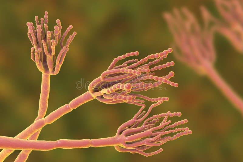
Free with trial Fungi Penicillium roqueforti, a fungus used in the production of blue cheeses, 3D illustration. Conidiophore illustrations Fungi Penicillium roqueforti
Free with trial Fungi Penicillium which cause food spoilage and are used for production of the first antibiotic penicillin. 3D illustration showing spores conidia and conidiophore. Conidiophore illustrations Fungi Penicillium which cause food spoilage and are used for production of the first antibiotic penicillin
Free with trial Fungi Penicillium which cause food spoilage and are used for production of the first antibiotic penicillin. 3D illustration showing spores conidia and conidiophore. Conidiophore illustrations Fungi Penicillium which cause food spoilage and are used for production of the first antibiotic penicillin
Free with trial Fungi Penicillium which cause food spoilage and are used for production of the first antibiotic penicillin. 3D illustration showing spores conidia and conidiophore. Conidiophore illustrations Fungi Penicillium which cause food spoilage and are used for production of the first antibiotic penicillin
Free with trial Reproductive Structures of Penicillium. Life cycle. Conidiophore vectors Reproductive Structures of Penicillium.
Free with trial Fungi Penicillium roqueforti, a fungus used in the production of blue cheeses, 3D illustration. Conidiophore illustrations Fungi Penicillium roqueforti
Free with trial Fungi Penicillium producing penicillin antibiotic, 3D illustration. Conidiophore illustrations Fungi Penicillium producing penicillin antibiotic
Free with trial Mandarin with mold. Photo and 3D illustration of microscopic fungi Penicillium which cause food spoilage and produce antibiotic penicillin. Conidiophore illustrations Mandarin with mold. Microscopic fungi Penicillium which cause food spoilage. Mandarin with mold. Photo and 3D illustration of microscopic fungi Penicillium which cause food spoilage and produce antibiotic penicillin
Free with trial Coloring page with structure of Penicillium. Mycelium with conidiophore and conidium isolated on white background. Conidiophore vectors Coloring page with structure of Penicillium. Mycelium with conidiophore and conidium
Free with trial Penicillium mold fungi, 3D illustration and photo of colonies grown on nutrient medium, fungus, microbial, penicillin, science, food, green, spore, growth, scientific, microbiology, fungal, mycology, mycosis, mould, health, cheese, macro, antibiotic, conidiophore, eumycota, nature, source, conidia, first, fleming, colony, morphology, agar, petri, dish, plate, sabouraud, dextrose. Conidiophore illustrations Penicillium mold fungi, illustration and photo of colony grown on nutrient medium. Penicillium mold fungi, 3D illustration and photo of colonies grown on nutrient medium, fungus, microbial, penicillin, science, food, green, spore, growth, scientific, microbiology, fungal, mycology, mycosis, mould, health, cheese, macro, antibiotic, conidiophore, eumycota, nature, source, conidia, first, fleming, colony, morphology, agar, petri, dish, plate, sabouraud, dextrose
Free with trial Penicillium mold fungi, 3D illustration and photo of colonies grown on nutrient medium, fungus, microbial, penicillin, science, food, green, spore, growth, scientific, microbiology, fungal, mycology, mycosis, mould, health, cheese, macro, antibiotic, conidiophore, eumycota, nature, source, conidia, first, fleming, colony, morphology, agar, petri, dish, plate, sabouraud, dextrose, black, background. Conidiophore illustrations Penicillium mold fungi, illustration and photo of colony grown on nutrient medium. Penicillium mold fungi, 3D illustration and photo of colonies grown on nutrient medium, fungus, microbial, penicillin, science, food, green, spore, growth, scientific, microbiology, fungal, mycology, mycosis, mould, health, cheese, macro, antibiotic, conidiophore, eumycota, nature, source, conidia, first, fleming, colony, morphology, agar, petri, dish, plate, sabouraud, dextrose, black, background
Free with trial Penicillium mold fungi, 3D illustration and photo of colonies grown on nutrient medium, fungus, microbial, penicillin, science, food, green, spore, growth, scientific, microbiology, fungal, mycology, mycosis, mould, health, cheese, macro, antibiotic, conidiophore, eumycota, nature, source, conidia, first, fleming, colony, morphology, agar, petri, dish, plate, sabouraud, dextrose, black, background. Conidiophore illustrations Penicillium mold fungi, illustration and photo of colony grown on nutrient medium. Penicillium mold fungi, 3D illustration and photo of colonies grown on nutrient medium, fungus, microbial, penicillin, science, food, green, spore, growth, scientific, microbiology, fungal, mycology, mycosis, mould, health, cheese, macro, antibiotic, conidiophore, eumycota, nature, source, conidia, first, fleming, colony, morphology, agar, petri, dish, plate, sabouraud, dextrose, black, background
Free with trial Penicillium mold fungi, 3D illustration and photo of colonies grown on nutrient medium, fungus, microbial, penicillin, science, food, green, spore, growth, scientific, microbiology, fungal, mycology, mycosis, mould, health, cheese, macro, antibiotic, conidiophore, eumycota, nature, source, conidia, first, fleming, colony, morphology, agar, petri, dish, plate, sabouraud, dextrose. Conidiophore illustrations Penicillium mold fungi, illustration and photo of colony grown on nutrient medium. Penicillium mold fungi, 3D illustration and photo of colonies grown on nutrient medium, fungus, microbial, penicillin, science, food, green, spore, growth, scientific, microbiology, fungal, mycology, mycosis, mould, health, cheese, macro, antibiotic, conidiophore, eumycota, nature, source, conidia, first, fleming, colony, morphology, agar, petri, dish, plate, sabouraud, dextrose
Free with trial Penicillium mold fungi, 3D illustration and photo of colonies grown on nutrient medium, fungus, microbial, penicillin, science, food, green, spore, growth, scientific, microbiology, fungal, mycology, mycosis, mould, health, cheese, macro, antibiotic, conidiophore, eumycota, nature, source, conidia, first, fleming, colony, morphology, agar, petri, dish, plate, sabouraud, dextrose. Conidiophore illustrations Penicillium mold fungi, illustration and photo of colony grown on nutrient medium. Penicillium mold fungi, 3D illustration and photo of colonies grown on nutrient medium, fungus, microbial, penicillin, science, food, green, spore, growth, scientific, microbiology, fungal, mycology, mycosis, mould, health, cheese, macro, antibiotic, conidiophore, eumycota, nature, source, conidia, first, fleming, colony, morphology, agar, petri, dish, plate, sabouraud, dextrose
Free with trial Penicillium mold fungi, 3D illustration and photo of colonies grown on nutrient medium, fungus, microbial, penicillin, science, food, green, spore, growth, scientific, microbiology, fungal, mycology, mycosis, mould, health, cheese, macro, antibiotic, conidiophore, eumycota, nature, source, conidia, first, fleming, colony, morphology, agar, petri, dish, plate, sabouraud, dextrose. Conidiophore illustrations Penicillium mold fungi, illustration and photo of colony grown on nutrient medium. Penicillium mold fungi, 3D illustration and photo of colonies grown on nutrient medium, fungus, microbial, penicillin, science, food, green, spore, growth, scientific, microbiology, fungal, mycology, mycosis, mould, health, cheese, macro, antibiotic, conidiophore, eumycota, nature, source, conidia, first, fleming, colony, morphology, agar, petri, dish, plate, sabouraud, dextrose
Free with trial Penicillium mold fungi, 3D illustration and photo of colonies grown on nutrient medium, fungus, microbial, penicillin, science, food, green, spore, growth, scientific, microbiology, fungal, mycology, mycosis, mould, health, cheese, macro, antibiotic, conidiophore, eumycota, nature, source, conidia, first, fleming, colony, morphology, agar, petri, dish, plate, sabouraud, dextrose, black, background. Conidiophore illustrations Penicillium mold fungi, illustration and photo of colony grown on nutrient medium. Penicillium mold fungi, 3D illustration and photo of colonies grown on nutrient medium, fungus, microbial, penicillin, science, food, green, spore, growth, scientific, microbiology, fungal, mycology, mycosis, mould, health, cheese, macro, antibiotic, conidiophore, eumycota, nature, source, conidia, first, fleming, colony, morphology, agar, petri, dish, plate, sabouraud, dextrose, black, background
Free with trial Fungi Penicillium which cause food spoilage and are used for production of the first antibiotic penicillin. 3D illustration showing spores conidia and conidiophore. Conidiophore illustrations Fungi Penicillium which cause food spoilage and are used for production of the first antibiotic penicillin
Free with trial Fungi Penicillium which cause food spoilage and are used for production of the first antibiotic penicillin. 3D illustration showing spores conidia and conidiophore. Conidiophore illustrations Fungi Penicillium which cause food spoilage and are used for production of the first antibiotic penicillin
Free with trial Fungi Penicillium which cause food spoilage and are used for production of the antibiotic penicillin. 3D illustration showing spores conidia and conidiophore, mold, fungus, microbial, science, green, growth, scientific, microbiology, fungal, mycology, mycosis, mould, health, cheese, macro, eumycota, nature, naturemycology, chrysogenum, source, formation, notatum, first, fleming. Conidiophore illustrations Fungi Penicillium which cause food spoilage and are used for production of the penicillin. Fungi Penicillium which cause food spoilage and are used for production of the antibiotic penicillin. 3D illustration showing spores conidia and conidiophore, mold, fungus, microbial, science, green, growth, scientific, microbiology, fungal, mycology, mycosis, mould, health, cheese, macro, eumycota, nature, naturemycology, chrysogenum, source, formation, notatum, first, fleming
Free with trial Penicillium mold fungi, 3D illustration and photo of colonies grown on nutrient medium, fungus, microbial, penicillin, science, food, green, spore, growth, scientific, microbiology, fungal, mycology, mycosis, mould, health, cheese, macro, antibiotic, conidiophore, eumycota, nature, source, conidia, first, fleming, colony, morphology, agar, petri, dish, plate, sabouraud, dextrose. Conidiophore illustrations Penicillium mold fungi, illustration and photo of colony grown on nutrient medium. Penicillium mold fungi, 3D illustration and photo of colonies grown on nutrient medium, fungus, microbial, penicillin, science, food, green, spore, growth, scientific, microbiology, fungal, mycology, mycosis, mould, health, cheese, macro, antibiotic, conidiophore, eumycota, nature, source, conidia, first, fleming, colony, morphology, agar, petri, dish, plate, sabouraud, dextrose
Free with trial Penicillium mold fungi, 3D illustration and photo of colonies grown on nutrient medium, fungus, microbial, penicillin, science, food, green, spore, growth, scientific, microbiology, fungal, mycology, mycosis, mould, health, cheese, macro, antibiotic, conidiophore, eumycota, nature, source, conidia, first, fleming, colony, morphology, agar, petri, dish, plate, sabouraud, dextrose, black, background. Conidiophore illustrations Penicillium mold fungi, illustration and photo of colony grown on nutrient medium. Penicillium mold fungi, 3D illustration and photo of colonies grown on nutrient medium, fungus, microbial, penicillin, science, food, green, spore, growth, scientific, microbiology, fungal, mycology, mycosis, mould, health, cheese, macro, antibiotic, conidiophore, eumycota, nature, source, conidia, first, fleming, colony, morphology, agar, petri, dish, plate, sabouraud, dextrose, black, background
Free with trial Fungi Penicillium which cause food spoilage and are used for production of the first antibiotic penicillin. 3D illustration showing spores conidia and conidiophore. Conidiophore illustrations Fungi Penicillium which cause food spoilage and are used for production of the first antibiotic penicillin
Free with trial Fungi Penicillium which cause food spoilage and are used for production of the first antibiotic penicillin. 3D illustration showing spores conidia and conidiophore. Conidiophore illustrations Fungi Penicillium which cause food spoilage and are used for production of the first antibiotic penicillin
Free with trial 3d illustration of an Aspergillus fumigatus co conidiophore and spores germinating. Conidiophore illustrations Aspergillus fumigatus
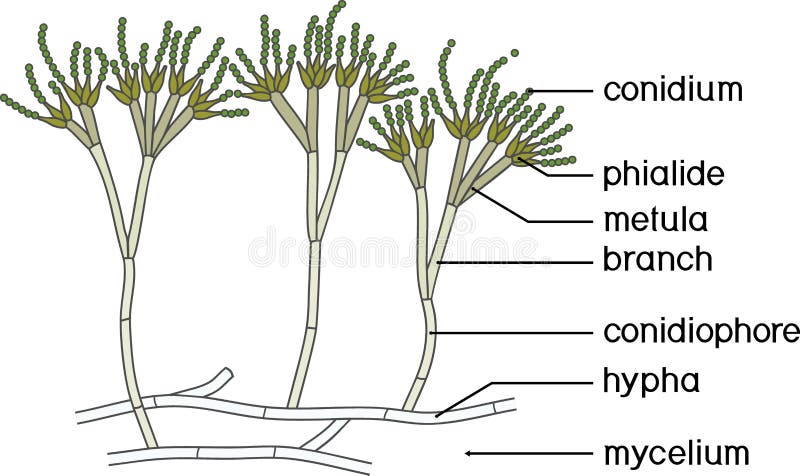
Free with trial Penicillium anatomy. Structure of a Microscopic fungi that use in food and drug production. Part of a Fungus. Close-up of a Metula, Sterigma, Conidia, Hypha. vector illustration isolated on white background. Conidiophore vectors Penicillium anatomy. Structure of a Microscopic fungi
Free with trial Penicillium Slide blue mold, mycelium and conidiophores. Mold Microscopy. Conidiophore vectors Penicillium Slide blue mold, mycelium and conidiophores.
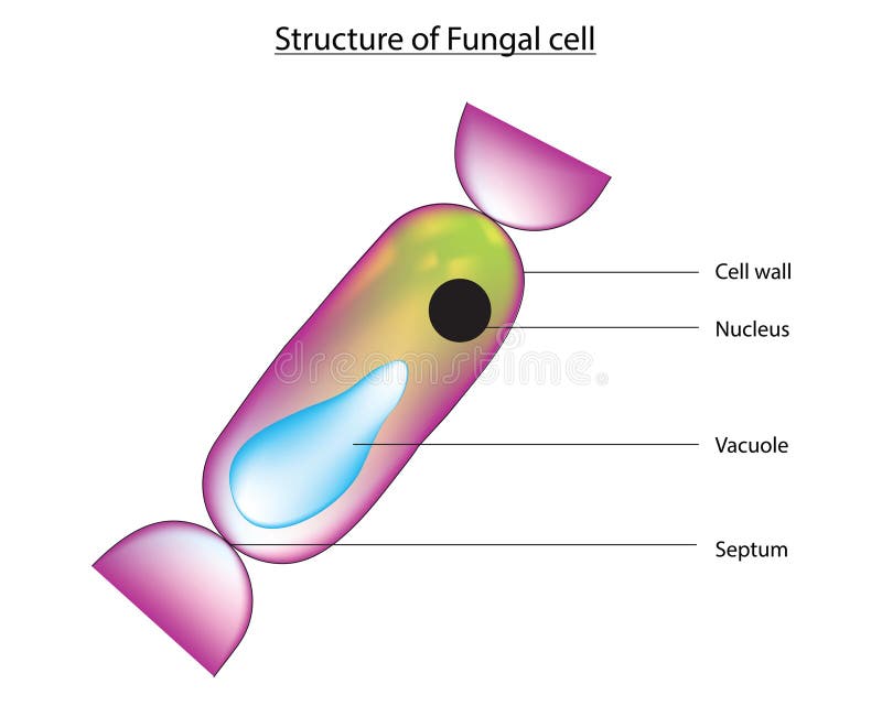
Free with trial Anatomy of typical fungi cell. Fungal Hyphae and Cell Structure. Vector diagram. Conidiophore vectors Fungi cell structure. Anatomy of typical fungi cell. Fungal Hyphae and Cell Structure. Vector diagram
Free with trial Mucor and Penicillium isolated on white background. Conidiophore vectors Mucor and Penicillium
Free with trial Microscopic fungi Hormographiella, scientific illustration. Occasionally cause pulmonary infections in immunocompromised patients, endocarditis following valve replacement, eye and skin infections. Conidiophore illustrations Microscopic fungi Hormographiella, scientific illustration
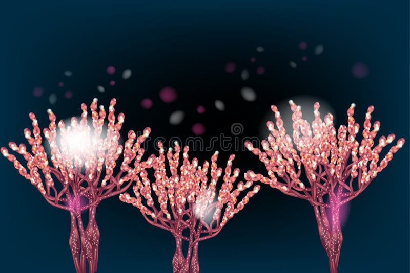
Free with trial Fungi Penicillium roqueforti, a fungus used in the production of blue cheeses, 3D illustration. Conidiophore illustrations Fungi Penicillium roqueforti
Free with trial Fungi Penicillium roqueforti, a fungus used in the production of blue cheeses, 3D illustration. Conidiophore illustrations Fungi Penicillium roqueforti
Free with trial Mold Alternaria alternata, allergic fungus, 3D illustration. Alternaria is the causative agent of plant diseases, is common indoor mold and causes allergy, asthma, onychomycosis, sinusitis. Conidiophore illustrations Mold Alternaria alternata, allergic fungus
Free with trial Depiction of a saprophytic fungus actively sporulating on a shriveled, decomposing fruit surface. Conidiophore illustrations Microscopic Mold Colonies in Artistic Detail. Depiction of a saprophytic fungus actively sporulating on a shriveled, decomposing fruit surface.
Free with trial Fungi Penicillium producing penicillin antibiotic, 3D illustration. Conidiophore illustrations Fungi Penicillium producing penicillin antibiotic
Free with trial Mandarin with mold. Photo and 3D illustration of microscopic fungi Penicillium which cause food spoilage and produce antibiotic penicillin. Conidiophore illustrations Mandarin with mold. Microscopic fungi Penicillium which cause food spoilage. Mandarin with mold. Photo and 3D illustration of microscopic fungi Penicillium which cause food spoilage and produce antibiotic penicillin
Free with trial Mold Alternaria alternata, allergic fungus, 3D illustration and photo of colony on agar plate. Alternaria is the causative agent of plant diseases, is common indoor mold and causes allergy. Conidiophore illustrations Mold Alternaria alternata, illustration and photo of colony on nutrient medium. Mold Alternaria alternata, allergic fungus, 3D illustration and photo of colony on agar plate. Alternaria is the causative agent of plant diseases, is common indoor mold and causes allergy
Free with trial Mold Alternaria alternata, allergic fungus, 3D illustration. Alternaria is the causative agent of plant diseases, is common indoor mold and causes allergy, asthma, onychomycosis, sinusitis. Conidiophore illustrations Mold Alternaria alternata, allergic fungus
Free with trial Fungi Penicillium roqueforti, a fungus used in the production of blue cheeses, 3D illustration. Conidiophore illustrations Fungi Penicillium roqueforti
Free with trial Mold Alternaria alternata, allergic fungus, 3D illustration. Alternaria is the causative agent of plant diseases, is common indoor mold and causes allergy, asthma, onychomycosis, sinusitis. Conidiophore illustrations Mold Alternaria alternata, allergic fungus
Free with trial Mold Alternaria alternata, allergic fungus, 3D illustration. Alternaria is the causative agent of plant diseases, is common indoor mold and causes allergy, asthma, onychomycosis, sinusitis. Conidiophore illustrations Mold Alternaria alternata, allergic fungus
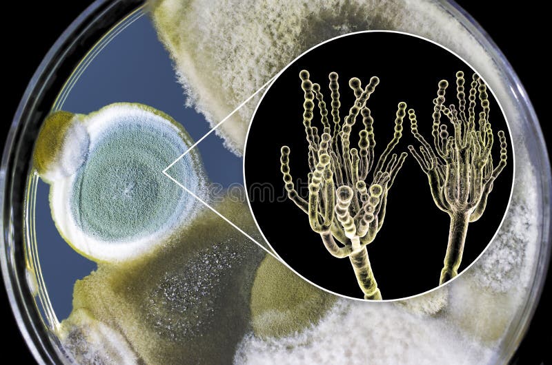
Free with trial Penicillium mold fungi, 3D illustration and photo of colonies grown on nutrient medium. Conidiophore illustrations Penicillium mold fungi, illustration and photo of colony grown on nutrient medium
Free with trial Mold Alternaria alternata, allergic fungus, 3D illustration. Alternaria is the causative agent of plant diseases, is common indoor mold and causes allergy, asthma, onychomycosis, sinusitis. Conidiophore illustrations Mold Alternaria alternata, allergic fungus
Free with trial Microscopic fungi Scopulariopsis brevicaulis, 3D illustration. Fungus that infects nails, causes subcutaneous and invasive infections, endocarditis, sinusitis, disseminated infection. Conidiophore illustrations Microscopic fungi Scopulariopsis brevicaulis, 3D illustration
Free with trial Mold Alternaria alternata, allergic fungus, 3D illustration and photo of colony on agar plate. Alternaria is the causative agent of plant diseases, is common indoor mold and causes allergy. Conidiophore illustrations Mold Alternaria alternata, illustration and photo of colony on nutrient medium. Mold Alternaria alternata, allergic fungus, 3D illustration and photo of colony on agar plate. Alternaria is the causative agent of plant diseases, is common indoor mold and causes allergy
Free with trial Mold Alternaria alternata, allergic fungus, 3D illustration and photo of colony on agar plate. Alternaria is the causative agent of plant diseases, is common indoor mold and causes allergy. Conidiophore illustrations Mold Alternaria alternata, illustration and photo of colony on nutrient medium. Mold Alternaria alternata, allergic fungus, 3D illustration and photo of colony on agar plate. Alternaria is the causative agent of plant diseases, is common indoor mold and causes allergy
Free with trial Mold Alternaria alternata, allergic fungus, 3D illustration and photo of colony on agar plate. Alternaria is the causative agent of plant diseases, is common indoor mold and causes allergy. Conidiophore illustrations Mold Alternaria alternata, illustration and photo of colony on nutrient medium. Mold Alternaria alternata, allergic fungus, 3D illustration and photo of colony on agar plate. Alternaria is the causative agent of plant diseases, is common indoor mold and causes allergy
Free with trial Mold Alternaria alternata, allergic fungus, 3D illustration and photo of colony on agar plate. Alternaria is the causative agent of plant diseases, is common indoor mold and causes allergy. Conidiophore illustrations Mold Alternaria alternata, illustration and photo of colony on nutrient medium. Mold Alternaria alternata, allergic fungus, 3D illustration and photo of colony on agar plate. Alternaria is the causative agent of plant diseases, is common indoor mold and causes allergy
Free with trial Microscopic fungi Scopulariopsis brevicaulis, 3D illustration. Fungus that infects nails, causes subcutaneous and invasive infections, endocarditis, sinusitis, disseminated infection. Conidiophore illustrations Microscopic fungi Scopulariopsis brevicaulis, 3D illustration
Free with trial Penicillium mold fungi, 3D illustration and photo of colonies grown on nutrient medium. Conidiophore illustrations Penicillium mold fungi, illustration and photo of colony grown on nutrient medium
Free with trial Emmonsia microscopic pathogenic fungi, 3D illustration. The causative agent of adiaspiromycosis pulmonary disease in small animals, and also lung and disseminated infections in HIV-positive patients. Conidiophore illustrations Emmonsia microscopic pathogenic fungi, 3D illustration
Free with trial Microscopic fungi Scopulariopsis brevicaulis, 3D illustration. Fungus that infects nails, causes subcutaneous and invasive infections, endocarditis, sinusitis, disseminated infection. Conidiophore illustrations Microscopic fungi Scopulariopsis brevicaulis, 3D illustration
Free with trial Scanning electron microscopy of Aspergillus fumigatus conidiophore showing vesicle, metulae, phialides, and conidia chain architecture. Conidiophore illustrations Fungal Conidiophore Vesicle Metula Phialide SEM. Scanning electron microscopy of Aspergillus fumigatus conidiophore showing vesicle, metulae, phialides, and. Scanning electron microscopy of Aspergillus fumigatus conidiophore showing vesicle, metulae, phialides, and conidia chain architecture
Free with trial Artistic study of fungal reproduction through conidiophore growth in a humid, decaying environment. Conidiophore illustrations Fungal Sporulation on Organic Substrate. Artistic study of fungal reproduction through conidiophore growth in a humid, decaying environment.
Free with trial A fungal colony forms radiating sporophores on a rotting fruit, evoking both scientific clarity and aesthetic composition. Conidiophore illustrations Fruiting Bodies of Mold Fungus Emerging. A fungal colony forms radiating sporophores on a rotting fruit, evoking both scientific clarity and aesthetic composition.
Free with trial Mature conidiophores of a mold fungus rise from a mold-covered substrate, dispersing spores into the air. Conidiophore illustrations Aspergillus Blooming from Decomposing Host. Mature conidiophores of a mold fungus rise from a mold-covered substrate, dispersing spores into the air.
Free with trial Illustrated spore-bearing heads of Aspergillus emerging from a decayed fruit, shown in stylized precision. Conidiophore illustrations Spore Heads of Aspergillus on Rotting Fruit. Illustrated spore-bearing heads of Aspergillus emerging from a decayed fruit, shown in stylized precision.
Free with trial Microscopic view of blue penicillium fungus showing branching hyphae and spore structures, representing mycology, antibiotic production, and mold. Conidiophore illustrations Microscopic view of blue penicillium fungus showing branching hyphae and spore structures, representing mycology
Free with trial Dirty, damp towel showing visible black and green mold spots, paired with a microscopic inset of fungal hyphae and spores, representing household contamination. Conidiophore illustrations Dirty, damp towel showing visible black and green mold spots, paired with a microscopic inset of fungal hyphae and spores
Free with trial Laboratory microscope view of blue-stained penicillium fungus showing characteristic conidiophores and spore chains, representing mycology. Conidiophore illustrations Laboratory microscope view of blue-stained penicillium fungus showing characteristic conidiophores and spore chains
Free with trial Hand-drawn pen illustration of Rhizopus fungi on black paper, highlighting hyphal structure and conidia, the causative agent of mucormycosis. Conidiophore illustrations Hand-drawn pen illustration of Rhizopus fungi
Free with trial Mucor is a genus of fungi commonly found in soil, plants, decaying organic matter, and indoor environments. These fungi belong to the phylum Mucoromycota and are characterized by their rapid growth and ability to produce large quantities of asexual spores called sporangiospores. Mucor species are often opportunistic pathogens, meaning they primarily infect individuals with weakened immune systems or underlying health conditions. Mucormycosis, the infection caused by these fungi, can manifest in various forms depending on the site of infection. Rhinocerebral mucormycosis affects the sinuses and brain, while pulmonary mucormycosis affects the lungs. Other types include cutaneous mucormycosis (skin) and gastrointestinal mucormycosis. Conidiophore vectors Mucor
Free with trial Lung adiaspiromycosis, a rare respiratory infection caused by the fungus Emmonsia spp. , characterized by the presence of enlarged encapsulated fungal spores within lung tissues, 3D illustration. Conidiophore illustrations Lung adiaspiromycosis, 3D illustration. Lung adiaspiromycosis, a rare respiratory infection caused by the fungus Emmonsia spp., characterized by the presence of enlarged encapsulated fungal spores within lung tissues, 3D illustration.
Free with trial Lung adiaspiromycosis, a rare respiratory infection caused by the fungus Emmonsia spp. , characterized by the presence of enlarged encapsulated fungal spores within lung tissues, 3D illustration. Conidiophore illustrations Lung adiaspiromycosis, 3D illustration. Lung adiaspiromycosis, a rare respiratory infection caused by the fungus Emmonsia spp., characterized by the presence of enlarged encapsulated fungal spores within lung tissues, 3D illustration.
Free with trial Lung adiaspiromycosis, a rare respiratory infection caused by the fungus Emmonsia spp. , characterized by the presence of enlarged encapsulated fungal spores within lung tissues, 3D illustration. Conidiophore illustrations Lung adiaspiromycosis, 3D illustration. Lung adiaspiromycosis, a rare respiratory infection caused by the fungus Emmonsia spp., characterized by the presence of enlarged encapsulated fungal spores within lung tissues, 3D illustration.
Free with trial Lung adiaspiromycosis, a rare respiratory infection caused by the fungus Emmonsia spp. , characterized by the presence of enlarged encapsulated fungal spores within lung tissues, 3D illustration. Conidiophore illustrations Lung adiaspiromycosis, 3D illustration. Lung adiaspiromycosis, a rare respiratory infection caused by the fungus Emmonsia spp., characterized by the presence of enlarged encapsulated fungal spores within lung tissues, 3D illustration.
Free with trial The anatomy of Aspergillus is adapted for efficient nutrient uptake, reproduction, and dispersal in diverse environments. The ability of Aspergillus to produce abundant spores and colonize a wide range of habitats contributes to its importance as both a beneficial organism in industrial applications (e. g. , production of enzymes and antibiotics) and a potential human pathogen. Conidiophore vectors Aspergillus anatomy. The anatomy of Aspergillus is adapted for efficient nutrient uptake, reproduction, and dispersal in diverse environments. The ability of Aspergillus to produce abundant spores and colonize a wide range of habitats contributes to its importance as both a beneficial organism in industrial applications (e.g., production of enzymes and antibiotics) and a potential human pathogen.
Free with trial Fungal cells, like those of other eukaryotic organisms, have complex structures that enable them to carry out various functions. Fungal cells have a rigid cell wall surrounding the plasma membrane. The composition of the cell wall varies depending on the type of fungus but often includes chitin, glucans, and other polysaccharides. The cell wall provides structural support and protection against environmental stresses. Conidiophore vectors Fungal cell structure. Fungal cells, like those of other eukaryotic organisms, have complex structures that enable them to carry out various functions. Fungal cells have a rigid cell wall surrounding the plasma membrane. The composition of the cell wall varies depending on the type of fungus but often includes chitin, glucans, and other polysaccharides. The cell wall provides structural support and protection against environmental stresses.
Free with trial Emmonsia microscopic pathogenic fungi, adiaspore stage, 3D illustration. The causative agent of adiaspiromycosis pulmonary disease in small animals and rarely in humans. Conidiophore illustrations Emmonsia microscopic pathogenic fungi, adiaspore stage, 3D illustration
Free with trial Emmonsia microscopic pathogenic fungi, adiaspore stage, 3D illustration. The causative agent of adiaspiromycosis pulmonary disease in small animals and rarely in humans. Conidiophore illustrations Emmonsia microscopic pathogenic fungi, adiaspore stage, 3D illustration
Free with trial Emmonsia microscopic pathogenic fungi, adiaspore stage, 3D illustration. The causative agent of adiaspiromycosis pulmonary disease in small animals and rarely in humans. Conidiophore illustrations Emmonsia microscopic pathogenic fungi, adiaspore stage, 3D illustration
Free with trial Emmonsia microscopic pathogenic fungi, adiaspore stage, 3D illustration. The causative agent of adiaspiromycosis pulmonary disease in small animals and rarely in humans. Conidiophore illustrations Emmonsia microscopic pathogenic fungi, adiaspore stage, 3D illustration
Free with trial Lung adiaspiromycosis, a rare respiratory infection caused by the fungus Emmonsia spp. , characterized by the presence of enlarged encapsulated fungal spores within lung tissues, 3D illustration. Conidiophore illustrations Lung adiaspiromycosis and close-up view of Emmonsia fungi, 3D illustration. Lung adiaspiromycosis, a rare respiratory infection caused by the fungus Emmonsia spp., characterized by the presence of enlarged encapsulated fungal spores within lung tissues, 3D illustration.
Free with trial Lung adiaspiromycosis, a rare respiratory infection caused by the fungus Emmonsia spp. , characterized by the presence of enlarged encapsulated fungal spores within lung tissues, 3D illustration. Conidiophore illustrations Lung adiaspiromycosis and close-up view of Emmonsia fungi, 3D illustration. Lung adiaspiromycosis, a rare respiratory infection caused by the fungus Emmonsia spp., characterized by the presence of enlarged encapsulated fungal spores within lung tissues, 3D illustration.
Free with trial Lung adiaspiromycosis, a rare respiratory infection caused by the fungus Emmonsia spp. , characterized by the presence of enlarged encapsulated fungal spores within lung tissues, 3D illustration. Conidiophore illustrations Lung adiaspiromycosis and close-up view of Emmonsia fungi, 3D illustration. Lung adiaspiromycosis, a rare respiratory infection caused by the fungus Emmonsia spp., characterized by the presence of enlarged encapsulated fungal spores within lung tissues, 3D illustration.
Free with trial Lung adiaspiromycosis, a rare respiratory infection caused by the fungus Emmonsia spp. , characterized by the presence of enlarged encapsulated fungal spores within lung tissues, 3D illustration. Conidiophore illustrations Lung adiaspiromycosis and close-up view of Emmonsia fungi, 3D illustration. Lung adiaspiromycosis, a rare respiratory infection caused by the fungus Emmonsia spp., characterized by the presence of enlarged encapsulated fungal spores within lung tissues, 3D illustration.
Free with trial Electron microscope view of Aspergillus fungus. Detailed cellular structure with branching hyphae, conidiophores. Microbiology, biology, medicine, scientific research, spore, mycosis. Conidiophore illustrations Electron microscope view of Aspergillus fungus. Detailed cellular structure with branching hyphae, conidiophores. Microbiology
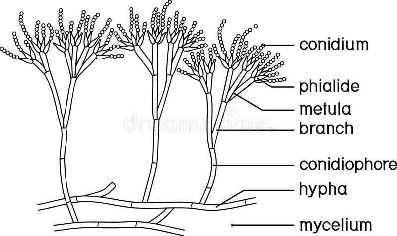
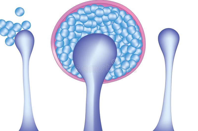
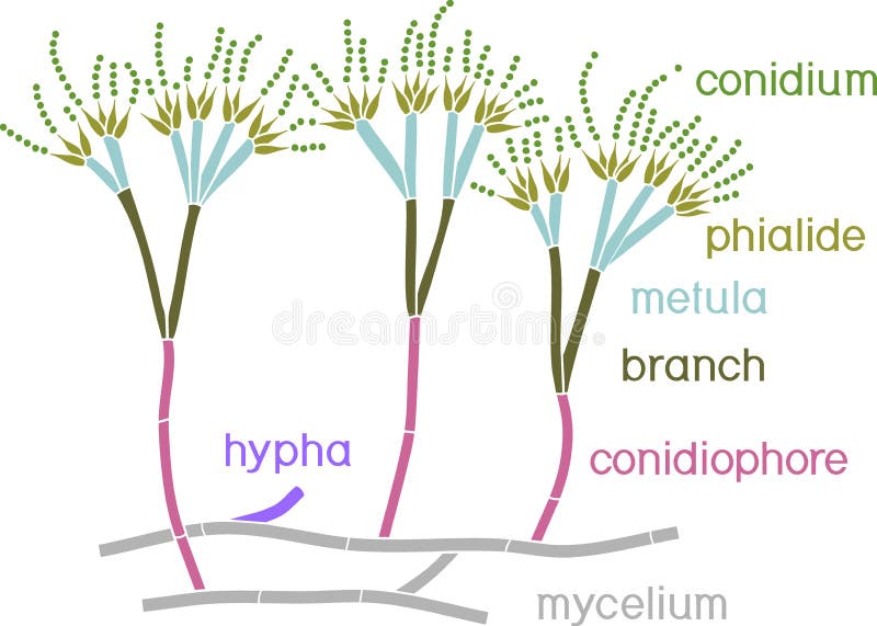
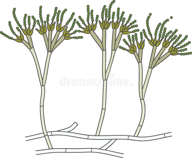
Free with trial Structure of Penicillium. Mycelium with conidiophore and conidium isolated on white background. Conidiophore vectors Structure of Penicillium. Mycelium with conidiophore and conidium
Free with trial Structure of Penicillium. Mycelium with conidiophore and conidium isolated on white background. Conidiophore vectors Structure of Penicillium. Mycelium with conidiophore and conidium
Free with trial Structure of Penicillium. Mycelium with conidiophore and conidium isolated on white background. Conidiophore vectors Structure of Penicillium. Mycelium with conidiophore and conidium
Free with trial Structure of Penicillium. Mycelium with conidiophore and conidium isolated on white background. Conidiophore vectors Structure of Penicillium. Mycelium with conidiophore and conidium