Free with trial Anatomy of blood vessels from artery through capillaries and vein. Endothelial cells vectors Anatomy of blood vessels
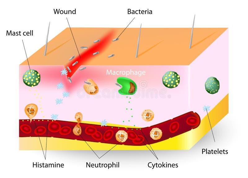
Free with trial Inflammation. innate immune system. Vector diagram. Endothelial cells vectors Inflammation. innate immune system
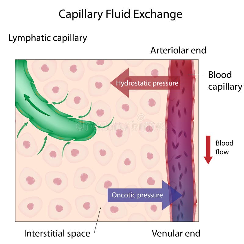
Free with trial Mechanism of capillary fluid exchange between the blood and body tissues, eps10. Endothelial cells vectors Capillary Fluid Exchange
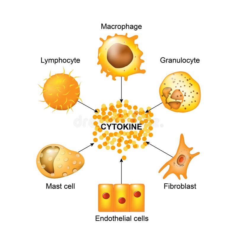
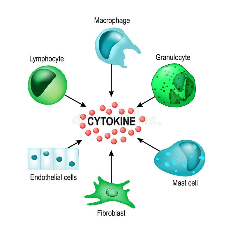
Free with trial Cytokines are produced by macrophages, lymphocytes, mast cells, endothelial cells and fibroblasts. Cytokines include chemokines, interferons, interleukins, lymphokines, and tumour necrosis factors, but not hormones or growth factors. Endothelial cells vectors Cells that produce cytokines. Cytokines are produced by macrophages, lymphocytes, mast cells, endothelial cells and fibroblasts. Cytokines include chemokines, interferons, interleukins, lymphokines, and tumour necrosis factors, but not hormones or growth factors
Free with trial Components of blood, white blood cells, and granulocytes of human immune defense mechanism. Endothelial cells vectors Collection of all 12 immune cells of human and they are dendritic cells, eosinophils, natural killer cell, mast and b cell, t, and. Components of blood, white blood cells, and granulocytes of human immune defense mechanism
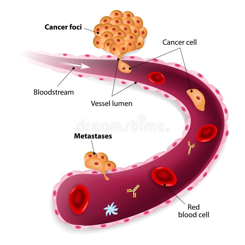
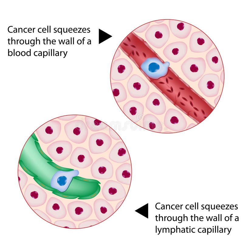
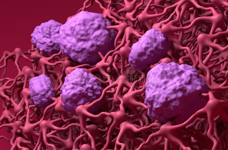
Free with trial Cancer cell squeezes through blood vessel during Metastases. Endothelial cells vectors Cancer cells, cancer foci and Metastases. Cancer cell squeezes through blood vessel during Metastases
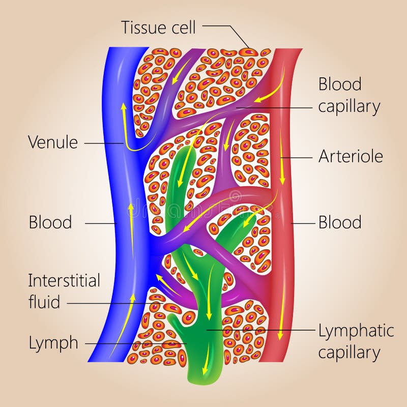
Free with trial The lymph system, relationship of lymphatic capillaries to tissue cells and blood capillaries, vector medical illustration. Endothelial cells vectors Lymphatic and Blood Capillaries. The lymph system, relationship of lymphatic capillaries to tissue cells and blood capillaries, vector medical illustration
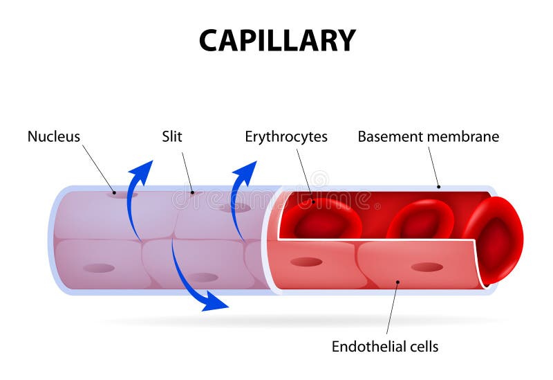
Free with trial Capillary for blood medical illusration works. Endothelial cells illustrations Capillary
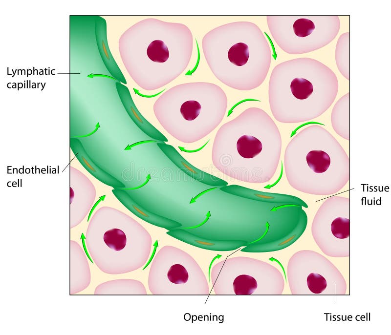
Free with trial Uptake of tissue fluid by lymphatic capillary, eps8. Endothelial cells vectors Lymphatic capillary
Free with trial Capillary. blood vessel. labelled. Vector Diagram. Endothelial cells vectors Capillary. blood vessel. labelled
Free with trial Cytokines are produced by macrophages, lymphocytes, mast cells, endothelial cells and fibroblasts. Cytokines include chemokines, interferons, interleukins, lymphokines, and tumour necrosis factors, but not hormones or growth factors. Endothelial cells vectors Cytokine. Vector concept. Cytokines are produced by macrophages, lymphocytes, mast cells, endothelial cells and fibroblasts. Cytokines include chemokines, interferons, interleukins, lymphokines, and tumour necrosis factors, but not hormones or growth factors
Free with trial Collection of all the blood cells vector illsutaion. Neutrophil, eosinophil, macrophage, dendritic cell natural killer cell, blood cells vector design in isolated background. Endothelial cells illustrations Collection of all the blood cells vector illsutaion. Neutrophil, eosinophil, macrophage, dendritic cell natural killer cell, blood
Free with trial Properties of cytokines. signal transduction between cells. endocrine, paracrine and autocrine secretion. Endothelial cells vectors Properties of cytokines
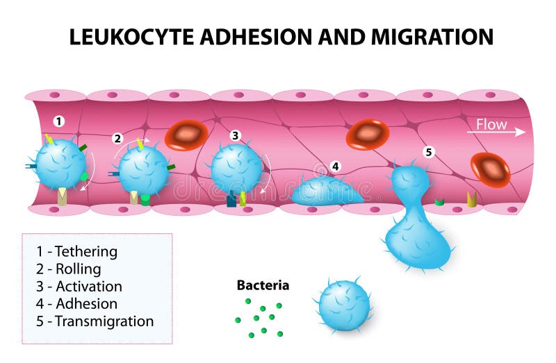
Free with trial Leukocyte adhesion and migration. After activation by chemotactic agents, the leukocytes change shape. The leukocytes then crawl and undergo diapedesis by interacting with platelet-endothelial cell adhesion molecules. Endothelial cells vectors Leukocyte adhesion and migration
Free with trial Macrophages are important cells of the immune system that are formed in response to an infection or accumulating damaged or dead cells. When macrophages are exposed to inflammatory stimuli, they secrete cytokines such as tumor necrosis factor (TNF), IL-1, IL-6, IL-8, and IL-12. Although monocytes and macrophages are the main sources of these cytokines, they are also produced by activated lymphocytes, endothelial cells, and fibroblasts. Endothelial cells illustrations Macrophage releasing cytokines. Macrophages are important cells of the immune system that are formed in response to an infection or accumulating damaged or dead cells. When macrophages are exposed to inflammatory stimuli, they secrete cytokines such as tumor necrosis factor (TNF), IL-1, IL-6, IL-8, and IL-12. Although monocytes and macrophages are the main sources of these cytokines, they are also produced by activated lymphocytes, endothelial cells, and fibroblasts.
Free with trial Primary cancer in the pancreas spreads through blood circulation to the lungs (secondary cancer), eps8. Endothelial cells vectors Metastatic cancer. Primary cancer in the pancreas spreads through blood circulation to the lungs (secondary cancer), eps8
Free with trial Cancer cell squeezes through blood and lymph capillary during metastasis, eps8. Endothelial cells vectors Cancer cell squeezes through vessel. Cancer cell squeezes through blood and lymph capillary during metastasis, eps8
Free with trial Changes in the hepatic associated with hepatic fibrosis. Normal liver and liver injury. Endothelial cells vectors Normal liver and liver injury.
Free with trial Fluorescence Microscope image of Bovine Pulmonary Artery Endothelial Cells BPAE stained for mitochondria, phalloidin, and nuclei undergoing mitosis. Endothelial cells illustrations Fluorescence Microscope image of human cells undergoing mitosis. Fluorescence Microscope image of Bovine Pulmonary Artery Endothelial Cells BPAE stained for mitochondria, phalloidin, and nuclei undergoing mitosis
Free with trial Pathogenesis of atherosclerosis. Cholesterol plaque, and thrombus formation. Cells structure vascular smooth muscle cells, T-lymphocyte, endothelial cells, monocyte, Foam cell, Macrophage, and Cholesterol. Cardiovascular disease. Vector illustration for medical, educational, and science use. Endothelial cells vectors Pathogenesis of atherosclerosis
Free with trial Pericyte anatomy. Structure of Blood vessel. Cross section of capillary with Basement membrane, Capillary lumen, Endothelial cells, and Rouget cells. Pericytes wrap around the endothelial cells that line the capillaries and venules that embedded in basement membrane. Endothelial cells vectors Pericyte anatomy. Structure of Blood vessel. Cross section of capillary
Free with trial Cancer cell squeezes through blood vessel during Metastases. Endothelial cells vectors Cancer cells, cancer foci and Metastases. Cancer cell squeezes through blood vessel during Metastases
Free with trial Illustration of formation of tumor cells through abnormal cell division, and cancer associated other processes like angiogenesis and metastasis. Endothelial cells vectors Angiogenesis of blood vessels in tumor. Illustration of formation of tumor cells through abnormal cell division, and cancer associated other processes like angiogenesis and metastasis.
Free with trial Cancer. Tumor development from Abnormal Cell growth, Lymphangiogenesis and Angiogenesis to Metastasis. Spread cancer cells from an initial site to a different tissues. Cancer invasion. Tumor growth. Vector diagram. Endothelial cells vectors Cancer. Tumor development. Angiogenesis and Metastasis. Cancer. Tumor development from Abnormal Cell growth, Lymphangiogenesis and Angiogenesis to Metastasis. Spread cancer cells from an initial site to a different tissues. Cancer invasion. Tumor growth. Vector diagram
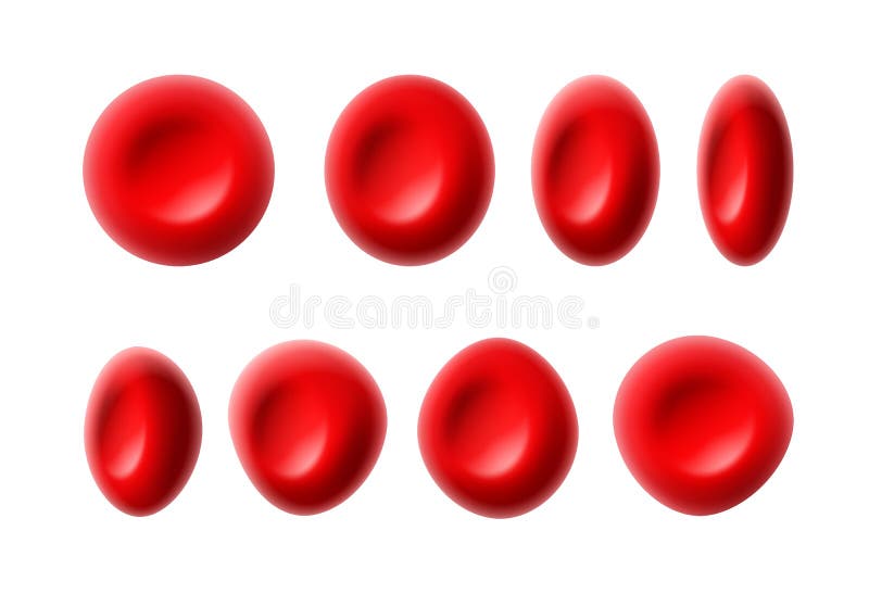
Free with trial Vector set of red blood cells or erythrocytes on white background. Endothelial cells vectors Red blood cells
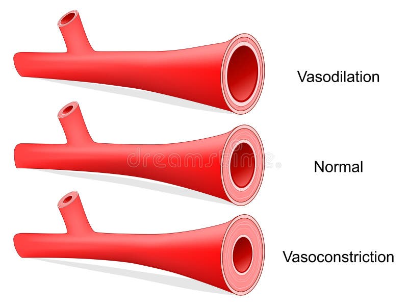
Free with trial Vasoconstriction and Vasodilation of an artery. Lumen of vein. Cross section of the blood vessel with red blood cells. comparison of normal, constricted, and dilated blood vessels. Vector illustration. Endothelial cells vectors Vasoconstriction and Vasodilation
Free with trial The Starling equation is an equation that illustrates the role of hydrostatic and oncotic forces (the so-called Starling forces) in the movement of fluid across capillary membranes. Capillary fluid movement may occur as a result of three processes: Diffusion, filtration and pinocytosis. Endothelial cells illustrations Capillary diffusion. The Starling equation is an equation that illustrates the role of hydrostatic and oncotic forces (the so-called Starling forces) in the movement of fluid across capillary membranes. Capillary fluid movement may occur as a result of three processes: Diffusion, filtration and pinocytosis
Free with trial Acute proliferative glomerulonephritis. disorder of the glomeruli, and small blood vessels in the kidneys. infections disease. Vector illustration for biology, educational, medical and science use. Endothelial cells vectors Acute proliferative glomerulonephritis
Free with trial Glomerulus. liquid enters to the glomerulus in Browman`s capsule goes down by the loop of henle to collecting duct in the kidneys. Vector illustration for biology, educational, medical and science use. Endothelial cells vectors Glomerulus. part of kidney. Glomerulus. liquid enters to the glomerulus in Browman`s capsule goes down by the loop of henle to collecting duct in the kidneys. Vector illustration for biology, educational, medical and science use
Free with trial Cytokines releasing cells list for immune system response outline diagram. Labeled educational scheme with macrophage, granulocyte, endothelial, fibroblast, mast and lymphocyte vector illustration. Endothelial cells vectors Cytokines releasing cells list for immune system response outline diagram
Free with trial Red blood cells erythrocytes in interior of arterial or capillary blood vessel Showing endothelial cells and blood flow or stream Human anatomy model 3D visualization. Endothelial cells illustrations Red blood cells erythrocytes in interior of arterial or capillary blood vessel Showing endothelial cells and blood flow or stream
Free with trial Red blood cells erythrocytes in interior of arterial or capillary blood vessel Showing endothelial cells and blood flow or stream Human anatomy model 3D visualization. Endothelial cells illustrations Red blood cells erythrocytes in interior of arterial or capillary blood vessel Showing endothelial cells and blood flow or stream
Free with trial Red blood cells erythrocytes in interior of arterial or capillary blood vessel Showing endothelial cells and blood flow or stream Human anatomy model 3D. Endothelial cells illustrations Red blood cells erythrocytes in interior of arterial or capillary blood vessel Showing endothelial cells and blood flow or stream
Free with trial Resistin is a hormone from adipose tissue, regulator of inflammation, autoimmune processes, obesity and insulin resistance. Atherosclerosis and its related complications. Target cells for resistin: macrophage, PBMC, osteoclast, osteoblast, plasma and endothelial cells, adipocyte fat cells. Endothelial cells vectors Resistin is a hormone from adipose tissue, regulator of inflammation, autoimmune processes, obesity and insulin resistance
Free with trial Collection of immune cells of human body including neutrophil, monocyte, dendritic cell. Endothelial cells illustrations Cells of the Immune system. List of immune cells- dendritic, Mast, Neutrophil, Macrophage, Cell, Phagocytosis, Natural Killer, B. Collection of immune cells of human body including neutrophil, monocyte, dendritic cell
Free with trial Cytokines are produced by macrophage, lymphocyte, mast cell, endothelial cells and fibroblast. Cell signaling by Interleukins, Chemokines, Tumor necrosis factor. Cytokine storm. Vector illustration. Endothelial cells vectors Cytokines are produced by macrophage, lymphocyte, mast cell, endothelial cell and fibroblast. Cytokines are produced by macrophage, lymphocyte, mast cell, endothelial cells and fibroblast. Cell signaling by Interleukins, Chemokines, Tumor necrosis factor. Cytokine storm. Vector illustration
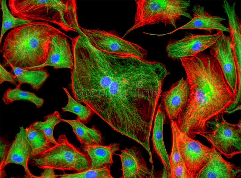
Free with trial Bovine pulmonary artery endothelial cells in culture. Blue: nuclei; green: microfilaments; red: mitochondria. Endothelial cells illustrations Bovine pulmonary arthery cells. Bovine pulmonary artery endothelial cells in culture. Blue: nuclei; green: microfilaments; red: mitochondria
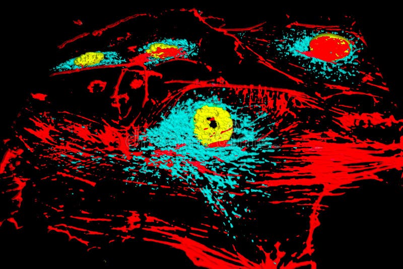
Free with trial Bovine pulmonary artery endothelial cells in culture. Yellow: nuclei; red: microfilaments; blue: mitochondria. Endothelial cells illustrations Bovine pulmonary arthery cells. Bovine pulmonary artery endothelial cells in culture. Yellow: nuclei; red: microfilaments; blue: mitochondria
Free with trial Red blood cells erythrocytes in interior of arterial or capillary blood vessel. Showing endothelial cells and blood flow or stream. Human anatomy model 3D visualization. Endothelial cells illustrations Red blood cells
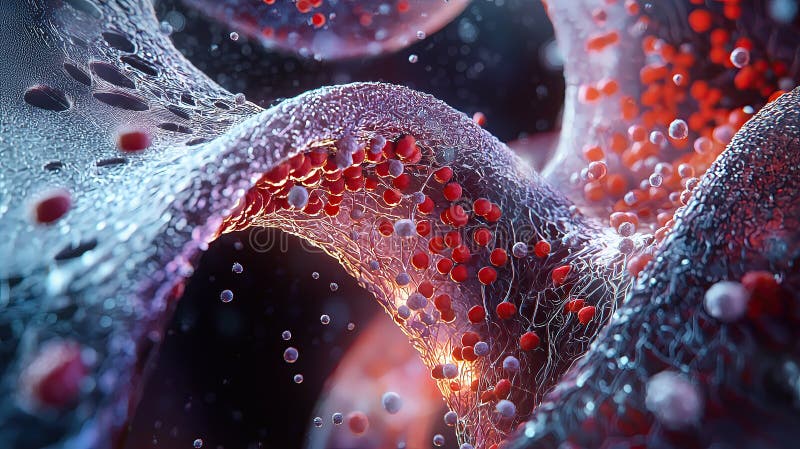
Free with trial A hyperrealistic, high-resolution anatomical cross-section of the human microcirculation system, showcasing the flow of red and white blood cells through plasma-filled capillaries. The highly detailed vessel walls reveal the delicate endothelial structures and subtle textures of the vascular lining, emphasizing the complexity of microcirculatory dynamics. Soft subsurface lighting enhances depth and realism, illuminating the biological textures with scientific accuracy. The color palette consists of rich reds, soft blues, and translucent plasma tones, creating an immersive medical visualization. The perspective provides a microscopic yet anatomically precise representation of the circulatory system, making it perfect for medical education, research, and artistic interpretation of human physiology. Endothelial cells illustrations Anatomical Cross-Section of the Human Microcirculation System
Free with trial Multisystem disorder and Cardiovascular kidney metabolic syndrome as disease related to a group of organs as kidneys heart pancreas and Adipose cells. Endothelial cells illustrations Multisystem disorder
Free with trial Vector set of red blood cells or erythrocytes isolated on white background. Endothelial cells vectors Red blood cells
Free with trial Atherosclerosis. Cholesterol level. Cross section of a Artery with red blood cells, HDL, LDL, and atheromatous plaques. Good and bad cholesterol. Vector poster. Isometric Flat illustration. Endothelial cells vectors Atherosclerosis. Cholesterol level. Cross section of a Artery with red blood cells, HDL, LDL, and atheromatous plaques
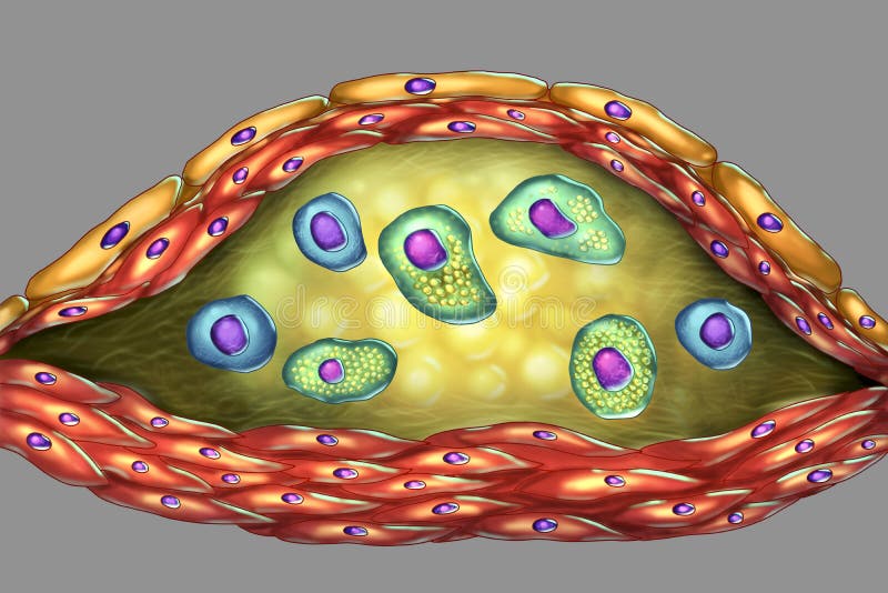
Free with trial Structure of atherosclerotic plaque. Illustration showing necrotic center, foam cells, T-lymphocytes inside of cholesterol plaque with walls made of smooth muscle cells and endothelium of blood vessel. Endothelial cells illustrations Atherosclerotic plaque in human artery. Structure of atherosclerotic plaque. Illustration showing necrotic center, foam cells, T-lymphocytes inside of cholesterol plaque with walls made of smooth muscle cells and endothelium of blood vessel
Free with trial Structure of atherosclerotic plaque. Illustration showing necrotic center, foam cells, T-lymphocytes inside of cholesterol plaque with walls made of smooth muscle cells and endothelium of blood vessel. Endothelial cells illustrations Structure of atherosclerotic plaque
Free with trial Bovine pulmonary artery epitheliial cells, fluorescent staining and modelling by surface rendering software. Red - nuclei, blue - microfilaments, yellow - mitochondria. Endothelial cells illustrations BPAE cells. Bovine pulmonary artery epitheliial cells, fluorescent staining and modelling by surface rendering software. Red - nuclei, blue - microfilaments, yellow - mitochondria.
Free with trial Microfilaments, mitochondria and nuclei in bovine pulmonary arthery endothelial BPAE cells. Endothelial cells illustrations BPAE
Free with trial Structure of atherosclerotic plaque. Illustration showing necrotic center, foam cells, T-lymphocytes inside of cholesterol plaque with walls made of smooth muscle cells and endothelium of blood vessel. Endothelial cells illustrations Structure of atherosclerotic plaque
Free with trial Structure of atherosclerotic plaque. Illustration showing necrotic center, foam cells, T-lymphocytes inside of cholesterol plaque with walls made of smooth muscle cells and endothelium of blood vessel. Endothelial cells illustrations Atherosclerotic plaque in human artery. Structure of atherosclerotic plaque. Illustration showing necrotic center, foam cells, T-lymphocytes inside of cholesterol plaque with walls made of smooth muscle cells and endothelium of blood vessel
Free with trial Lymphatic system, vessel and endothelial cells. Human lymph nodes and ducts, flow and fluid exchange illustration for clinic or medical poster. Anatomical banner for education isolated flat vector. Endothelial cells vectors Lymphatic vessel concept. lymphatic system, vessel and endothelial cells. Human lymph nodes and ducts, flow and fluid exchange illustration for clinic or medical poster. Anatomical banner for education isolated flat vector.
Free with trial Lymphatic system, vessel and endothelial cells concept. Human lymph nodes and ducts illustration for clinic, hospital or medical poster. Anatomical banner for education or science isolated flat vector. Endothelial cells vectors Lymphatic vessel concept. lymphatic system, vessel and endothelial cells concept. Human lymph nodes and ducts illustration for clinic, hospital or medical poster. Anatomical banner for education or science isolated flat vector
Free with trial Lymphatic system, vessel and endothelial cells. Human lymph nodes and ducts, flow and fluid exchange illustration for clinic or medical poster. Anatomical banner for education isolated flat vector. Endothelial cells vectors Lymphatic vessel concept. lymphatic system, vessel and endothelial cells. Human lymph nodes and ducts, flow and fluid exchange illustration for clinic or medical poster. Anatomical banner for education isolated flat vector.
Free with trial Lymphatic system, vessel and endothelial cells. Human lymph nodes and ducts, flow and fluid exchange illustration for clinic or medical poster. Anatomical banner for education isolated flat vector. Endothelial cells vectors Lymphatic vessel concept. lymphatic system, vessel and endothelial cells. Human lymph nodes and ducts, flow and fluid exchange illustration for clinic or medical poster. Anatomical banner for education isolated flat vector.
Free with trial Lymphatic system, vessel and endothelial cells concept. Human lymph nodes and ducts illustration for clinic, hospital or medical poster. Anatomical banner for education or science isolated flat vector. Endothelial cells vectors Lymphatic vessel concept. lymphatic system, vessel and endothelial cells concept. Human lymph nodes and ducts illustration for clinic, hospital or medical poster. Anatomical banner for education or science isolated flat vector
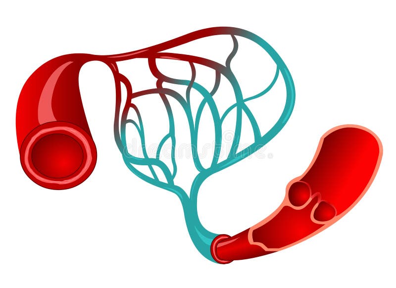
Free with trial Blood flows away from a body's heart via arteries, which branch and narrow into arterioles, and then branch further still into capillaries. Endothelial cells illustrations Blood capillaries. Blood flows away from a body's heart via arteries, which branch and narrow into arterioles, and then branch further still into capillaries.
Free with trial Hemoglobins improperly forms long chains giving cell its sickle shape and prevents adequate uptake of oxygen and nutrien. Endothelial cells vectors Hemoglobins improperly forms long chains giving cell its sickle
Free with trial Cancer cells using angiogenesis to grow and spread through the body. Digital illustration, 3D render. Endothelial cells illustrations Angiogenesis in tumor growth. Cancer cells using angiogenesis to grow and spread through the body. Digital illustration, 3D render
Free with trial Scientific Designing of Blood Vessels Structure. Capilary Blood Flow in Circulatory System. Colorful Symbols. Vector Illustration. Endothelial cells vectors Blood Vessels Structure
Free with trial Human bloodstream - didactic board of anatomy of blood system of human circulation sanguine, cardiovascular, vascular and venous system, construction of the veins and arteries. Endothelial cells illustrations BOARD Construction of the veins and arteries. Human bloodstream - didactic board of anatomy of blood system of human circulation sanguine, cardiovascular, vascular and venous system, construction of the veins and arteries
Free with trial Scientific Designing of Blood Vessels Structure. Capilary Blood Flow in Circulatory System. Colorful Symbols. Vector Illustration. Endothelial cells vectors Blood Vessels Structure
Free with trial Vasoconstriction, Vasodilation, and normal artery. Comparison of normal, constricted, and dilated blood vessels. Vector illustration. Endothelial cells vectors Vasoconstriction, Vasodilation, and normal artery. Comparison of normal, constricted, and dilated blood vessels
Free with trial The blood-brain barrier (BBB) is a crucial immunological feature of the human central nervous system (CNS). Composed of many cell types, the BBB is both a structural and functional roadblock to microorganisms, such as bacteria, fungi, viruses or parasites, that may be circulating in the bloodstream. Endothelial cells vectors Blood Brain Barrier (BBB) science vector illustration. The blood-brain barrier (BBB) is a crucial immunological feature of the human central nervous system (CNS). Composed of many cell types, the BBB is both a structural and functional roadblock to microorganisms, such as bacteria, fungi, viruses or parasites, that may be circulating in the bloodstream
Free with trial Interleukin 8 IL8 or chemokine C-X-C motif ligand 8, CXCL8 is a chemokine produced by macrophages and other cell types such as epithelial cells, airway smooth muscle cells and endothelial cells. 3D cartoon model isolated with differently colored secondary structure elements, white background. Endothelial cells illustrations Structure of human interleukin-8 homodimer. Interleukin 8 IL8 or chemokine C-X-C motif ligand 8, CXCL8 is a chemokine produced by macrophages and other cell types such as epithelial cells, airway smooth muscle cells and endothelial cells. 3D cartoon model isolated with differently colored secondary structure elements, white background
Free with trial Pathogenesis of atherosclerotic plaque in blood vessels thrombi response to injure medical poster. Endothelial cells vectors Pathogenesis of atherosclerotic plaque in blood vessels
Free with trial A dark blue and red background with cracks lava texture neurons artery for wallpaper, abstract background with glowing lines,. Endothelial cells vectors A dark blue and red background with cracks lava texture neurons artery for wallpaper, abstract background with glowing lines
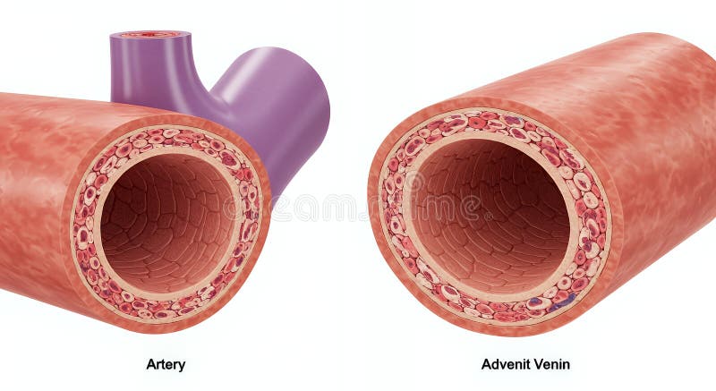
Free with trial The image shows a cross-sectional illustration comparing the layered structures of an artery and a vein. Both are depicted with their characteristic anatomy. Endothelial cells illustrations Illustration comparing the cross section of an artery and vein. The image shows a cross-sectional illustration comparing the layered structures of an artery and a vein. Both are depicted with their characteristic anatomy
Free with trial Thisis a highly selective semipermeable border of endothelial cells that prevents solutes in the circulating blood from non-selectively crossing into the extracellular fluid of the central nervous system where neurons reside. Endothelial cells illustrations Blood-brain barrier BBB in the human brain - closeup view 3d illustration. Thisis a highly selective semipermeable border of endothelial cells that prevents solutes in the circulating blood from non-selectively crossing into the extracellular fluid of the central nervous system where neurons reside.
Free with trial Lymphatic system of capillaries and vessels in complex with blood vessels. Lymph circulation scientific illustration. Vector illustration. Endothelial cells vectors Lymphatic system of capillaries and vessels in complex with blood vessels. Lymph circulation scientific illustration.
Free with trial A vivid visualization of the cardiovascular system shows arteries, veins, and flowing blood. Endothelial cells illustrations Vibrant cardiovascular system visualization showing arteries, veins, and the continuous flow of blood. A vivid visualization of the cardiovascular system shows arteries, veins, and flowing blood
Free with trial This detailed medical illustration vividly depicts the process of atherosclerosis, a leading cause of cardiovascular disease. Observe the gradual buildup of plaque within arteries, a complex process involving lipid accumulation, inflammation, and cellular changes. The illustration highlights the critical stages of plaque formation, from initial fatty deposits to advanced plaque rupture,. Endothelial cells illustrations Understanding Atherosclerosis A Detailed Visual Guide to Plaque Formation Arterial Blockage and Cardiovascular Disease. This detailed medical illustration vividly depicts the process of atherosclerosis, a leading cause of cardiovascular disease. Observe the gradual buildup of plaque within arteries, a complex process involving lipid accumulation, inflammation, and cellular changes. The illustration highlights the critical stages of plaque formation, from initial fatty deposits to advanced plaque rupture,
Free with trial ACE2 receptor location of 2019-nCoV. Angiotensin converting enzyme. Endothelial cells vectors ACE2 receptor location
Free with trial A bio-printer is utilized in a laboratory to construct a vascular graft. The process involves layering endothelial cells and incorporating regenerative signaling molecules to form new blood vessels. Endothelial cells illustrations Bio-printer creating vascular graft with endothelial cells and signaling molecules in lab setting. A bio-printer is utilized in a laboratory to construct a. A bio-printer is utilized in a laboratory to construct a vascular graft. The process involves layering endothelial cells and incorporating regenerative signaling molecules to form new blood vessels.
Free with trial Cytokines cells, labeled with fibroblast, granulocyte, macrophage, lymphocyte, endothelial cells, mast cell. Signaling protein, immune system and blood cell functions. Healthcare Biology Education. Endothelial cells vectors Cytokines cells, labeled with fibroblast, granulocyte, macrophage, lymphocyte, endothelial cells, mast cell. Signaling protein
Free with trial Cytokines. Proteins for cell signaling. Cytokines are produced by macrophage, lymphocyte, mast cell, endothelial cells and fibroblast. Cytokine storm. Vector illustration. Endothelial cells vectors Cytokines are produced by macrophage, lymphocyte, mast cell, endothelial cells and fibroblast
Free with trial Cytokines are produced by macrophage, lymphocyte, mast cell, endothelial cells, fibroblast and Granulocyte. Cytokines include chemokines, interferons, interleukins, lymphokines, and tumour necrosis factors, but not hormones or growth factors. Isometric Flat vector illustration. Endothelial cells vectors Cytokines that produced by macrophage, lymphocyte, mast cell, endothelial cells, fibroblast and Granulocyte. Cytokines are produced by macrophage, lymphocyte, mast cell, endothelial cells, fibroblast and Granulocyte. Cytokines include chemokines, interferons, interleukins, lymphokines, and tumour necrosis factors, but not hormones or growth factors. Isometric Flat vector illustration
Free with trial Detailed illustration of a cross-section through a capillary, showcasing the endothelial cells lining the vessel wall, the presence of erythrocytes (red blood cells) within the lumen, and the basement membrane supporting the capillary structure. Various cellular components and features, including intercellular clefts and the nuclei of the cells, are also depicted. Endothelial cells illustrations Cross-section of a capillary showing endothelial cells, erythrocytes, and other cellular components. Detailed illustration of a cross-section through a capillary, showcasing the endothelial cells lining the vessel wall, the presence of erythrocytes (red blood cells) within the lumen, and the basement membrane supporting the capillary structure. Various cellular components and features, including intercellular clefts and the nuclei of the cells, are also depicted