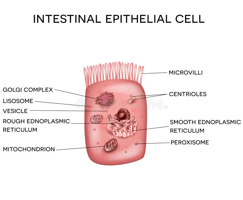
Free with trial Enterocytes. intestinal absorptive cells. Intestinal epithelial cell with microvilli. Epithelial intestinal cell vectors Enterocytes. intestinal absorptive cells
Free with trial Small intestine lining anatomy, a fold of the intestinal lining, villi and epithelial cell with microvilli detailed illustrations. Epithelial intestinal cell vectors Small intestine lining
Free with trial Intestinal villi anatomy, small intestine lining, villi and epithelial cells with microvilli detailed illustration. Epithelial intestinal cell vectors Intestinal villi anatomy
Free with trial Gastrointestinal system small intestine detailed wall anatomy. Small intestine villi and epithelial cell with microvilli detailed illustration. Epithelial intestinal cell vectors Gastrointestinal system small intestine anatomy. Gastrointestinal system small intestine detailed wall anatomy. Small intestine villi and epithelial cell with microvilli detailed illustration.
Free with trial Intestinal villi and microvilli detailed anatomy on a white background. Epithelial intestinal cell vectors Intestinal villi and microvilli
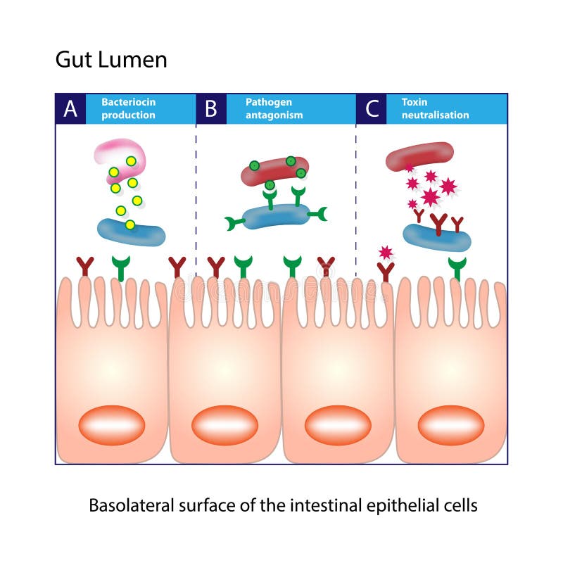
Free with trial Intestinal mucosa immunity cells medical vector illustration. Epithelial intestinal cell vectors Intestinal mucosa immunity
Free with trial Set of human cells, eps8, gradient and mesh printing compatible. Epithelial intestinal cell vectors Human cell collection. Set of human cells, eps8, gradient and mesh printing compatible
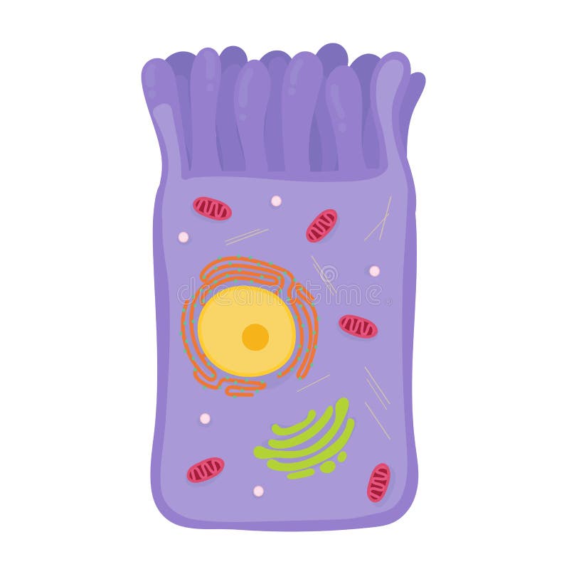
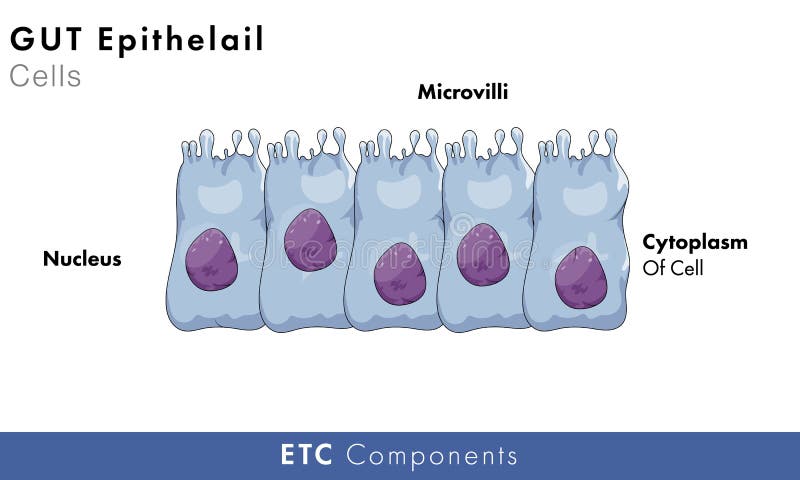
Free with trial Intestinal epithelial cell with microvilli, detailed bright illustation. Epithelial intestinal cell illustrations Intestinal epithelial cell
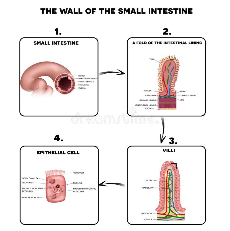
Free with trial Small intestine wall anatomy, a fold of the intestinal lining, villi and epithelial cell with microvilli detailed illustrations. Epithelial intestinal cell vectors Small intestine wall anatomy
Free with trial Wall of small intestine with villi and epithelial cells Enterocyte, Goblet and Paneth cell. Epithelial intestinal cell vectors Small intestine with villi and epithelial cells
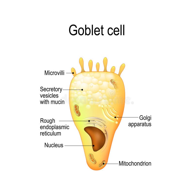
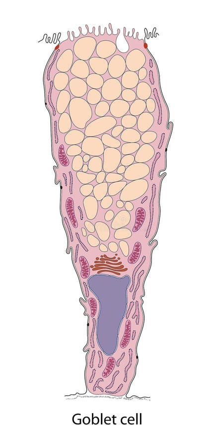
Free with trial Goblet cell. simple columnar epithelial cell for secrete mucus. They are found inside the trachea, bronchi, small and large intestine, and conjunctiva in the eyes. Structure cell nucleus and other organelles. Epithelial intestinal cell vectors Goblet cell. Structure cell nucleus and other organelles. Goblet cell. simple columnar epithelial cell for secrete mucus. They are found inside the trachea, bronchi, small and large intestine, and conjunctiva in the eyes. Structure cell nucleus and other organelles.
Free with trial Stem cells vector illustration. Medical labeled diagram with all kind cells. Example of blood, neurons, cardiac, bone, intestinal, epithelial, fat, liver and muscle. Epithelial intestinal cell vectors Stem cells vector illustration. Medical labeled diagram with all kind cells
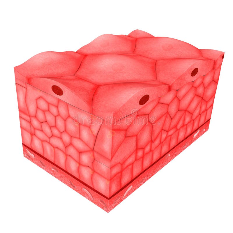
Free with trial Epithelial tissue covers the whole surface of the body. It is made up of cells closely packed and ranged in one or more layers. This tissue is specialised to form the covering or lining of all internal and external body surfaces. Epithelial tissue that occurs on surfaces on the interior of the body is known as endothelium. Epithelial cells are packed tightly together, with almost no intercellular spaces and only a small amount of intercellular substance. Epithelial tissue, regardless of the type, is usually separated from the underlying tissue by a thin sheet of connective tissue; basement membrane. The basement membrane provides structural support for the epithelium and also binds it to neighbouring structures. Epithelial intestinal cell illustrations Epithelial Tissues. Epithelial tissue covers the whole surface of the body. It is made up of cells closely packed and ranged in one or more layers. This tissue is specialised to form the covering or lining of all internal and external body surfaces. Epithelial tissue that occurs on surfaces on the interior of the body is known as endothelium. Epithelial cells are packed tightly together, with almost no intercellular spaces and only a small amount of intercellular substance. Epithelial tissue, regardless of the type, is usually separated from the underlying tissue by a thin sheet of connective tissue; basement membrane. The basement membrane provides structural support for the epithelium and also binds it to neighbouring structures.
Free with trial Epithelial tissue covers the whole surface of the body. It is made up of cells closely packed and ranged in one or more layers. This tissue is specialised to form the covering or lining of all internal and external body surfaces. Epithelial tissue that occurs on surfaces on the interior of the body is known as endothelium. Epithelial cells are packed tightly together, with almost no intercellular spaces and only a small amount of intercellular substance. Epithelial tissue, regardless of the type, is usually separated from the underlying tissue by a thin sheet of connective tissue; basement membrane. The basement membrane provides structural support for the epithelium and also binds it to neighbouring structures. Epithelial intestinal cell illustrations Epithelial Tissues. Epithelial tissue covers the whole surface of the body. It is made up of cells closely packed and ranged in one or more layers. This tissue is specialised to form the covering or lining of all internal and external body surfaces. Epithelial tissue that occurs on surfaces on the interior of the body is known as endothelium. Epithelial cells are packed tightly together, with almost no intercellular spaces and only a small amount of intercellular substance. Epithelial tissue, regardless of the type, is usually separated from the underlying tissue by a thin sheet of connective tissue; basement membrane. The basement membrane provides structural support for the epithelium and also binds it to neighbouring structures.
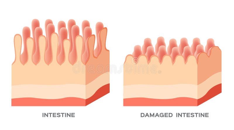
Free with trial Celiac disease Small intestine lining damage. Healthy villi and damaged villi. Small intestine, a fold of the intestinal lining and villi. Epithelial intestinal cell vectors Celiac disease Small intestine lining damage
Free with trial Celiac disease affected small intestine villi on a white background. Healthy villi and unhealthy villi with damaged cells. Epithelial intestinal cell vectors Celiac disease
Free with trial Celiac disease Small intestine lining damage. good and damaged villi. leaky gut progression on white. Epithelial intestinal cell vectors Celiac disease Small intestine lining damage. good and damaged villi . leaky gut progression
Free with trial Celiac disease Small intestine lining damage. good and damaged villi. leaky gut progression on white background. Epithelial intestinal cell vectors Celiac disease Small intestine lining damage. good and damaged villi . leaky gut progression
Free with trial 3d Rendering of leaky gut, in intestine with celiac disease and gluten sensitivity these tight junctions come apart. Epithelial intestinal cell illustrations 3d Rendering of leaky gut, in intestine with celiac disease and gluten sensitivity these tight junctions come apart.
Free with trial Intestinal cell system, human medical anatomy closeup. Vector flat style cartoon illustration isolated on white background. Epithelial intestinal cell vectors Intestinal cell system, human medical anatomy closeup
Free with trial Small intestine lining detailed anatomy, a fold of the intestinal lining, villi and epithelial cell with microvilli. Epithelial intestinal cell vectors Small intestine lining
Free with trial Human cell set: bone osteocyte, Muscle Myocyte, nerve neuron and photoreceptor, epithelial Enterocytes hemocyte red and white blood cells, reproductive sperm, Adipocyte. Types of Cells on a white background. Medical icons collection for Poster. vector. Epithelial intestinal cell vectors Cell set. collection of icons. Human cell set: bone osteocyte, Muscle Myocyte, nerve neuron and photoreceptor, epithelial Enterocytes hemocyte red and white blood cells, reproductive sperm, Adipocyte. Types of Cells on a white background. Medical icons collection for Poster. vector
Free with trial Gut lumen. Enterocytes, or intestinal absorptive cells. Small intestine. Columnar epithelial cells. Epithelial intestinal cell vectors Gut Lumen. Columnar epithelial cells. Gut lumen. Enterocytes, or intestinal absorptive cells. Small intestine. Columnar epithelial cells
Free with trial Intestinal villi in the surface area of intestinal walls. Folds, villus, microvilli, epithelial and goblet cells. Small intestine anatomical poster. Digestive system medical flat vector illustration. Epithelial intestinal cell vectors Intestinal villi anatomy. Intestinal villi in the surface area of intestinal walls. Folds, villus, microvilli, epithelial and goblet cells. Small intestine anatomical poster. Digestive system medical flat vector illustration.
Free with trial Stem cell application. Embryonic Origin of Tissues and Major Organs. endoderm, mesoderm, and ectoderm. generating specialized tissues from embryonic stem cells and prospects for their applications. Epithelial intestinal cell vectors Stem cell
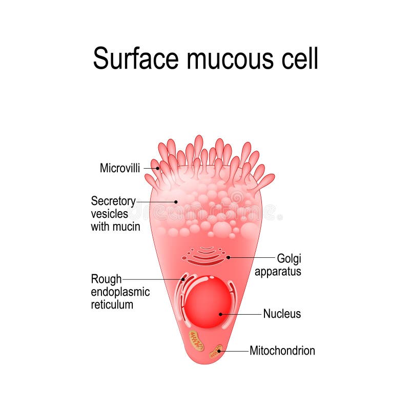
Free with trial Surface mucous cell is a foveolar mucus-producing cell that covering the inside of the stomach. Structure cell: golgi apparatus, secretory vesicle, mucin, nucleus, mitochondrion, microvilli. Epithelial intestinal cell vectors Surface mucous cell
Free with trial Intestinal villi. Small intestine lining. Epithelial intestinal cell vectors Intestinal villi
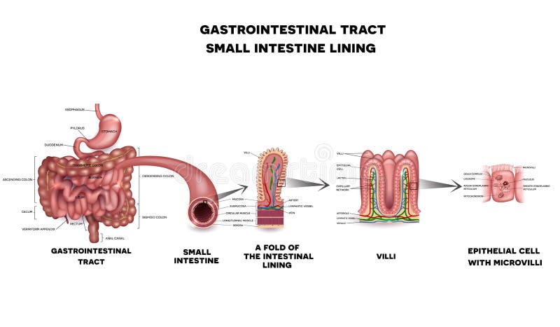
Free with trial Gastrointestinal tract anatomy. Intestinal villi, small intestine lining, epithelial cells with microvilli detailed illustration. Epithelial intestinal cell vectors Gastrointestinal tract detailed anatomy. Gastrointestinal tract anatomy. Intestinal villi, small intestine lining, epithelial cells with microvilli detailed illustration.
Free with trial Hormones of the gastrointestinal tract and Enteroendocrine cell. Enterocyte. Human endocrine system. Vector illustration for medical, education and science use. Epithelial intestinal cell vectors Hormones of the gastrointestinal tract and Enteroendocrine cell. Enterocyte. Human endocrine system
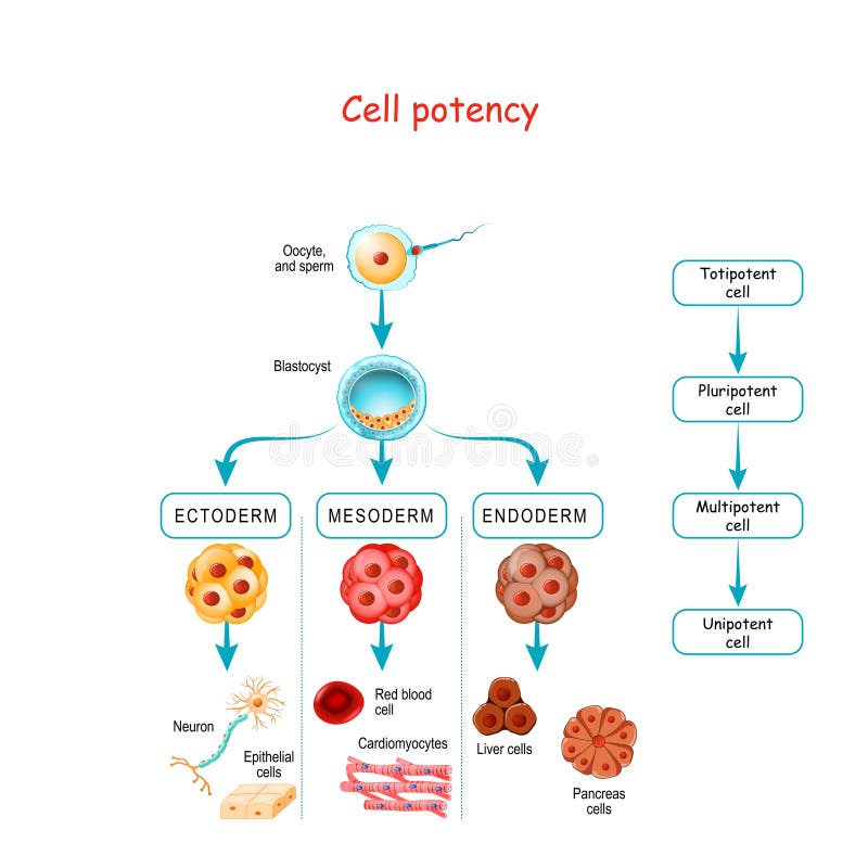
Free with trial Cell potency. From Totipotent to Pluripotent, Multipotent, and Unipotent cell. endoderm, mesoderm and ectoderm. Development of fertilized egg and blastocyst to human fetus. Epithelial intestinal cell vectors Cell potency. From Totipotent to Pluripotent, Multipotent, and Unipotent cell
Free with trial A fold of the intestinal lining on a white background. Epithelial intestinal cell vectors A fold of the intestinal lining
Free with trial Zonulin is a protein that increases the permeability of tight junctions between cells of the wall of the gastrointestinal tract. digestive system. intestinal cells with Zonulin receptors, normal and Faulty tight junctions. Intestinal permeability. Gut barrier dysfunction. Vector poster. Epithelial intestinal cell vectors Intestinal cells with Zonulin receptors, normal and Faulty tight
Free with trial Intestinal villi. Human intestine. 3d illustration. Epithelial intestinal cell illustrations Intestinal villi. Human intestine.
Free with trial Intestinal villi, mucosa intestinal. Bacteria and microbes in intestines. Microscopic villi and capillary. Human intestine, chronic disease. Hepatitis viruses, influenza, cell infections. 3D Rendering. Epithelial intestinal cell illustrations Intestinal villi, mucosa intestinal. Bacteria and microbes in intestines. Microscopic villi and capillary. Human
Free with trial Intestinal villus, Intestinal villus refers to any one of the small, finger-shaped outgrowths of the epithelial lining of the wall of the intestine. Clusters of projections are referred as Intestinal villi. Epithelial intestinal cell illustrations Villus
Free with trial Normal Gastrointestinal tract and small intestine detailed anatomy. Epithelial intestinal cell vectors Normal Gastrointestinal tract
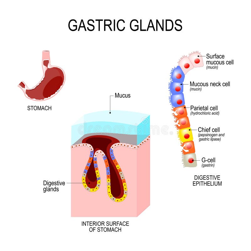
Free with trial Structure of the stomach: interior surface and cells of digestive epithelium. Human anatomy. Vector illustration for medical, educational and science use. Epithelial intestinal cell vectors Structure of the stomach: interior surface and cells of digestive epithelium
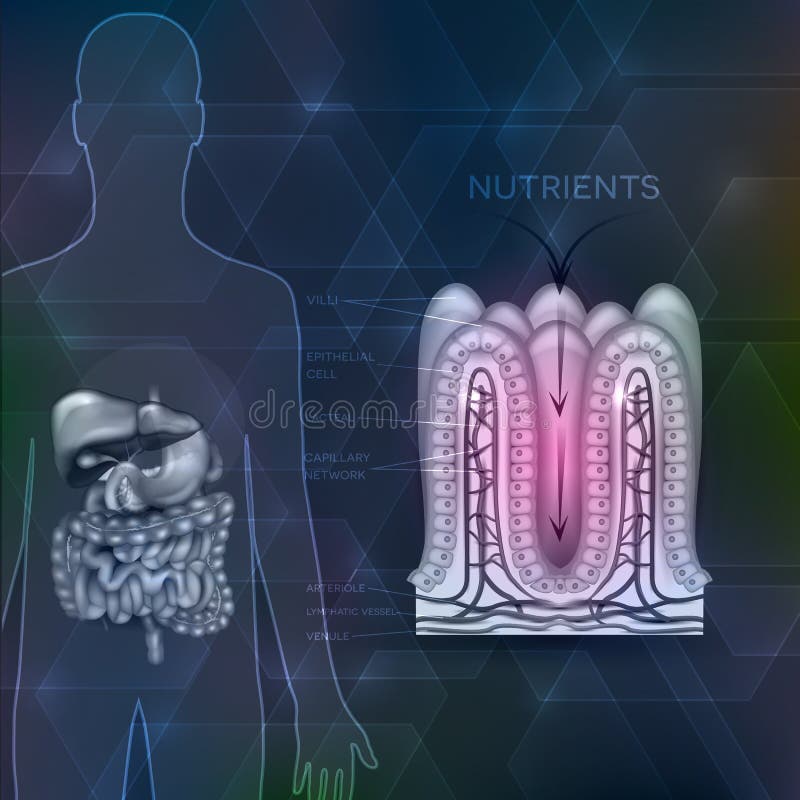
Free with trial Absorption of nutrients in the small intestine. Healthy and damaged villi. Medical vector illustration. Epithelial intestinal cell vectors Absorption of nutrients in the small intestine
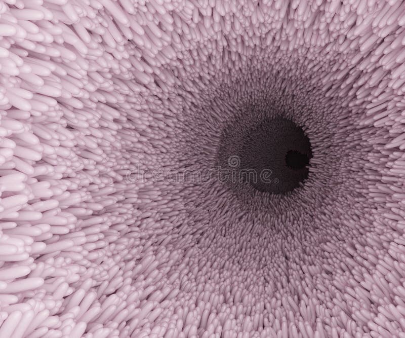
Free with trial Intestine microvilli close up 3d render. Gut microbiome and digestive system of human. High quality 3d illustration. Epithelial intestinal cell illustrations Intestine microvilli close up 3d render. Gut microbiome and digestive system of human
Free with trial Enterocyte. intestinal absorptive epithelial cell with microvilli. Structure of the enterocyte. Infographics. Vector illustration on white background. Epithelial intestinal cell vectors Enterocyte. Structure of the intestinal absorptive epithelial cell with microvilli. Enterocyte. intestinal absorptive epithelial cell with microvilli. Structure of the enterocyte. Infographics. Vector illustration on white background
Free with trial The illustration is about cell in the human body. This is the intestinal cell. Epithelial intestinal cell vectors The intestinal cell
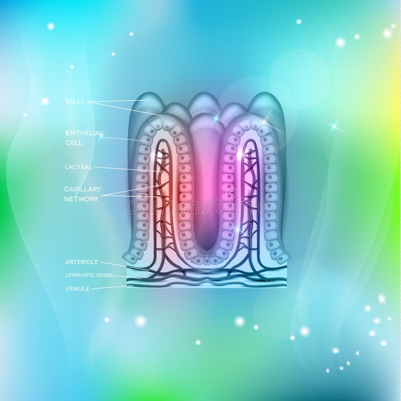
Free with trial Intestinal villi anatomy, epithelial cells with microvilli and capillary network detailed illustration, 2d. Epithelial intestinal cell illustrations Intestinal villi anatomy, epithelial cells with microvilli and capillary network detailed illustration
Free with trial Simple epithelial cell of kidney tubule under microscope. Epithelial intestinal cell illustrations Simple epithelial cell of kidney
Free with trial Intestinal epithelial cells with capillary network. Enterocyte and Goblet cell. Vector illustration. Epithelial intestinal cell vectors Intestinal villi. Microvilli. Intestinal epithelium. Villi absorb nutrients from the food. Intestinal epithelial cells with capillary network. Enterocyte and Goblet cell. Vector illustration.
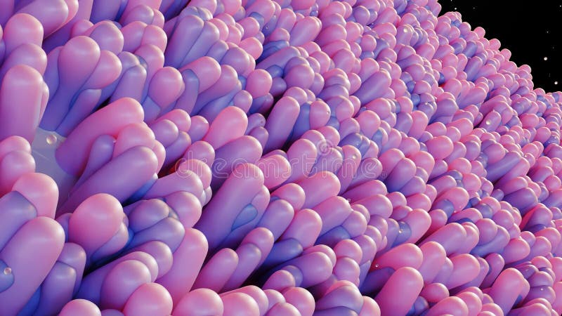
Free with trial Intestinal villi, Red microvilli in a intestinal tract. close-up, Microbiology, anatomy, biology, science, medicine, medical and healthcare concepts, Digestive system, epithelial cells, 3D Render. Epithelial intestinal cell illustrations Intestinal villi, Red microvilli in a intestinal tract. Digestive system, epithelial cells. intestinal villi, Red microvilli in a intestinal tract. close-up, Microbiology, anatomy, biology, science, medicine, medical and healthcare concepts, Digestive system, epithelial cells, 3D Render
Free with trial Intestinal villi in the surface of intestinal. Epithelial and goblet cells. Healthy digestive system, with microorganisms maintenaning intestinal cells. Flat vector illustration. Epithelial intestinal cell illustrations Intestinal villi in the surface of intestinal. Epithelial and goblet cells.
Free with trial Intestinal villi, Red microvilli in a intestinal tract. close-up, Microbiology, anatomy, biology, science, medicine, medical and healthcare concepts, Digestive system, epithelial cells, 3D Render. Epithelial intestinal cell illustrations Intestinal villi, Red microvilli in a intestinal tract. Digestive system, epithelial cells. intestinal villi, Red microvilli in a intestinal tract. close-up, Microbiology, anatomy, biology, science, medicine, medical and healthcare concepts, Digestive system, epithelial cells, 3D Render
Free with trial The intestinal epithelium is the single cell layer that form the luminal surface of intestine (colon) of the gastrointestinal tract 3d rendering. Epithelial intestinal cell illustrations The intestinal epithelium is the single cell layer that form the luminal surface of intestine (colon)
Free with trial Small intestine lining anatomy, a fold of the intestinal lining, villi and epithelial cell with microvilli detailed illustrations. Epithelial intestinal cell vectors Small intestine lining
Free with trial Intestine lining detailed anatomy, a fold of the intestinal lining, villi and epithelial cell with microvilli. Epithelial intestinal cell vectors Intestine anatomy. Intestine lining detailed anatomy, a fold of the intestinal lining, villi and epithelial cell with microvilli
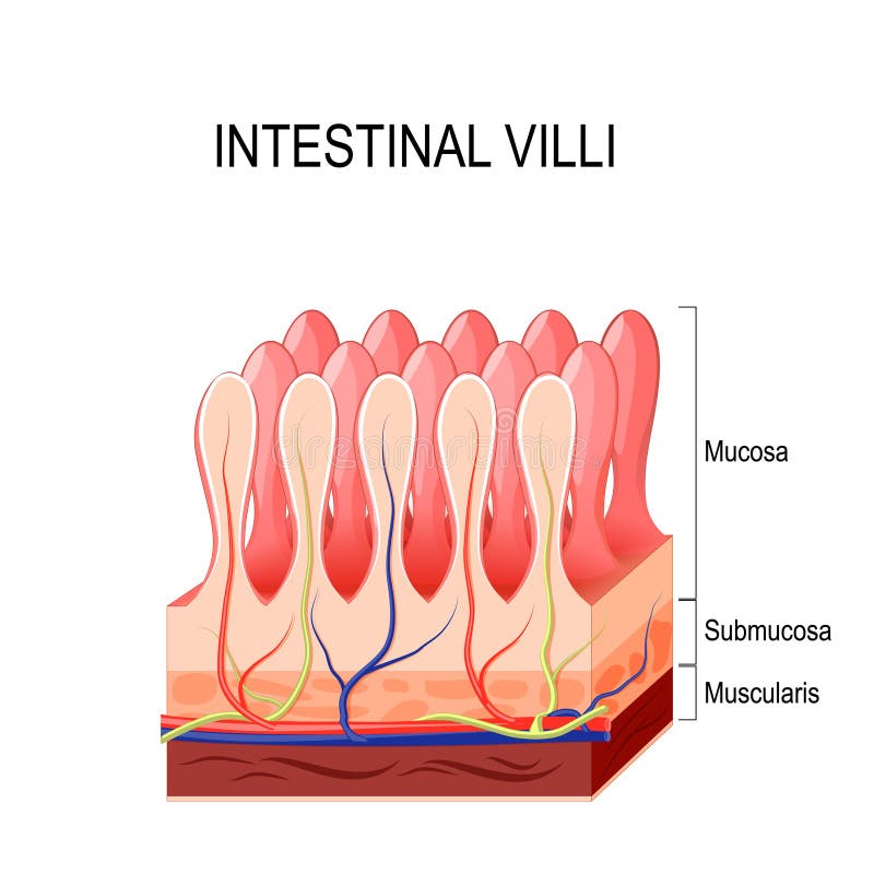
Free with trial Intestinal villi diagram. Surface area of intestinal walls. Small intestine cross section, fold, villus, microvilli and epithelial cells. Digestive system medical flat vector illustration for clinic. Epithelial intestinal cell vectors Intestinal villi concept. Intestinal villi diagram. Surface area of intestinal walls. Small intestine cross section, fold, villus, microvilli and epithelial cells. Digestive system medical flat vector illustration for clinic
Free with trial Intestinal villi in the surface area of intestinal walls. Folds, villus, microvilli, epithelial and goblet cells. Small intestine anatomical poster. Digestive system medical flat vector illustration. Epithelial intestinal cell vectors Intestinal villi anatomy. Intestinal villi in the surface area of intestinal walls. Folds, villus, microvilli, epithelial and goblet cells. Small intestine anatomical poster. Digestive system medical flat vector illustration.
Free with trial Intestinal villi in the surface area of intestinal walls. Folds, villus, microvilli, epithelial and goblet cells. Small intestine anatomical poster. Digestive system medical flat vector illustration. Epithelial intestinal cell vectors Intestinal villi anatomy. Intestinal villi in the surface area of intestinal walls. Folds, villus, microvilli, epithelial and goblet cells. Small intestine anatomical poster. Digestive system medical flat vector illustration.
Free with trial 3d rendering of the intestinal epithelium is the single cell layer that form the luminal surface of intestine (colon) of the gastrointestinal tract. Epithelial intestinal cell illustrations 3d rendering of the intestinal epithelium cells. 3d rendering of the intestinal epithelium is the single cell layer that form the luminal surface of intestine (colon) of the gastrointestinal tract
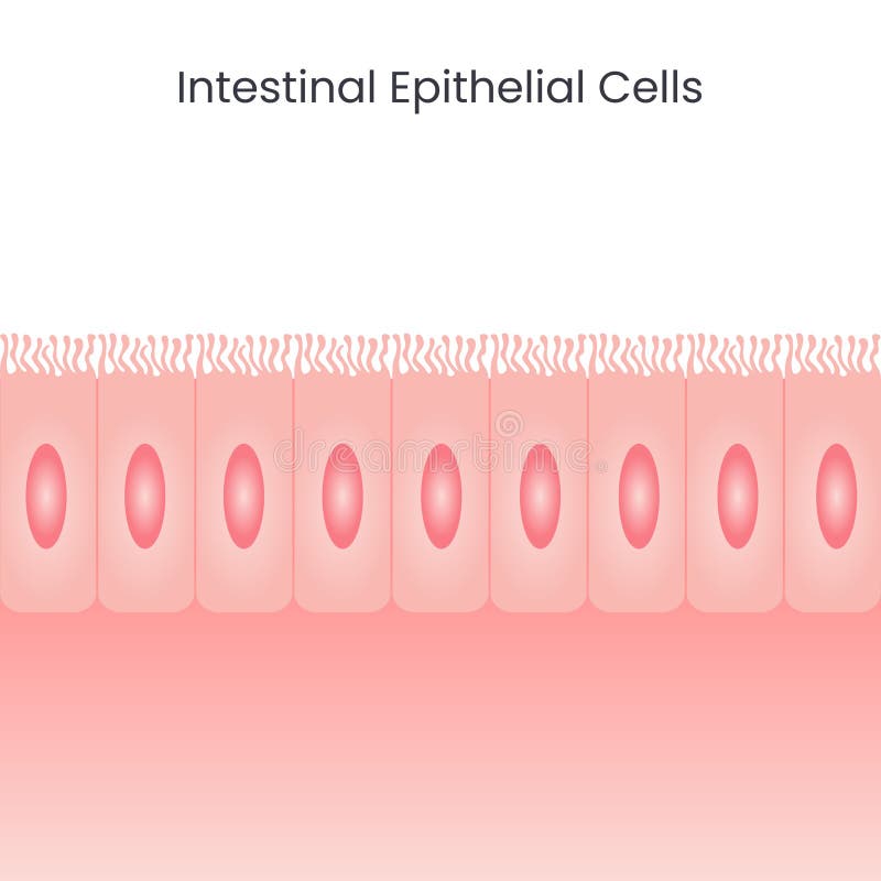
Free with trial Vector illustration science background of the epithelial cells that line the large and small intestines. Epithelial intestinal cell vectors Intestinal Epithelial Cells background. vector illustration science background of the epithelial cells that line the large and small intestines
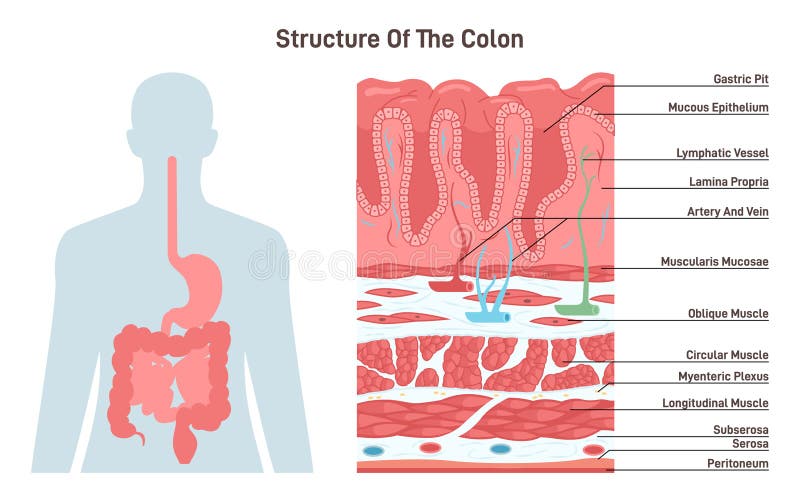
Free with trial Structure of the colon. Human digestive system anatomy. Intestinal villi in the surface of intestinal. Epithelial and goblet cells. Healthy digestive system. Flat vector illustration. Epithelial intestinal cell vectors Structure of the colon. Human digestive system anatomy. Intestinal villi
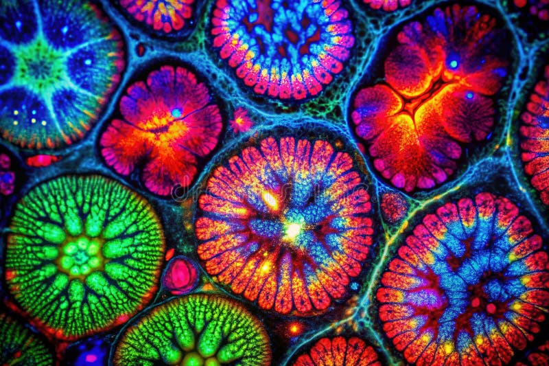
Free with trial This detailed immunofluorescence microscopy image showcases the intricate network of tight junctions within the intestinal mucosa. The image, captured under optimized studio lighting, presents a clear visualization of protein expression within the epithelial cells lining the intestine. The precise focus and depth of field highlight the structural components of the tissue, including the cellular. Epithelial intestinal cell illustrations Visualizing Intestinal Mucosa Tight Junctions Immunofluorescence Microscopy Reveals Protein Expression and Tissue. This detailed immunofluorescence microscopy image showcases the intricate network of tight junctions within the intestinal mucosa. The image, captured under optimized studio lighting, presents a clear visualization of protein expression within the epithelial cells lining the intestine. The precise focus and depth of field highlight the structural components of the tissue, including the cellular
Free with trial Intestinal epithelium showing functions of enterocytes, dendritic cell, goblet cell, neuroendocrine cell, stem cell, mucosa, and mucus. Source: Kong, S. , Zhang, Y. H. , & Zhang, W. 2018. Regulation of intestinal epithelial cells properties and functions by amino acids. BioMed research international, 2018. Epithelial intestinal cell illustrations Intestinal epithelium showing functions of cells. Intestinal epithelium showing functions of enterocytes, dendritic cell, goblet cell, neuroendocrine cell, stem cell, mucosa, and mucus. Source: Kong, S., Zhang, Y. H., & Zhang, W. 2018. Regulation of intestinal epithelial cells properties and functions by amino acids. BioMed research international, 2018.
Free with trial Small intestine anatomy. Layers of the small bowel Mucosa, Submucosa, muscularis externa, serosa. Cross section of Intestinal villi. Close-up of Stem cell, Goblet and Paneth cells, Enterocyte. Detailed Vector poster. Epithelial intestinal cell vectors Small intestine anatomy. Layers and cells. Small intestine anatomy. Layers of the small bowel Mucosa, Submucosa, muscularis externa, serosa. Cross section of. small intestine anatomy. Layers of the small bowel Mucosa, Submucosa, muscularis externa, serosa. Cross section of Intestinal villi. Close-up of Stem cell, Goblet and Paneth cells, Enterocyte. Detailed Vector poster
Free with trial Intestinal villi anatomy on a beautiful abstract technology background. Epithelial intestinal cell vectors Intestinal villi blue technology background. Intestinal villi anatomy on a beautiful abstract technology background
Free with trial Paneth cell it is the epithelium cells of the small intestine. their granules consist of anti-microbial compounds for important in immunity. Structure of the Paneth cell nucleus, mitochondria and other organelles. Epithelial intestinal cell vectors Paneth cell
Free with trial Parietal cell of stomach wall, located in the gastric glands secretes hydrochloric acid. Secreting and resting parietal cell. Epithelial intestinal cell vectors Parietal cell function. Parietal cell of stomach wall, located in the gastric glands secretes hydrochloric acid. Secreting and resting parietal cell.
Free with trial Intestinal villi anatomy, small intestine lining. organ / white background. Epithelial intestinal cell vectors Intestinal villi anatomy, small intestine lining . organ
Free with trial Parietal cell of the stomach wall, located in the gastric glands secretes hydrochloric acid. Secreting and resting parietal cell. Epithelial intestinal cell vectors Parietal cell of stomach wall, functions. Parietal cell of the stomach wall, located in the gastric glands secretes hydrochloric acid. Secreting and resting parietal cell.
Free with trial Intestinal lining anatomy, absorption of nutrients, human silhouette with digestive organs. Epithelial intestinal cell vectors Intestinal lining
Free with trial Anatomy and structure of gut epithelial cells vector illustration graphic design concept. Epithelial intestinal cell vectors Anatomy and structure of gut epithelial cells vector illustration
Free with trial Intestinal villi anatomy on a beautiful light blue mesh background. Epithelial intestinal cell vectors Intestinal villi background. Intestinal villi anatomy on a beautiful light blue mesh background
Free with trial Intestinal villus. Different between Villi and microvilli. Cross section of a duodenum with submucosa, mucosa and muscularis layers. Vertical section of a villus with Lymph capillary or Lacteal and Blood vessels. Close-up of a Columnar epithelium, and enterocytes. Vector illustration. Epithelial intestinal cell vectors Intestinal villus. Different between Villi and microvilli
Free with trial Intestinal villi, mucosa intestinal. Bacteria and microbes in intestines. Microscopic villi and capillary. Human intestine, chronic disease. Hepatitis viruses, influenza, cell infections. 3D Rendering. Epithelial intestinal cell illustrations Intestinal villi, mucosa intestinal. Bacteria and microbes in intestines. Microscopic villi and capillary. Human
Free with trial 3D illustration close-up Intestinal villi. Intestine lining. Microscopic villi and capillary. Human intestine. Concept of a healthy or diseased intestine. Epithelial intestinal cell illustrations 3D illustration close-up Intestinal villi. Intestine lining. Microscopic villi and capillary. Human intestine. Concept
Free with trial 3D illustration close-up Intestinal villi. Intestine lining. Microscopic villi and capillary. Human intestine. Concept of a healthy or diseased intestine. Epithelial intestinal cell illustrations 3D illustration close-up Intestinal villi. Intestine lining. Microscopic villi and capillary. Human intestine. Concept
Free with trial Intestinal villi, mucosa intestinal. Bacteria and microbes in intestines. Microscopic villi and capillary. Human intestine, chronic disease. Hepatitis viruses, influenza, cell infections. 3D Rendering. Epithelial intestinal cell illustrations Intestinal villi, mucosa intestinal. Bacteria and microbes in intestines. Microscopic villi and capillary. Human
Free with trial Intestinal villi. Human intestine. 3d illustration. Epithelial intestinal cell illustrations Intestinal villi. Human intestine.
Free with trial Goblet cells are a type of intestinal mucosal epithelial cell, which serves as the primary site for nutrient digestion and mucosal absorption. The primary function of goblet cells is to synthesize and secrete mucus. As the primary secretory cell in the superficial epithelium of large airways, goblet cells secrete. Epithelial intestinal cell illustrations Diagram of goblet cell. Cells are located in the gastric glands into stomach. Goblet cells are a type of intestinal mucosal epithelial cell, which serves as the primary site for nutrient digestion and mucosal absorption. The primary function of goblet cells is to synthesize and secrete mucus. As the primary secretory cell in the superficial epithelium of large airways, goblet cells secrete
Free with trial 3d illustration Intestinal villi. Intestine lining. Microscopic villi and capillary. Human intestine. Viral infection causing chronic disease. Hepatitis viruses, influenza virus, cell infect organism. Epithelial intestinal cell illustrations 3d illustration Intestinal villi. Intestine lining. Microscopic villi and capillary. Human intestine. Viral infection
Free with trial 3D illustration close-up Intestinal villi. Intestine lining. Microscopic villi and capillary. Human intestine. Concept of a healthy or diseased intestine. Epithelial intestinal cell illustrations 3D illustration close-up Intestinal villi. Intestine lining. Microscopic villi and capillary. Human intestine. Concept