Free with trial Knee joint with bones anatomy illustration on transparent body cartilage layers. Extremity anatomy illustrations Knee anatomy with highlighted bone and cartilage on medical concept design. Knee joint with bones anatomy illustration on transparent body cartilage layers
Free with trial A detailed, macro view of a human foot, specifically the heel and ankle area, with blue lines drawn to mark veins. Extremity anatomy illustrations Close-up of Human Foot with Blue. A detailed, macro view of a human foot, specifically the heel and ankle area, with blue lines drawn to mark veins.
Free with trial Knee pain showing person holding painful knee joint with glowing red inflammation knee pain joint pain. High. Extremity anatomy illustrations Person holding painful knee joint with glowing red inflammation knee pain joint pain
Free with trial Knee joint bones anatomy, 3D illustration showing femur, tibia, fibula, and patella in normal alignment. Extremity anatomy illustrations Knee joint bones anatomy, 3D illustration
Free with trial X-ray view of human feet bones and joints. Medical illustration shows skeletal structure for anatomy study. Digital art depicting bone details, against dark backdrop. Extremity anatomy illustrations X-ray view of human feet bones and joints. Medical illustration shows skeletal structure for anatomy study. Digital art depicting
Free with trial Tibia and fibula bones anatomy, 3D illustration. Bones making knee joint together with femur and patella. Extremity anatomy illustrations Tibia and fibula bones anatomy, 3D illustration
Free with trial Tibia and fibula bones anatomy, 3D illustration. Bones making knee joint together with femur and patella. Extremity anatomy illustrations Tibia and fibula bones anatomy, 3D illustration
Free with trial Concept Sciatic Nerve Pain, Lower Back, Anatomy, Nervous System, Pain Management Sciatic Nerve Pain Concept Focus on Lower Back. Extremity anatomy illustrations Sciatic Nerve Pain Concept - Focus on Lower Back. Concept Sciatic Nerve Pain, Lower Back, Anatomy. Concept Sciatic Nerve Pain, Lower Back, Anatomy, Nervous System, Pain Management Sciatic Nerve Pain Concept Focus on Lower Back
Free with trial Hand X-Ray Shows The Skeleton for Medical Analysis, Featuring Detailed Anatomy PNG high resolution. Extremity anatomy illustrations Hand X-Ray Shows The Skeleton for Medical Analysis, Featuring Detailed Anatomy
Free with trial A minimalist line drawing of a human foot. Clean and simple design showcasing foot anatomy. Perfect for medical illustrations or anatomical studies. Black and white drawing. Extremity anatomy illustrations Simple Foot Anatomy Drawing Illustration Line Art Human Body Foot Health Medical Design Ankle Sole Toe Digital Art Black. A minimalist line drawing of a human foot. Clean and simple design showcasing foot anatomy. Perfect for medical illustrations or anatomical studies. Black and white drawing.
Free with trial Anatomy illustration of human ankle joints showing inflammation and pain. Red areas indicate discomfort and swelling, in bones and cartilage. Medical view. Extremity anatomy illustrations Anatomy illustration of human ankle joints showing inflammation and pain. Red areas indicate discomfort and swelling in bones and. Anatomy illustration of human ankle joints showing inflammation and pain. Red areas indicate discomfort and swelling, in bones and cartilage. Medical view.
Free with trial Detailed medical illustration comparing varus leg alignment with normal leg alignment. Genu Varum Comparison with Normal Lower Limb Anatomy. Extremity anatomy vectors Detailed medical illustration comparing varus leg alignment with normal leg alignment. Genu Varum Comparison with Normal Lower
Free with trial This image shows a close-up of a human foot with visible veins, set against a transparent background. The foot is depicted in a realistic and detailed manner, showcasing its anatomy and structure. The image is suitable for educational, informative, and visual purposes in the fields of medicine, healthcare, and science. Extremity anatomy vectors A close-up of a human foot with visible veins on transparent background. This image shows a close-up of a human foot with visible veins, set against a transparent background. The foot is depicted in a realistic and detailed manner, showcasing its anatomy and structure. The image is suitable for educational, informative, and visual purposes in the fields of medicine, healthcare, and science.
Free with trial A detailed, translucent X-ray rendering of a human foot and ankle, showcasing the intricate bone structure. The image highlights the tarsals, metatarsals, phalanges, tibia, and fibula, presented against a clean, gradient background. This visualization is ideal for medical, educational, or scientific purposes, illustrating the complex anatomy of the lower extremity and its supporting framework. Extremity anatomy illustrations Anatomical X-ray of Human Foot and Ankle. A detailed, translucent X-ray rendering of a human foot and ankle, showcasing the intricate bone structure. The image highlights the tarsals, metatarsals, phalanges, tibia, and fibula, presented against a clean, gradient background. This visualization is ideal for medical, educational, or scientific purposes, illustrating the complex anatomy of the lower extremity and its supporting framework.
Free with trial Hand Surgery Word Cloud. A visual collection of key concepts in orthopedic microsurgery, focusing on trauma, nerve repair, arthritis, and functional recovery of the upper extremity. Extremity anatomy illustrations Hand Surgery Word Cloud. Core Concepts in Upper Extremity Care and Reconstructive Techniques. Hand Surgery Word Cloud. A visual collection of key concepts in orthopedic microsurgery, focusing on trauma, nerve repair, arthritis, and functional recovery of the upper extremity
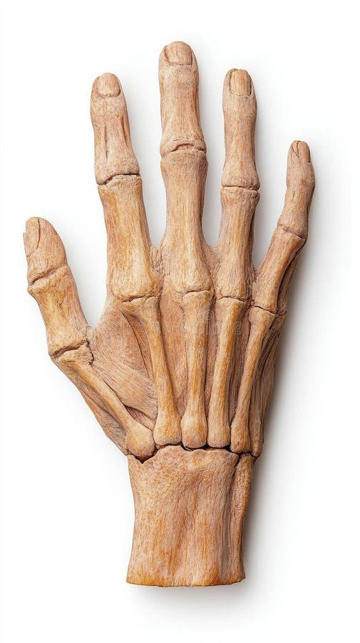
Free with trial AI image: A hyperrealistic rendering of an open hand with detailed skin texture, subtly gesturing against a clean, white backdrop. The visual representation conveys themes of offering, connection , dynamic background flow. Extremity anatomy illustrations AI image, Open hand gesturing against white space human anatomy detailed skin fingers palm. AI image: A hyperrealistic rendering of an open hand with detailed skin texture, subtly gesturing against a clean, white backdrop. The visual representation conveys themes of offering, connection , dynamic background flow
Free with trial Detailed 3D rendering of a human hand skeleton against a soft, neutral background. The image showcases the intricate bone structure and articulation of the hand, suitable for medical illustration, educational materials, or anatomical studies. The warm lighting accentuates the texture and form of the bones. Extremity anatomy illustrations Human Hand Skeleton Anatomy. Detailed 3D rendering of a human hand skeleton against a soft, neutral background. The image showcases the intricate bone structure and articulation of the hand, suitable for medical illustration, educational materials, or anatomical studies. The warm lighting accentuates the texture and form of the bones.
Free with trial A detailed line art illustration showcasing the intricate network of bones, tendons, and ligaments in a human foot. Perfect for medical, anatomical, or educational purposes. Extremity anatomy vectors Human Foot Anatomy, Detailed Line Art Illustration. A detailed line art illustration showcasing the intricate network of bones, tendons, and ligaments in a human foot. Perfect for medical, anatomical, or educational purposes.
Free with trial A labeled diagram from behind and front. A detailed medical illustration. Extremity anatomy vectors Right Thigh Bone- Femur. Bone of the lower extremity diagram. A labeled diagram from behind and front. A detailed medical illustration.
Free with trial This image showcases an x-ray-like view of an ankle joint with highlighted inflammation, suggesting pain or injury. The red area pinpoints the location of the discomfort, likely affecting the bones and ligaments of the ankle. The visual aids in understanding the anatomy of the affected area. Extremity anatomy illustrations Ankle Joint Pain. This image showcases an x-ray-like view of an ankle joint with highlighted inflammation, suggesting pain or injury. The red area pinpoints the location of the discomfort, likely affecting the bones and ligaments of the ankle. The visual aids in understanding the anatomy of the affected area.
Free with trial A detailed 3D rendering of a human foot, showcasing the intricate bone structure, muscles, and ligaments isolated on a white background. Extremity anatomy illustrations Human foot anatomy model isolated on white background. A detailed 3D rendering of a human foot, showcasing the intricate bone structure, muscles, and ligaments isolated on a white background
Free with trial Detailed illustration of human hand skeleton. Extremity anatomy illustrations Human hand skeletal system anatomy. Detailed illustration of human hand skeleton
Free with trial Detailed mechanical prosthetic arm with realistic exposed muscles and tendons rests on a surface, combining anatomy and robotics. Extremity anatomy illustrations Highly detailed mechanical prosthetic arm. detailed mechanical prosthetic arm with realistic exposed muscles and tendons rests on a surface, combining anatomy and robotics.
Free with trial Human hand bones shown with inflamed joints. Medical illustration displays pain points in fingers and wrist. Digital art depicts skeletal anatomy showing skeletal structure of hand. Extremity anatomy illustrations Human hand bones shown with inflamed joints. Medical illustration displays pain points in fingers and wrist. Digital art depicts
Free with trial This is a detailed illustration of a human foot skeleton, showcasing the intricate structure of the skeletal system. The image features a gray foot skeleton with a transparent background, allowing for clear visibility of the bones. The skeleton is composed of multiple bones, including the phalanges, metatarsal, tarsal, calcaneus, talus, cuboid, navicular, and cuneiform. This image is ideal for medical, scientific, and educational purposes, providing a comprehensive view of the human foot anatomy. Extremity anatomy vectors A detailed illustration of a human foot skeleton on transparent background. This is a detailed illustration of a human foot skeleton, showcasing the intricate structure of the skeletal system. The image features a gray foot skeleton with a transparent background, allowing for clear visibility of the bones. The skeleton is composed of multiple bones, including the phalanges, metatarsal, tarsal, calcaneus, talus, cuboid, navicular, and cuneiform. This image is ideal for medical, scientific, and educational purposes, providing a comprehensive view of the human foot anatomy.
Free with trial A detailed anatomical X-ray rendering of the human foot skeleton against a black background. Extremity anatomy illustrations Anatomical X-ray of Human Foot Skeleton on Black Background anatomy bone. A detailed anatomical X-ray rendering of the human foot skeleton against a black background
Free with trial 3D illustration of human foot bones with visible ligaments and tendons against a dark blue background. Extremity anatomy illustrations Human foot bones with ligaments and tendons in a dark blue background anatomy. 3D illustration of human foot bones with visible ligaments and tendons against a dark blue background
Free with trial A detailed 3D rendering of a human foot skeleton, showcasing bones and joints in a side profile. Extremity anatomy illustrations Detailed human foot skeleton with bones and joints in side view human anatomy tarsals. A detailed 3D rendering of a human foot skeleton, showcasing bones and joints in a side profile
Free with trial 3D illustration of a human foot skeleton with a glowing laser beam targeting an inflamed joint. Extremity anatomy illustrations 3D medical illustration of a human foot with glowing laser treatment anatomy bone. 3D illustration of a human foot skeleton with a glowing laser beam targeting an inflamed joint
Free with trial 3D illustration of human foot bones with glowing orange energy lines and bokeh. Clear details and vibrant colo. Extremity anatomy illustrations Human foot bones anatomy with glowing energy lines and bokeh background skeleton. 3D illustration of human foot bones with glowing orange energy lines and bokeh. Clear details and vibrant colo
Free with trial 3D illustration of human foot skeleton with glowing yellow pain points on a dark reflective surface. Extremity anatomy illustrations Human foot skeleton with glowing pain points and reflective surface bones anatomy. 3D illustration of human foot skeleton with glowing yellow pain points on a dark reflective surface
Free with trial A close-up view of a human hand with clearly visible blue veins on the skin's surface against a white background. Extremity anatomy illustrations Close up of a human hand with prominent blue veins visible on the skin human anatomy. A close-up view of a human hand with clearly visible blue veins on the skin's surface against a white background
Free with trial 3D illustration of a human foot skeleton with glowing blue bones against a dark background. Extremity anatomy illustrations Human foot skeleton anatomy with glowing blue bones and dark background image photo. 3D illustration of a human foot skeleton with glowing blue bones against a dark background
Free with trial Labeled human foot skeleton with glowing blue lines and data points, set against wave patterns. Extremity anatomy illustrations Labeled human foot skeleton with digital wave patterns and data points anatomy bones. Labeled human foot skeleton with glowing blue lines and data points, set against wave patterns
Free with trial Human foot bones with glowing red heat and light, indicating intense pain in the arch area. Extremity anatomy illustrations Human foot bones glowing red with intense heat and pain in the arch anatomy skeleton. Human foot bones with glowing red heat and light, indicating intense pain in the arch area
Free with trial 3D medical illustration of a human ankle joint with glowing yellow synovial fluid highlighting inflammation. Extremity anatomy illustrations 3D medical illustration of a human ankle joint with glowing synovial fluid foot anatomy. 3D medical illustration of a human ankle joint with glowing yellow synovial fluid highlighting inflammation
Free with trial 3D rendering of human foot bones with glowing joints and abstract purple and blue light. Extremity anatomy illustrations Human foot anatomy with glowing joints and abstract energy light effects bones skeleton. 3D rendering of human foot bones with glowing joints and abstract purple and blue light
Free with trial This detailed anatomical photograph showcases the lower extremities of a human skeleton, offering a clear and precise view of the key bones. The femur, tibia, and fibula, crucial for support and movement, are presented in their anatomical relationship. The patella, or kneecap, is also prominently displayed, highlighting its role in joint mechanics. The image provides a valuable learning tool. Extremity anatomy illustrations Human Lower Extremity Skeleton Anatomy Detailed View of Femur Tibia Fibula and Patella. This detailed anatomical photograph showcases the lower extremities of a human skeleton, offering a clear and precise view of the key bones. The femur, tibia, and fibula, crucial for support and movement, are presented in their anatomical relationship. The patella, or kneecap, is also prominently displayed, highlighting its role in joint mechanics. The image provides a valuable learning tool
Free with trial A high-resolution image of a human hand skeleton is perfect for education, medicine, and art Its detailed depiction of bone structure and anatomy offers a captivating visual for presentations, studies, or creative projects Download this striking image to explore the intricacies of skeletal anatomy AI Generative. Extremity anatomy illustrations Explore a detailed, high-resolution image of a hand skeleton Perfect for educational resources, medical presentations, anatomical. A high-resolution image of a human hand skeleton is perfect for education, medicine, and art Its detailed depiction of bone structure and anatomy offers a captivating visual for presentations, studies, or creative projects Download this striking image to explore the intricacies of skeletal anatomy AI Generative
Free with trial This depiction showcases the skeletal structure of a human hand, revealing the arrangement of bones and. Extremity anatomy illustrations Detailed view of a human hand skeleton highlighting bone structure and anatomy against a black background. This depiction showcases the skeletal structure of a human hand, revealing the arrangement of bones and
Free with trial Medical illustration showing the veins of the legs. Extremity anatomy vectors Human legs showing veins anatomy. Medical illustration showing the veins of the legs
Free with trial A realistic human hand skeleton with visible bones, joints, and wrist structure against a clean white background. Extremity anatomy illustrations Human hand skeleton bones isolated on white background anatomical view anatomy. A realistic human hand skeleton with visible bones, joints, and wrist structure against a clean white background
Free with trial A realistic illustration in black and white depicting a human hand protruding from a mound of snow, creating a striking and surreal winter scene. The hand is detailed, showing the natural anatomy of fingers and palm, and appears to be emerging from the icy surface amidst a cold environment. Extremity anatomy illustrations Illustration of a human hand emerging from snow in black and white style. A realistic illustration in black and white depicting a human hand protruding from a mound of snow, creating a striking and surreal winter scene. The hand is detailed, showing the natural anatomy of fingers and palm, and appears to be emerging from the icy surface amidst a cold environment
Free with trial 3D anatomical illustration of a human foot showing inflamed tendons and ligaments glowing with heat. Extremity anatomy illustrations 3D anatomical view of inflamed tendons and ligaments in a human foot anatomy bone. 3D anatomical illustration of a human foot showing inflamed tendons and ligaments glowing with heat
Free with trial A detailed close-up image of a human thumb against a black background. The focus is on the texture of the skin and the nail structure, providing a clear view of the human anatomy. Extremity anatomy illustrations Close-up of a human thumb highlighting the detail of the skin and nail structure. A detailed close-up image of a human thumb against a black background. The focus is on the texture of the skin and the nail structure, providing a clear view of the human anatomy
Free with trial X-ray style illustration of a human wrist highlighting the carpal bones. The bones are depicted in yellow, with a red glow indicating pain or inflammation at the carpal tunnel area. The diagram shows the articulation between the radius, ulna, and the carpal bones, which are associated with wrist joint pain, possibly related to carpal tunnel syndrome. The background is black, emphasizing the skeletal structure and highlighting the affected area. Extremity anatomy illustrations Wrist joint pain illustration x-ray carpal tunnel syndrome anatomy diagram. X-ray style illustration of a human wrist highlighting the carpal bones. The bones are depicted in yellow, with a red glow indicating pain or inflammation at the carpal tunnel area. The diagram shows the articulation between the radius, ulna, and the carpal bones, which are associated with wrist joint pain, possibly related to carpal tunnel syndrome. The background is black, emphasizing the skeletal structure and highlighting the affected area.
Free with trial A medical illustration shows two human legs with their skeletal structures illuminated. The left leg's bones are a pale white, while the right leg's bones, particularly the knee and shin, glow with intense red and orange, indicating pain or inflammation. Extremity anatomy illustrations Human leg bones glowing with pain and health on black background anatomy joint. A medical illustration shows two human legs with their skeletal structures illuminated. The left leg's bones are a pale white, while the right leg's bones, particularly the knee and shin, glow with intense red and orange, indicating pain or inflammation
Free with trial A vibrant graphic illustrates the lower leg, highlighting the muscular and skeletal systems. The focus is on the anatomy, depicting the interplay of muscles and bones in a striking manner. Extremity anatomy illustrations Detailed graphic representation of a man\'s leg showcasing muscular and skeletal systems in an anatomical display. A vibrant graphic illustrates the lower leg, highlighting the muscular and skeletal systems. The focus is on the anatomy, depicting the interplay of muscles and bones in a striking manner
Free with trial An isolated image shows a caucasian human arm and hand with fingers extended upwards, creating a waving gesture, showcasing skin and anatomy. Extremity anatomy illustrations Isolated image of a caucasian human arm and hand with fingers extended upwards in a waving gesture. an isolated image shows a caucasian human arm and hand with fingers extended upwards, creating a waving gesture, showcasing skin and anatomy
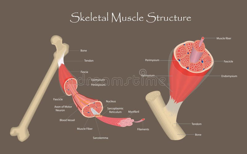
Free with trial Detailed anatomical illustration of skeletal muscle structure and its attachment to bone with labels. Detailed skeletal muscle structure anatomy illustration. Extremity anatomy vectors Detailed anatomical illustration of skeletal muscle structure and its attachment to bone with labels.
Free with trial A high-contrast side view of human foot anatomy reveals intricate bone structures, including the ankle joint and toes, set against a stark black backdrop. Extremity anatomy illustrations Detailed medical X-ray scan showing the side profile of human foot bones including ankle joint and toes in high contrast against a. A high-contrast side view of human foot anatomy reveals intricate bone structures, including the ankle joint and toes, set against a stark black backdrop
Free with trial Femoral Endovascular Procedure. Illustration depicting the femoral arteries and popliteal arteries. Angioplasty femoral artery. Extremity anatomy vectors Femoral Endovascular Procedure. Angioplasty femoral artery. Femoral Endovascular Procedure. Illustration depicting the femoral arteries and popliteal arteries. Angioplasty femoral artery
Free with trial Illustration of a human foot with the toes colored blue to represent coldness or discomfort, isolated on a white background. Extremity anatomy illustrations Close-up of a human foot with toes highlighted in blue indicating cold or pain. Illustration of a human foot with the toes colored blue to represent coldness or discomfort, isolated on a white background
Free with trial It is a black and white outline image of a hand and a foot, representing human extremities and basic body parts. Vector sketch line art draw using ai tool. Extremity anatomy vectors Hand and foot drawing. It is a black and white outline image of a hand and a foot, representing human extremities and basic body parts. Vector sketch line art draw using ai tool.
Free with trial A leg exhibits prominent varicose veins. A small card labeled FIG 2 VARICOSE VEIN PRESENTATION rests beside the affected area for medical reference. Extremity anatomy illustrations A portrait of lower limb varicose veins near a card, showcasing a presentation. A leg exhibits prominent varicose veins. A small card labeled FIG 2 VARICOSE VEIN PRESENTATION rests beside the affected area for medical reference.
Free with trial A black and white line drawing illustrates the bones and tendons of a human foot and ankle from the side, emphasizing anatomical detail. Extremity anatomy vectors Anatomical sketch of a human foot and ankle in profile view. A black and white line drawing illustrates the bones and tendons of a human foot and ankle from the side, emphasizing anatomical detail
Free with trial Leg with Bandage and Sport Shoe Recovery or Injury Illustration. Extremity anatomy vectors Leg with Bandage and Sport Shoe Recovery or Injury Illustration
Free with trial A detailed close up shows a foot with toes. The focus is on the big toe and its nail. The image highlights skin texture and anatomical details, suggesting themes of health, care, and the human body. Extremity anatomy illustrations Close up view of foot toes with focus on the big toe and its nail. A detailed close up shows a foot with toes. The focus is on the big toe and its nail. The image highlights skin texture and anatomical details, suggesting themes of health, care, and the human body
Free with trial Detailed top view of a dragonfly (order Odonata), vividly showcasing its anatomy. The transparent wings feature intricate venation patterns. The dragonfly's body is elongated and segmented with vibrant coloration, emphasizing green and black tones. Compound eyes are large and multifaceted, indicative of its excellent vision. Legs are slender and positioned forward. The background is plain white, isolating the dragonfly, allowing clear examination of its structure. The fine details are ideal for studying the morphology of this insect. Extremity anatomy illustrations Dragonfly Top View Macro Insect Isolated. Detailed top view of a dragonfly (order Odonata), vividly showcasing its anatomy. The transparent wings feature intricate venation patterns. The dragonfly's body is elongated and segmented with vibrant coloration, emphasizing green and black tones. Compound eyes are large and multifaceted, indicative of its excellent vision. Legs are slender and positioned forward. The background is plain white, isolating the dragonfly, allowing clear examination of its structure. The fine details are ideal for studying the morphology of this insect.
Free with trial Human foot sole illustration, anatomical detail on white. Extremity anatomy illustrations Illustration of human foot sole isolated on white background showing heel toes and skin texture with detailed dermatoglyphics for. Human foot sole illustration. human foot sole illustration, anatomical detail on white
Free with trial This studio shot showcases the intricate details of an aged hand, emphasizing the skin texture and the form shaped by time. The image captures the natural beauty and character of aging, making it suitable for themes related to healthcare, dermatology, or symbolic representations of time. Extremity anatomy illustrations Studio image capturing the detailed skin textures and shape of an aging hand. This studio shot showcases the intricate details of an aged hand, emphasizing the skin texture and the form shaped by time. The image captures the natural beauty and character of aging, making it suitable for themes related to healthcare, dermatology, or symbolic representations of time
Free with trial An intense close-up captures human feet, portraying a sense of vulnerability and raw texture. The obscured face suggests a hidden story, intensifying the mystery and tension within the image. Extremity anatomy illustrations Close-up perspective of human feet creating a dark and foreboding atmosphere. An intense close-up captures human feet, portraying a sense of vulnerability and raw texture. The obscured face suggests a hidden story, intensifying the mystery and tension within the image
Free with trial Detailed close-up of human finger joints against an isolated white background, ideal for medical illustrations and dermatological studies. Shows the intricate skin textures and anatomical structure. Extremity anatomy illustrations Close up of Human Finger Joints on Isolated White Backdrop for Medical Illustration and Dermatological Study. Detailed close-up of human finger joints against an isolated white background, ideal for medical illustrations and dermatological studies. Shows the intricate skin textures and anatomical structure
Free with trial Stunning Macro Photography of Spider Legs on Glossy Black Surface Highlighting Fine Details, Generated by AI. Extremity anatomy illustrations Stunning Macro Photography of Spider Legs on Glossy Black Surface Highlighting Fine Details
Free with trial An extreme macro photograph reveals the intricate beauty of iridescent beetle wing scales. The scales display a stunning spectrum of rainbow colors, from deep blues and greens to vibrant oranges and yellows, showcasing a detailed and complex natural texture. Extremity anatomy illustrations Extreme Macro of Iridescent Beetle Wing Scales, Rainbow Colors, Detailed Texture. An extreme macro photograph reveals the intricate beauty of iridescent beetle wing scales. The scales display a stunning spectrum of rainbow colors, from deep blues and greens to vibrant oranges and yellows, showcasing a detailed and complex natural texture.
Free with trial A close-up view of a leg with a noticeable scar running down it. Generative AI. Extremity anatomy illustrations Closeup of a leg with a scar. A close-up view of a leg with a noticeable scar running down it. Generative AI
Free with trial Incredible Close-up View of a Spider s Bristly Legs and Glossy Body in Stunning High Detail, Generated by AI. Extremity anatomy illustrations Incredible Close-up View of a Spider s Bristly Legs and Glossy Body in Stunning High Detail
Free with trial Detailed view of a persons fingertip and nail on a blurred background. Extremity anatomy illustrations Close-up of Fingertip with Nails Showing Detail. Detailed view of a persons fingertip and nail on a blurred background
Free with trial Extreme closeup of human fingertip showing sweat glands and ridges in detail. Extremity anatomy illustrations Closeup of Fingerprint with Sweat Glands Visible. Extreme closeup of human fingertip showing sweat glands and ridges in detail
Free with trial An illustration of a knee joint x-ray shows pain radiating in red, often used to represent knee injury or arthritis. Extremity anatomy illustrations Knee Pain X-ray Illustration. An illustration of a knee joint x-ray shows pain radiating in red, often used to represent knee injury or arthritis
Free with trial Podiatry concept. Feet treatment. Hardware medical pedicure. Nail cleaning apparatus. Patient on pedicure with pediatrician chiropodist. Clinic podology. Cosmetics of nails of legs. Vector design flat. Extremity anatomy vectors Podiatry concept. Feet treatment. Hardware medical pedicure.
Free with trial Intricate veining patterns are visible on a transparent insect wing against a black background. The wing showcases a complex network of interconnected veins, appearing delicate and fine, resembling a lace-like structure. Small, hair-like structures can be seen along the edge, highlighting the wing's texture. The wing's transparency against the dark background emphasizes its detailed structure and subtle variations in thickness and patterning. Extremity anatomy illustrations Extreme close-up of a transparent insect wing with intricate veining against a black background. Intricate veining patterns are visible on a transparent insect wing against a black background. The wing showcases a complex network of interconnected veins, appearing delicate and fine, resembling a lace-like structure. Small, hair-like structures can be seen along the edge, highlighting the wing's texture. The wing's transparency against the dark background emphasizes its detailed structure and subtle variations in thickness and patterning.
Free with trial A radiologist carefully examining an x-ray image of a human leg and pelvis, looking for any abnormalities. Medical diagnosis and healthcare concept in a clinical setting. Extremity anatomy illustrations Radiologist examining legs and pelvis x-ray for medical diagnosis at desk. A radiologist carefully examining an x-ray image of a human leg and pelvis, looking for any abnormalities. Medical diagnosis and healthcare concept in a clinical setting
Free with trial A detailed, glowing blue illustration of the human foot bones, rendered in an x-ray style against a black background. The image showcases the intricate structure of the foot, including the tibia, fibula, tarsals, metatarsals, and phalanges. Suitable for medical illustrations, educational materials, or healthcare related content. Extremity anatomy illustrations Foot Bones X-Ray Illustration. A detailed, glowing blue illustration of the human foot bones, rendered in an x-ray style against a black background. The image showcases the intricate structure of the foot, including the tibia, fibula, tarsals, metatarsals, and phalanges. Suitable for medical illustrations, educational materials, or healthcare related content.
Free with trial Detailed hand scan emphasizing skeletal structure and joint areas in vibrant red to depict discomfort or swelling, ideal for medical research and diagnostic illustra. Extremity anatomy illustrations Medical imaging of a human hand with highlighted bones and joints in red illustrating inflammation or pain in wrist and finger. Detailed hand scan emphasizing skeletal structure and joint areas in vibrant red to depict discomfort or swelling, ideal for medical research and diagnostic illustra
Free with trial X-ray image of human pelvis and leg bones. Extremity anatomy illustrations Human skeleton pelvis and leg bones. X-ray image of human pelvis and leg bones
Free with trial Small pearl resting on persons fingertips in a close-up view. Extremity anatomy illustrations Pearl Held Between Fingers Closeup Shot. Small pearl resting on persons fingertips in a close-up view