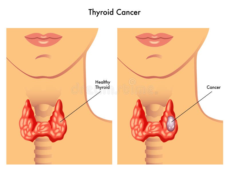
Free with trial Medical illustration of the effects of the thyroid cancer. Follicular cells vectors Thyroid cancer
Free with trial Female menstrual cycle, ovulation process and hormone levels, detailed medical illustration. Follicular cells illustrations Female menstrual cycle, ovulation process and hormone levels
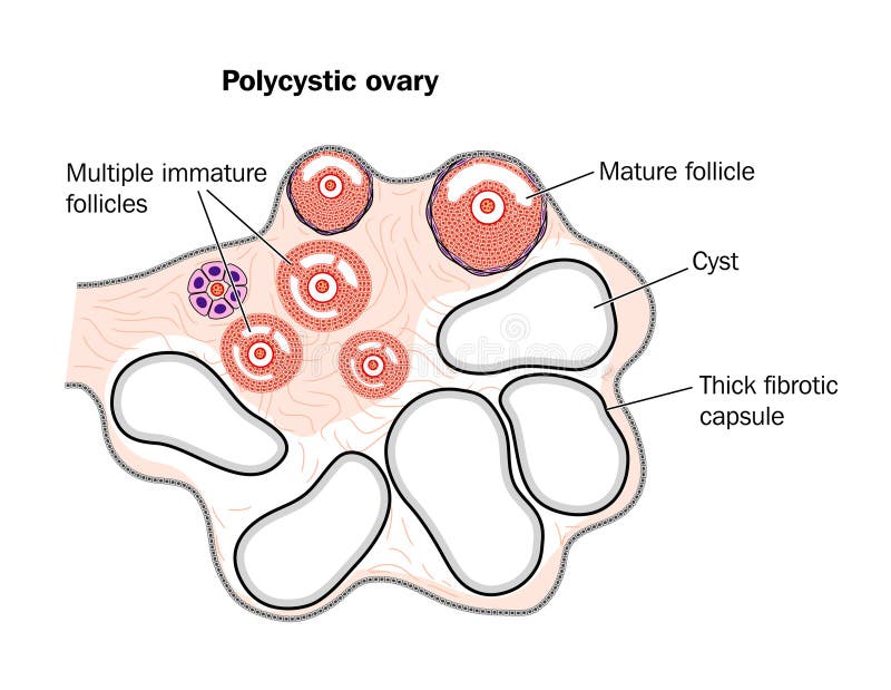
Free with trial Drawing (cross section) of a polycystic ovary showing multiple cysts. Follicular cells vectors Polycystic ovary
Free with trial Menstrual cycle graphic, detailed follicular development illustration, menstruation and ovulation days. Isolated on a white background. Follicular cells vectors Menstrual cycle graphic, follicules. Menstrual cycle graphic, detailed follicular development illustration, menstruation and ovulation days. Isolated on a white background.
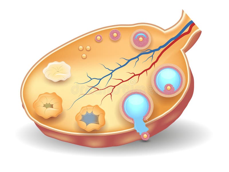
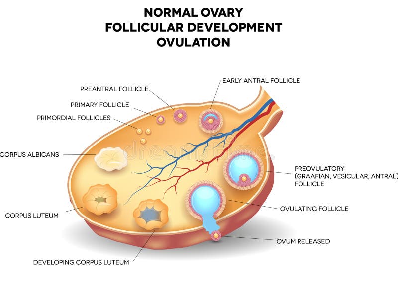
Free with trial Semi diagramatic representation of a normal ovary and the stages of ovulation. Follicular cells vectors Normal Ovary
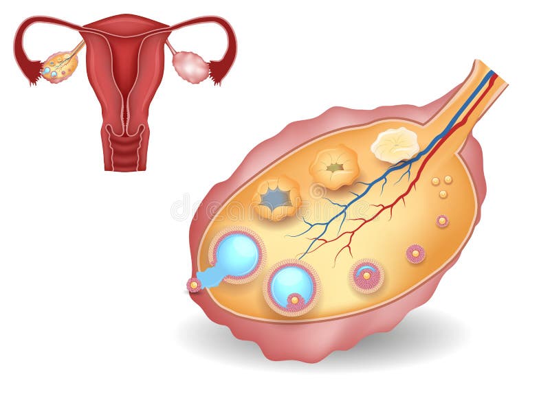
Free with trial Normal uterus and ovaries illustration. Healthy reproductive system organs. Follicular cells vectors Ovary, detailed follicular development and uterus. Normal uterus and ovaries illustration. Healthy reproductive system organs.
Free with trial Embryo development. Secondary oocyte ovulation, fertilization and development till blastocyst implantation. Follicular cells vectors Embryo development
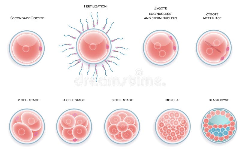
Free with trial Fertilised cell development. Stages from fertilization till morula cell. Follicular cells vectors Fertilised cell development. Stages from fertilization till morula cell.
Free with trial Female ovary structure, colorful medical illustration shows follicular development. Follicular cells illustrations Healthy ovary structure, follicular development. Female ovary structure, colorful medical illustration shows follicular development
Free with trial Normal ovary, follicular development and ovulation. Ovum is released from the ovarian follicles. Follicular cells illustrations Normal ovary, follicular development and ovulation
Free with trial Embryo development abstract blue background. Development till blastocyst implantation. Follicular cells vectors Embryo development blue background. Embryo development abstract blue background. Development till blastocyst implantation.
Free with trial A healthy spleen compared to that of one with lymphoma. Follicular cells vectors Lymphoma
Free with trial Thyroid gland anatomy and physiology. Structure of a human Thyroid gland. Follicular Cells, Basement Membrane, Blood Vessel, C-cell and Colloid. Hormones and endocrine function of thyroid gland. Triiodothyronine t3, thyroxine t4 and Calcitonin. Vector poster. Follicular cells vectors Thyroid gland anatomy and physiology
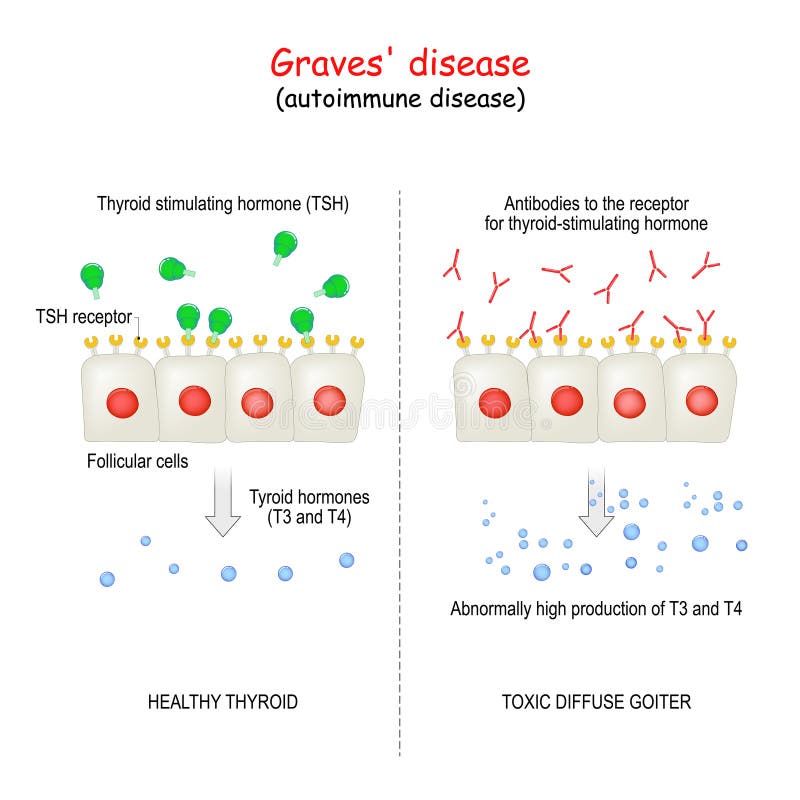
Free with trial Graves` disease. autoimmune disorder. toxic diffuse goiter and cell of healthy thyroid gland. explanation about Abnormally high production hormones of thyroid gland T3 and T4. Follicular cells vectors Graves` disease. autoimmune disorder
Free with trial Hodgkin's lymphoma. biopsy healthy human cells and lymphoma patient. Reed-Sternberg and other white blood cell. Illustration showing a classic Reed-Berezovsky-Sternberg cell. Follicular cells vectors Hodgkin's lymphoma
Free with trial Normal Female Ovary. Detailed follicular development. Follicular cells vectors Female Ovary
Free with trial Beautiful light blue menstrual cycle graphic wheel, reproductive system anatomy at the middle. Follicular cells vectors Beautiful light blue menstrual cycle graphic
Free with trial Thyroglobulin, a dimeric protein produced by the follicular cells of the thyroid and used entirely within the thyroid gland. The protein is a precursor of the thyroid hormones. 3d rendering. Follicular cells illustrations Thyroglobulin, a dimeric protein produced by the follicular cell
Free with trial Anatomy of the Thyroid Gland. Follicular cells. Follicular cells vectors Anatomy of the Thyroid Gland.
Free with trial Structure of the thyroid gland. Histological structure of Thryoid follicle thyroid or epithelial cells or thyrocytes. Follicular cells vectors Structure thyroid gland. Histological structure of Thryoid follicle thyroid or epithelial cells or thyrocytes
Free with trial Thyroid cancer with organs and tumors or cancerous cells 3D rendering illustration with male body contours. Anatomy, oncology, biomedical, medical, biology, science, healthcare concepts. Follicular cells illustrations Thyroid cancer with organs and tumors or cancerous cells 3D rendering illustration with male body contours. Anatomy, oncology
Free with trial Non-Hodgkin lymphoma also known as non-Hodgkin`s lymphoma, NHL, or sometimes just lymphoma is a cancer that starts in white blood cells called lymphocytes, which are part of the body`s immune system. NHL is a term that`s used for many different types of lymphoma that all share some of the same characteristics. Follicular cells illustrations T-cell lymphoma cells in the blood flow - isometric view 3d illustration. Non-Hodgkin lymphoma also known as non-Hodgkin`s lymphoma, NHL, or sometimes just lymphoma is a cancer that starts in white blood cells called lymphocytes, which are part of the body`s immune system. NHL is a term that`s used for many different types of lymphoma that all share some of the same characteristics.
Free with trial Woman in gynecology office concept. Patient visits clinic, medical appointment. Female egg cells collecting flat vector illustration. Oocyte and ovary anatomy. Doctors consultation. Laboratory testing. Follicular cells vectors Human cells collecting. Woman in gynecology office concept. Patient visits clinic, medical appointment. Female egg cells collecting flat vector illustration. Oocyte and ovary anatomy. Doctors consultation. Laboratory testing
Free with trial Ovarian cortex showing primary follicles with oocytes surrounded a single layer of follicular or granulosa cells. The zona pellucida is still absent. Follicular cells illustrations Ovary. Primary follicle. Ovarian cortex showing primary follicles with oocytes surrounded a single layer of follicular or granulosa cells. The zona pellucida is still absent
Free with trial Woman and man egg cells, embryo icon in tube. Sperm and ovum. Fertilisation, gynecology and genetic testing. Human sexual reproductive system. Pregnancy stage. Medical poster. IVF vector illustration. Follicular cells vectors Reproductive system concept. Woman and man egg cells, embryo icon in tube. Sperm and ovum. Fertilisation, gynecology and genetic testing. Human sexual reproductive system. Pregnancy stage. Medical poster. IVF vector illustration.
Free with trial Ovum Structure. Morphology of the human and animal ovule. Vector illustration. Didatic illustration. Follicular cells vectors Ovum Structure. Morphology of the ovule. Ovum Structure. Morphology of the human and animal ovule. Vector illustration. Didatic illustration.
Free with trial Woman egg cells icon set. Fertilisation, gynecology and genetic concept. Human sexual reproductive system. Pregnancy medical poster for clinic, lab and education. IVF flat vector isolated illustration. Follicular cells vectors Egg cell anatomy. Woman egg cells icon set. Fertilisation, gynecology and genetic concept. Human sexual reproductive system. Pregnancy medical poster for clinic, lab and education. IVF flat vector isolated illustration
Free with trial Woman egg cells in tube. Medical icon. Fertilisation, gynecology and genetic testing. Human sexual reproductive system concept. Pregnancy stage. Poster for clinic. IVF flat vector illustration. Follicular cells vectors In vitro fertilisation concept. Woman egg cells in tube. Medical icon. Fertilisation, gynecology and genetic testing. Human sexual reproductive system concept. Pregnancy stage. Poster for clinic. IVF flat vector illustration.
Free with trial Embryo development diagram. Insemination fertilization, ivf concept. Female, male egg cells. Doctors research, appointment in clinic. Human sexual reproductive system. Pregnancy vector illustration. Follicular cells vectors Embryo development concept. Embryo development diagram. Insemination fertilization, ivf concept. Female, male egg cells. Doctors research, appointment in clinic. Human sexual reproductive system. Pregnancy vector illustration.
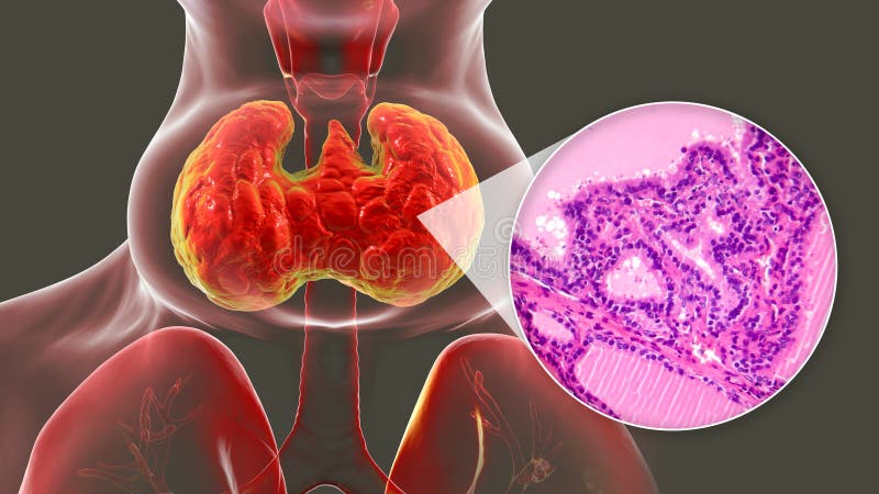
Free with trial A 3D illustration of a person with enlarged thyroid gland, alongside with a micrograph image of thyroid tissue affected by toxic goiter. Follicular cells illustrations Toxic goiter, 3D illustration and micrograph. A 3D illustration of a person with enlarged thyroid gland, alongside with a micrograph image of thyroid tissue affected by toxic goiter.
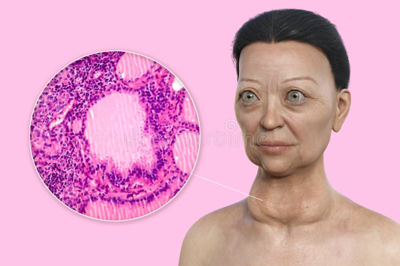
Free with trial A 3D illustration of a woman with Graves' disease having enlarged thyroid gland and exophthalmos, alongside a micrograph image of thyroid tissue affected by Graves' disease. Follicular cells illustrations A 3D illustration of a woman with Graves' disease alongside a micrograph image of thyroid tissue affected by. A 3D illustration of a woman with Graves' disease having enlarged thyroid gland and exophthalmos, alongside a micrograph image of thyroid tissue affected by Graves' disease.
Free with trial This detailed 3D rendering illustrates a cross-section of a human ovum, or egg cell, surrounded by its protective follicular cells. The vibrant red sphere represents the oocyte, while the intricate network of delicate, thread-like structures depicts the surrounding granulosa cells and the zona pellucida. The image showcases the complex cellular environment essential for ovulation and potential fertilization, making it ideal for educational, medical, and scientific applications. Follicular cells illustrations Microscopic View of a Human Ovum (Egg Cell) with Follicular Cells. This detailed 3D rendering illustrates a cross-section of a human ovum, or egg cell, surrounded by its protective follicular cells. The vibrant red sphere represents the oocyte, while the intricate network of delicate, thread-like structures depicts the surrounding granulosa cells and the zona pellucida. The image showcases the complex cellular environment essential for ovulation and potential fertilization, making it ideal for educational, medical, and scientific applications.
Free with trial Thyroid gland tissue under microscope showing follicular adenoma. Cells stained purple, pink. Histopathology of thyroid adenoma benign tumor. Tissue sample examined in laboratory for. Follicular cells illustrations Thyroid gland tissue under microscope showing follicular adenoma. Cells stained purple, pink. Histopathology of thyroid adenoma
Free with trial Detailed Microscopic View of Mammalian Ovary Histology Featuring Oocytes and Follicular Cells in a Beautiful Blue-Violet Stain. Generative AI. Follicular cells illustrations Detailed Microscopic View of Mammalian Ovary Histology Featuring Oocytes and Follicular Cells in a Beautiful BlueViolet Stain
Free with trial Close-up of microscopic lymph node tissue with follicular pattern. Abnormal cells in cancer diagnosis exam. Tissue analysis for oncology, histology study. Lymphoma diagnosis treatment. Follicular cells illustrations Close-up of microscopic lymph node tissue with follicular pattern. Abnormal cells in cancer diagnosis exam. Tissue analysis for
Free with trial Light micrograph of thyroid gland follicular adenoma. Microscopic photo shows histology of thyroid tissue. Benign lesion with cells, follicles. Medical, scientific research, biology. Follicular cells illustrations Light micrograph of thyroid gland follicular adenoma. Microscopic photo shows histology of thyroid tissue. Benign lesion with
Free with trial An informative and clear depiction of a human oocyte, bringing attention to its cellular structure and surrounding cells in contrast to a white background. Follicular cells illustrations A detailed illustration showcasing a human oocyte, emphasizing its intricate structure and associated follicular cells against a. An informative and clear depiction of a human oocyte, bringing attention to its cellular structure and surrounding cells in contrast to a white background.
Free with trial An informative and clear depiction of a human oocyte, bringing attention to its cellular structure and surrounding cells in contrast to a white background. Follicular cells illustrations A detailed illustration showcasing a human oocyte, emphasizing its intricate structure and associated follicular cells against a. An informative and clear depiction of a human oocyte, bringing attention to its cellular structure and surrounding cells in contrast to a white background.
Free with trial An informative and clear depiction of a human oocyte, bringing attention to its cellular structure and surrounding cells in contrast to a white background. Follicular cells illustrations A detailed illustration showcasing a human oocyte, emphasizing its intricate structure and associated follicular cells against a. An informative and clear depiction of a human oocyte, bringing attention to its cellular structure and surrounding cells in contrast to a white background.
Free with trial An informative and clear depiction of a human oocyte, bringing attention to its cellular structure and surrounding cells in contrast to a white background. Follicular cells illustrations A detailed illustration showcasing a human oocyte, emphasizing its intricate structure and associated follicular cells against a. An informative and clear depiction of a human oocyte, bringing attention to its cellular structure and surrounding cells in contrast to a white background.
Free with trial This microscopic image reveals the intricate cellular architecture of follicular lymphoma within a lymph node tissue section. The image showcases the characteristic features of this type of non-Hodgkin lymphoma, highlighting the presence of centroblasts, the abnormal B lymphocytes central to the disease. These cells are often large and have prominent nucleoli. Also visible are tingible body. Follicular cells illustrations Microscopic Anatomy of Follicular Lymphoma Centroblasts Tingible Body Macrophages and Lymph Node Tissue. This microscopic image reveals the intricate cellular architecture of follicular lymphoma within a lymph node tissue section. The image showcases the characteristic features of this type of non-Hodgkin lymphoma, highlighting the presence of centroblasts, the abnormal B lymphocytes central to the disease. These cells are often large and have prominent nucleoli. Also visible are tingible body
Free with trial This vibrant, detailed photomicrograph showcases a microscopic section of lymph node tissue affected by follicular lymphoma. The image highlights the characteristic features of this type of lymphoma, specifically focusing on the presence of centroblasts and tingible body macrophages. Centroblasts, large, atypical lymphoid cells, are readily apparent within the follicular structures. Their. Follicular cells illustrations Detailed Microscopic View of Follicular Lymphoma Centroblasts and Tingible Body Macrophages in Lymph Node Tissue. This vibrant, detailed photomicrograph showcases a microscopic section of lymph node tissue affected by follicular lymphoma. The image highlights the characteristic features of this type of lymphoma, specifically focusing on the presence of centroblasts and tingible body macrophages. Centroblasts, large, atypical lymphoid cells, are readily apparent within the follicular structures. Their
Free with trial A 3D render shows a single hair follicle trapped below glowing skin cells. The follicle is curved in a spiral shape against a peach background. Follicular cells illustrations Understanding Follicle Obstruction With a Minimalist 3D Render Highlighting a Hair Follicle Beneath Glowing Skin Cells. A 3D render shows a single hair follicle trapped below glowing skin cells. The follicle is curved in a spiral shape against a peach background.
Free with trial A 3D render shows a hair follicle trapped under glowing skin cells. Follicular cells illustrations Exploring Follicle Obstruction and Its Effects on Hair Growth in a Minimalistic Design With Glowing Skin Cells and a. A 3D render shows a hair follicle trapped under glowing skin cells.
Free with trial A 3D render shows a single hair follicle trapped under glowing skin cells. The hair is curved in a spiral against a soft peach background. Follicular cells illustrations Concept of Follicle Obstruction With a Spiral Hair Structure Under Skin Cells in a Minimalist 3D Render on a Peach. A 3D render shows a single hair follicle trapped under glowing skin cells. The hair is curved in a spiral against a soft peach background.
Free with trial Angioimmunoblastic T-cell lymphoma (AITL? ) is a mature T-cell lymphoma of blood or lymph vessel immunoblasts characterized by a polymorphous lymph node infiltrate showing a marked increase in follicular dendritic cells and high endothelial venules and systemic involvement. In rare cases, there may be infiltration of the peripheral blood. Wright x1000. Follicular cells illustrations Angioimmunoblastic T-cell lymphoma. Peripheral blood. Angioimmunoblastic T-cell lymphoma (AITL?) is a mature T-cell lymphoma of blood or lymph vessel immunoblasts characterized by a polymorphous lymph node infiltrate showing a marked increase in follicular dendritic cells and high endothelial venules and systemic involvement. In rare cases, there may be infiltration of the peripheral blood. Wright x1000.
Free with trial Angioimmunoblastic T-cell lymphoma (AITL? ) is a mature T-cell lymphoma of blood or lymph vessel immunoblasts characterized by a polymorphous lymph node infiltrate showing a marked increase in follicular dendritic cells and high endothelial venules and systemic involvement. In rare cases, there may be infiltration of the peripheral blood. Wright x1000. Follicular cells illustrations Angioimmunoblastic T-cell lymphoma. Peripheral blood. Angioimmunoblastic T-cell lymphoma (AITL?) is a mature T-cell lymphoma of blood or lymph vessel immunoblasts characterized by a polymorphous lymph node infiltrate showing a marked increase in follicular dendritic cells and high endothelial venules and systemic involvement. In rare cases, there may be infiltration of the peripheral blood. Wright x1000.
Free with trial Angioimmunoblastic T-cell lymphoma (AITL? ) is a mature T-cell lymphoma of blood or lymph vessel immunoblasts characterized by a polymorphous lymph node infiltrate showing a marked increase in follicular dendritic cells and high endothelial venules and systemic involvement. In rare cases, there may be infiltration of the peripheral blood. Wright x1000. Follicular cells illustrations Angioimmunoblastic T-cell lymphoma. Peripheral blood. Angioimmunoblastic T-cell lymphoma (AITL?) is a mature T-cell lymphoma of blood or lymph vessel immunoblasts characterized by a polymorphous lymph node infiltrate showing a marked increase in follicular dendritic cells and high endothelial venules and systemic involvement. In rare cases, there may be infiltration of the peripheral blood. Wright x1000.
Free with trial Angioimmunoblastic T-cell lymphoma (AITL? ) is a mature T-cell lymphoma of blood or lymph vessel immunoblasts characterized by a polymorphous lymph node infiltrate showing a marked increase in follicular dendritic cells and high endothelial venules and systemic involvement. In rare cases, there may be infiltration of the peripheral blood. Wright x1000. Follicular cells illustrations Angioimmunoblastic T-cell lymphoma. Peripheral blood. Angioimmunoblastic T-cell lymphoma (AITL?) is a mature T-cell lymphoma of blood or lymph vessel immunoblasts characterized by a polymorphous lymph node infiltrate showing a marked increase in follicular dendritic cells and high endothelial venules and systemic involvement. In rare cases, there may be infiltration of the peripheral blood. Wright x1000.
Free with trial Angioimmunoblastic T-cell lymphoma (AITL? ) is a mature T-cell lymphoma of blood or lymph vessel immunoblasts characterized by a polymorphous lymph node infiltrate showing a marked increase in follicular dendritic cells and high endothelial venules and systemic involvement. In rare cases, there may be infiltration of the peripheral blood. Wright x1000. Follicular cells illustrations Angioimmunoblastic T-cell lymphoma. Peripheral blood. Angioimmunoblastic T-cell lymphoma (AITL?) is a mature T-cell lymphoma of blood or lymph vessel immunoblasts characterized by a polymorphous lymph node infiltrate showing a marked increase in follicular dendritic cells and high endothelial venules and systemic involvement. In rare cases, there may be infiltration of the peripheral blood. Wright x1000.
Free with trial Acute myeloid leukemia AML is a type of blood cancer. It starts in your bone marrow, the soft inner parts of bones. AML usually begins in cells that turn into white blood cells, but it can start in other blood-forming cells, as well. Follicular cells illustrations White blood cells with Acute myeloid leukaemia (AML) cells - closeup view 3d illustration. Acute myeloid leukemia AML is a type of blood cancer. It starts in your bone marrow, the soft inner parts of bones. AML usually begins in cells that turn into white blood cells, but it can start in other blood-forming cells, as well.
Free with trial This captivating microscopic image showcases a follicular thyroid adenoma, a benign tumor of the thyroid gland. The high magnification reveals intricate details of the neoplastic follicular cells, a key component in diagnosing this type of thyroid lesion. The image highlights the characteristic architecture of the adenoma, typically composed of follicles filled with colloid, a protein-rich. Follicular cells illustrations Detailed Histological Examination of a Follicular Thyroid Adenoma A Microscopic View of Thyroid Gland Neoplasia. This captivating microscopic image showcases a follicular thyroid adenoma, a benign tumor of the thyroid gland. The high magnification reveals intricate details of the neoplastic follicular cells, a key component in diagnosing this type of thyroid lesion. The image highlights the characteristic architecture of the adenoma, typically composed of follicles filled with colloid, a protein-rich
Free with trial Microscopic view of thyroid follicular cells and histiocytes. Cellular material shows regular thyroid follicular epithelial cells in, a pinkish background with visible blood cells. Follicular cells illustrations Microscopic view of thyroid follicular cells and histiocytes. Cellular material shows regular thyroid follicular epithelial cells
Free with trial This captivating microscopic image reveals the fascinating detail of hair root cells, offering a close-up view of the follicle structure and root sheath. The intricate network of cells within the hair follicle and surrounding tissues is clearly visible, showcasing the complex biological processes responsible for hair growth. Observe the specialized cells that form the hair shaft, the dermal. Follicular cells illustrations Unveiling the Intricate Microstructure of Hair Root Cells A Detailed Look at Follicle Structure and Root Sheath. This captivating microscopic image reveals the fascinating detail of hair root cells, offering a close-up view of the follicle structure and root sheath. The intricate network of cells within the hair follicle and surrounding tissues is clearly visible, showcasing the complex biological processes responsible for hair growth. Observe the specialized cells that form the hair shaft, the dermal
Free with trial This image depicts a false color electron micrograph highlighting large golden cells, likely oocytes, interacting with surrounding smaller cells within a reddish biological matrix. The detailed visualization provides insight into complex reproductive cellular dynamics and structures labeled for educational purposes. Follicular cells illustrations Microscopic view of oocyte and follicular cells. This image depicts a false color electron micrograph highlighting large golden cells, likely oocytes, interacting with surrounding smaller cells within a reddish biological matrix. The detailed visualization provides insight into complex reproductive cellular dynamics and structures labeled for educational purposes
Free with trial This image depicts a false color electron micrograph highlighting large golden cells, likely oocytes, interacting with surrounding smaller cells within a reddish biological matrix. The detailed visualization provides insight into complex reproductive cellular dynamics and structures labeled for educational purposes. Follicular cells illustrations Microscopic view of oocyte and follicular cells. This image depicts a false color electron micrograph highlighting large golden cells, likely oocytes, interacting with surrounding smaller cells within a reddish biological matrix. The detailed visualization provides insight into complex reproductive cellular dynamics and structures labeled for educational purposes
Free with trial This image depicts a false color electron micrograph highlighting large golden cells, likely oocytes, interacting with surrounding smaller cells within a reddish biological matrix. The detailed visualization provides insight into complex reproductive cellular dynamics and structures labeled for educational purposes. Follicular cells illustrations Microscopic view of oocyte and follicular cells. This image depicts a false color electron micrograph highlighting large golden cells, likely oocytes, interacting with surrounding smaller cells within a reddish biological matrix. The detailed visualization provides insight into complex reproductive cellular dynamics and structures labeled for educational purposes
Free with trial Confocal microscopy of lymph node germinal center showing B cell zones stained for GL-7 and T follicular helper cells. Follicular cells illustrations Lymph Node Germinal Center B Cell Reaction. Confocal microscopy of lymph node germinal center showing B cell zones stained for GL-7 and T follicular helper cells
Free with trial Light microscopy of thyroid gland showing circular follicles filled with homogeneous colloid surrounded by follicular epithelial cells. Follicular cells illustrations Thyroid Follicle Colloid Storage Histology Section. Light microscopy of thyroid gland showing circular follicles filled with homogeneous colloid surrounded by. Light microscopy of thyroid gland showing circular follicles filled with homogeneous colloid surrounded by follicular epithelial cells
Free with trial A luminous, translucent butterfly appears suspended against a deep purple backdrop. Its wings are crafted with intricate, vein-like patterns, emitting a gentle pink-violet glow. The ethereal quality and delicate details give the butterfly a crystalline appearance, suggesting a high-tech or sci-fi aesthetic. Tiny points of light are scattered throughout the background, enhancing the sense of floating in a vast, cosmic space. Follicular cells illustrations A sci-fi-inspired depiction of a human thyroid gland, rendered in crystalline glass with precise follicular details, floating. A luminous, translucent butterfly appears suspended against a deep purple backdrop. Its wings are crafted with intricate, vein-like patterns, emitting a gentle pink-violet glow. The ethereal quality and delicate details give the butterfly a crystalline appearance, suggesting a high-tech or sci-fi aesthetic. Tiny points of light are scattered throughout the background, enhancing the sense of floating in a vast, cosmic space.
Free with trial Detailed illustration of Graafian follicle ovarian cell structure. Includes large oocyte, smaller surrounding cells within follicle wall. Vibrant colors clear anatomical detail suitable. Follicular cells illustrations Detailed illustration of Graafian follicle ovarian cell structure. Includes large oocyte, smaller surrounding cells within
Free with trial Non-Hodgkin lymphoma also known as non-Hodgkin`s lymphoma, NHL, or sometimes just lymphoma is a cancer that starts in white blood cells called lymphocytes, which are part of the body`s immune system. NHL is a term that`s used for many different types of lymphoma that all share some of the same characteristics. Follicular cells illustrations Dendritic cell recognize Non-hodgkin lymphoma (NHL) - closeup view 3d illustration. Non-Hodgkin lymphoma also known as non-Hodgkin`s lymphoma, NHL, or sometimes just lymphoma is a cancer that starts in white blood cells called lymphocytes, which are part of the body`s immune system. NHL is a term that`s used for many different types of lymphoma that all share some of the same characteristics.
Free with trial Acute myeloid leukemia AML is a type of blood cancer. It starts in your bone marrow, the soft inner parts of bones. AML usually begins in cells that turn into white blood cells, but it can start in other blood-forming cells, as well. Follicular cells illustrations Dendritic cell recognise Acute myeloid leukaemia (AML) cell - closeup view 3d illustration. Acute myeloid leukemia AML is a type of blood cancer. It starts in your bone marrow, the soft inner parts of bones. AML usually begins in cells that turn into white blood cells, but it can start in other blood-forming cells, as well.
Free with trial Non-Hodgkin lymphoma also known as non-Hodgkin`s lymphoma, NHL, or sometimes just lymphoma is a cancer that starts in white blood cells called lymphocytes, which are part of the body`s immune system. NHL is a term that`s used for many different types of lymphoma that all share some of the same characteristics. Follicular cells illustrations Monoclonal antibody treatment in Non-hodgkin lymphoma (NHL) - closeup view 3d illustration. Non-Hodgkin lymphoma also known as non-Hodgkin`s lymphoma, NHL, or sometimes just lymphoma is a cancer that starts in white blood cells called lymphocytes, which are part of the body`s immune system. NHL is a term that`s used for many different types of lymphoma that all share some of the same characteristics.
Free with trial Non-Hodgkin lymphoma also known as non-Hodgkin`s lymphoma, NHL, or sometimes just lymphoma is a cancer that starts in white blood cells called lymphocytes, which are part of the body`s immune system. NHL is a term that`s used for many different types of lymphoma that all share some of the same characteristics. Follicular cells illustrations Monoclonal antibody treatment in Non-hodgkin lymphoma (NHL) - isometric view 3d illustration. Non-Hodgkin lymphoma also known as non-Hodgkin`s lymphoma, NHL, or sometimes just lymphoma is a cancer that starts in white blood cells called lymphocytes, which are part of the body`s immune system. NHL is a term that`s used for many different types of lymphoma that all share some of the same characteristics.
Free with trial Acute myeloid leukemia AML is a type of blood cancer. It starts in your bone marrow, the soft inner parts of bones. AML usually begins in cells that turn into white blood cells, but it can start in other blood-forming cells, as well. Follicular cells illustrations Monoclonal antibody treatment in Acute myeloid leukaemia (AML) - closeup view 3d illustration. Acute myeloid leukemia AML is a type of blood cancer. It starts in your bone marrow, the soft inner parts of bones. AML usually begins in cells that turn into white blood cells, but it can start in other blood-forming cells, as well.
Free with trial Acute myeloid leukemia AML is a type of blood cancer. It starts in your bone marrow, the soft inner parts of bones. AML usually begins in cells that turn into white blood cells, but it can start in other blood-forming cells, as well. Follicular cells illustrations Monoclonal antibody treatment in Acute myeloid leukaemia (AML) - isometric view 3d illustration. Acute myeloid leukemia AML is a type of blood cancer. It starts in your bone marrow, the soft inner parts of bones. AML usually begins in cells that turn into white blood cells, but it can start in other blood-forming cells, as well.
Free with trial This captivating microscopic image reveals the complex and fascinating structure of a human hair follicle. The detailed close-up showcases the intricate network of cells, proteins, and tissues that form the foundation of hair growth. Observe the tightly packed keratin filaments, the nourishing blood vessels, and the delicate papilla at the base. This magnified view offers a unique perspective. Follicular cells illustrations Unveiling the Intricate Architecture of Human Hair A Microscopic Exploration of Hair Follicle Structure. This captivating microscopic image reveals the complex and fascinating structure of a human hair follicle. The detailed close-up showcases the intricate network of cells, proteins, and tissues that form the foundation of hair growth. Observe the tightly packed keratin filaments, the nourishing blood vessels, and the delicate papilla at the base. This magnified view offers a unique perspective
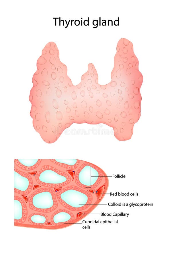
Free with trial The thyroid, or thyroid gland, is an endocrine gland in vertebrates. In humans, it is a butterfly-shaped gland located in the neck below the Adam's apple. It consists of two connected lobes. The lower two thirds of the lobes are connected by a thin band of tissue called the isthmus. Microscopically, the functional unit of the thyroid gland is the spherical thyroid follicle, lined with follicular cells (thyrocytes), and occasional parafollicular cells that surround a lumen containing colloid. Follicular cells illustrations Human thyroid - isometric view 3d illustration. The thyroid, or thyroid gland, is an endocrine gland in vertebrates. In humans, it is a butterfly-shaped gland located in the neck below the Adam's apple. It consists of two connected lobes. The lower two thirds of the lobes are connected by a thin band of tissue called the isthmus. Microscopically, the functional unit of the thyroid gland is the spherical thyroid follicle, lined with follicular cells (thyrocytes), and occasional parafollicular cells that surround a lumen containing colloid.
Free with trial Medical vector illustration of thyroid cancer showing abnormal malignant growth in the thyroid gland, leading to neck swelling, nodules, and potential spread to lymph nodes. Follicular cells vectors Thyroid cancer vector illustration. Medical vector illustration of thyroid cancer showing abnormal malignant growth in the thyroid gland, leading to neck swelling, nodules, and potential spread to lymph nodes.