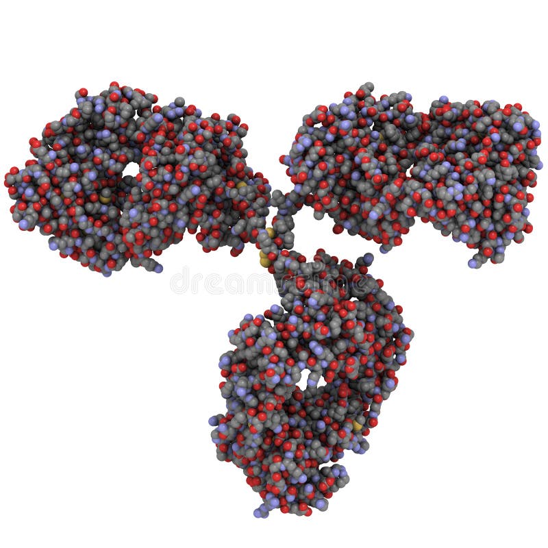
Free with trial Chemical structure of an immunoglobulin G (IgG, antibody) molecule. All-atom, space-filling representation. G protein vectors Immunoglobulin G (IgG, antibody) molecule
Free with trial Dendritic cells present antigens green to lymphocytes through their membran bound MHC-molecules violet. CD4 molecules light blue bind to other portions of the MHC, strengthening the interaction. After binding to the MHC-antigen complex, The T-cell receptor blue sends a signal cascade through an attached G-protein into the T-lymphocyte cell, that activates an immune response. G protein illustrations Activation of the immune response: antigen presenting cell activates T-lymphocytes (smaller c. Dendritic cells present antigens green to lymphocytes through their membran bound MHC-molecules violet. CD4 molecules light blue bind to other portions of the MHC, strengthening the interaction. After binding to the MHC-antigen complex, The T-cell receptor blue sends a signal cascade through an attached G-protein into the T-lymphocyte cell, that activates an immune response.
Free with trial Chemical structure of an immunoglobulin G (IgG1, antibody) molecule. Two space-filling, all atom views. G protein illustrations Immunoglobulin G (IgG1, antibody) molecule
Free with trial G protein coupled receptors gated ion channel. Structure of a G protein-coupled receptor (GPCR). Mechanism for the transport of ions. Cell membrane receptors for ligands bind. vector illustration. G protein vectors G protein coupled receptors gated ion channel. Vector illustration. G protein coupled receptors gated ion channel. Structure of a G protein-coupled receptor (GPCR). Mechanism for the transport of ions. Cell membrane receptors for ligands bind. vector illustration
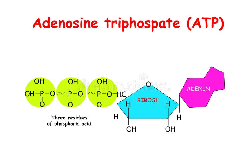
Free with trial Adenosine triphosphate ATP on white background. ATP provides energy to drive many processes in living cells, e. g. muscle contraction, nerve impulse propagation, chemical synthesis. vector. G protein vectors Adenosine triphosphate ATP on white background. ATP provides energy to drive many processes in living cells, e.g. muscle contrac
Free with trial Glucagon-like peptide-1 receptor. Close-up of GLP-1R with heterotrimeric G-protein and Ligands. GLP1R Receptor in the cell membrane. Weight loss therapy. Vector poster. G protein vectors Glucagon-like peptide-1 receptor. Close-up of GLP-1R with heterotrimeric G-protein and Ligands
Free with trial G Protein and Ion Channel Diagram in 3D. G protein vectors G Protein and Ion Channel Diagram
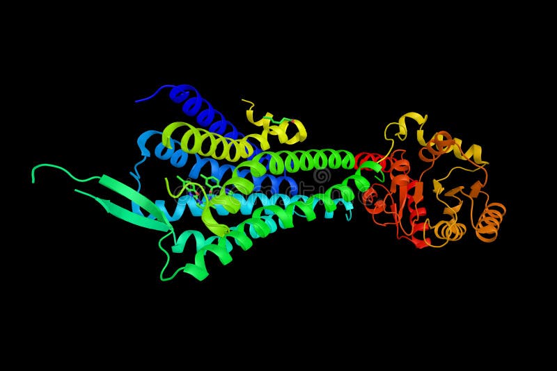
Free with trial Hypocretin (orexin) receptor 2, a G-protein coupled receptor expressed exclusively in the brain. Involved in the central feedback mechanism that regulates feeding behaviour. G protein illustrations Hypocretin (orexin) receptor 2, a G-protein coupled receptor exp
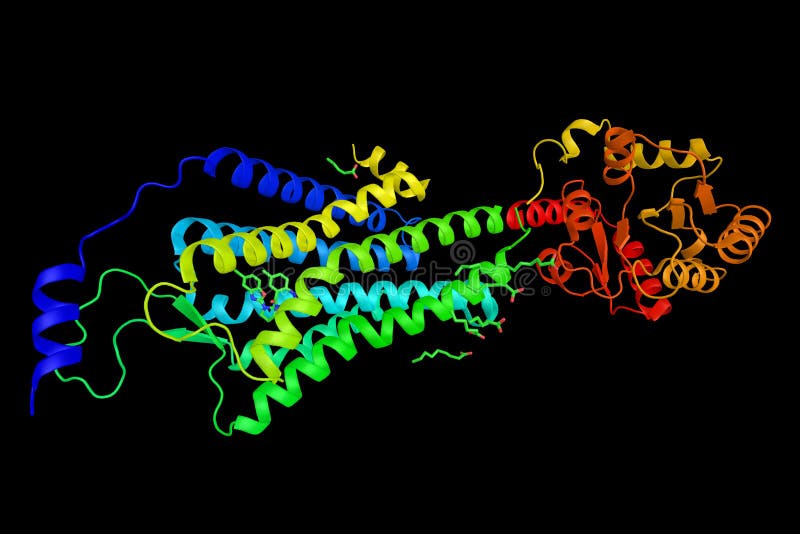
Free with trial Orexin receptor type 1, a G-protein coupled receptor that is heavily expressed in projections from the lateral hypothalamus and is involved in the regulation of feeding behaviour. 3d rendering. G protein illustrations Orexin receptor type 1, a G-protein coupled receptor that is heavily expressed in projections from the lateral hypothalamus and i
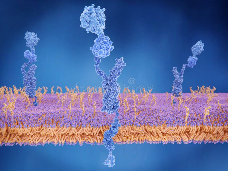
Free with trial The amyloid precursor protein APP is a complex protein with many functions. It is found on the surface of cells throughout the body. The intact protein binds to many stuctural proteins outside cells, such as heparin and laminin and sends signals through the G-protein system. G protein illustrations The amyloid precursor protein
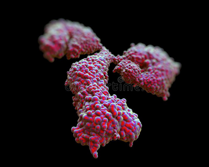
Free with trial Immunoglobulin G antibody. Molecular model of the antibody immunoglobulin G (IgG). This Y-shaped protein is produced by B-lymphocyte white blood cells as part of an immune response. Antibodies are used to fight off infections. The limbs (upper left & right) bind to foreign antigens such as the surface proteins of invading bacteria. Binding can neutralize toxins or invading organisms and prevent. G protein illustrations Immunoglobulin G antibody
Free with trial Immunoglobulin G antibody. Molecular model of the antibody immunoglobulin G (IgG). This Y-shaped protein is produced by B-lymphocyte white blood cells as part of an immune response. Antibodies are used to fight off infections. The limbs (upper left & right) bind to foreign antigens such as the surface proteins of invading bacteria. Binding can neutralize toxins or invading organisms and prevent. G protein illustrations Immunoglobulin G antibody
Free with trial Immunoglobulin G antibody. Molecular model of the antibody immunoglobulin G (IgG). This Y-shaped protein is produced by B-lymphocyte white blood cells as part of an immune response. Antibodies are used to fight off infections. The limbs (upper left & right) bind to foreign antigens such as the surface proteins of invading bacteria. Binding can neutralize toxins or invading organisms and prevent. G protein illustrations Immunoglobulin G antibody
Free with trial Immunoglobulin G antibody. Molecular model of the antibody immunoglobulin G (IgG). This Y-shaped protein is produced by B-lymphocyte white blood cells as part of an immune response. Antibodies are used to fight off infections. The limbs (upper left & right) bind to foreign antigens such as the surface proteins of invading bacteria. Binding can neutralize toxins or invading organisms and prevent. G protein illustrations Immunoglobulin G antibody
Free with trial Immunoglobulin G antibody. Molecular model of the antibody immunoglobulin G (IgG). This Y-shaped protein is produced by B-lymphocyte white blood cells as part of an immune response. Antibodies are used to fight off infections. The limbs (upper left & right) bind to foreign antigens such as the surface proteins of invading bacteria. Binding can neutralize toxins or invading organisms and prevent. G protein illustrations Immunoglobulin G antibody
Free with trial GABA B receptors are G protein-coupled receptors, also called metabotropic receptors. Binding of an agonist baclofen, red leads to a G-protein coupled C-AMP signaling pathway. Source: PDB entries 7eb2, 6r3q. G protein illustrations Activation of the GABA B receptor by the agonist baclofen. GABA B receptors are G protein-coupled receptors, also called metabotropic receptors. Binding of an agonist baclofen, red leads to a G-protein coupled C-AMP signaling pathway. Source: PDB entries 7eb2, 6r3q
Free with trial The adenosine A1 receptor is one member of the adenosine receptor group of G protein-coupled receptors with adenosine as endogenous ligand. G protein illustrations A1 Receptor
Free with trial The adenosine A1 receptor is one member of the adenosine receptor group of G protein-coupled receptors with adenosine as endogenous ligand. G protein illustrations A1 Receptor
Free with trial The adenosine A1 receptor is one member of the adenosine receptor group of G protein-coupled receptors with adenosine as endogenous ligand. G protein illustrations A1 Receptor
Free with trial The adenosine A1 receptor is one member of the adenosine receptor group of G protein-coupled receptors with adenosine as endogenous ligand. G protein illustrations A1 Receptor
Free with trial GABA B receptors are G protein-coupled receptors, also called metabotropic receptors. Binding of an agonist here baclofen, red leads to a G-protein coupled C-AMP signaling pathway. Source: PDB entries 7eb2, 6r3q. G protein illustrations Activation of the GABA B receptor by an agonist leads to a cAMP signal cascade. GABA B receptors are G protein-coupled receptors, also called metabotropic receptors. Binding of an agonist here baclofen, red leads to a G-protein coupled C-AMP signaling pathway. Source: PDB entries 7eb2, 6r3q
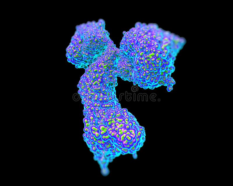
Free with trial IgG1, a subclass of Immunoglobulin G, an antibody. 3d model. G protein illustrations IgG1, a subclass of Immunoglobulin G, an antibody.
Free with trial Human k-opioid receptor, a member of a group of inhibitory G protein-coupled receptors with opioids as ligands. Distributed widely in the brain, and are found in the spinal cord and digestive tract. G protein illustrations Human k-opioid receptor, a member of a group of inhibitory G pro
Free with trial GABA B receptors are G protein-coupled receptors, also called metabotropic receptors. Binding of an agonist here baclofen, red leads to a G-protein coupled C-AMP signaling pathway. Source: PDB entries 7eb2, 6r3q. G protein illustrations Activation of the GABA B receptor by an agonist leads to a cAMP signal cascade. GABA B receptors are G protein-coupled receptors, also called metabotropic receptors. Binding of an agonist here baclofen, red leads to a G-protein coupled C-AMP signaling pathway. Source: PDB entries 7eb2, 6r3q
Free with trial Phosphatidylinositol-4-phosphate 3-kinase C2 domain-containing gamma polypeptide, a protein which belongs to the phosphoinositide 3-kinase (PI3K) family. 3d rendering. G protein illustrations Phosphatidylinositol-4-phosphate 3-kinase C2 domain-containing g
Free with trial Glycine Gly or G, is the amino acid. Structural chemical formula and molecule model. Vector illustration. G protein vectors Glycine Gly or G, is the amino acid. Structural chemical form
Free with trial Chemical structure of a molecule of glycine (gly, g). Glycine is the smallest amino acid found in proteins. It is achiral and functions as an inhibitory neurotransmitter. G protein illustrations Glycine (Gly, G) molecule. Chemical structure of a molecule of glycine (gly, g). Glycine is the smallest amino acid found in proteins. It is achiral and functions as an inhibitory neurotransmitter.
Free with trial Different pills stack in shape of letter G. suitable for medicine, healthcare and science themes. 3D illustration with blue background. G protein illustrations Different pills stack in shape of letter G
Free with trial Monoclonal antibody (Immunoglobulin G, IgG2a, mAb) molecule, chemical structure. Most current biotech drugs are monoclonal antibodies. Two surface representations. Heavy and light chains rendered in different colors. G protein vectors Monoclonal antibody (Immunoglobulin G, IgG2a, mAb) molecule, chemical structure. Most current biotech drugs are monoclonal
Free with trial The melanocortin receptor 4 is crucial for appetite, energy homeostasis and body-weight control in the central nervous system. Binding of the agonist setmelatonide to the melacortin receptor leads to a G-protein coupled C-AMP signaling pathway. Source: PDB entries 7aue, 6r3q,. Israeli, H. et al. 2021 Science. G protein illustrations Activation of the melanocortin receptor by an anti-obesity drug. The melanocortin receptor 4 is crucial for appetite, energy homeostasis and body-weight control in the central nervous system. Binding of the agonist setmelatonide to the melacortin receptor leads to a G-protein coupled C-AMP signaling pathway. Source: PDB entries 7aue, 6r3q,. Israeli, H. et al. 2021 Science
Free with trial Macrophages are important cells of the immune system that are formed in response to an infection or accumulating damaged or dead cells. A macrophage has the ability to locate and `eat` particles such as bacteria, viruses, fungi, and parasites. Macrophages are born from white blood cells called monocytes, which are produced by stem cells in our bone marrow. In biochemistry and pharmacology, a receptor is a protein molecule that receives chemical signals from outside a cell. When such chemical signals bind to a receptor, they cause some form of cellular/tissue response, e. g. a change in the electrical activity of a cell. G protein illustrations Macrophage
Free with trial Macrophages are important cells of the immune system that are formed in response to an infection or accumulating damaged or dead cells. A macrophage has the ability to locate and `eat` particles such as bacteria, viruses, fungi, and parasites. Macrophages are born from white blood cells called monocytes, which are produced by stem cells in our bone marrow. In biochemistry and pharmacology, a receptor is a protein molecule that receives chemical signals from outside a cell. When such chemical signals bind to a receptor, they cause some form of cellular/tissue response, e. g. a change in the electrical activity of a cell. G protein illustrations Macrophage
Free with trial Macrophages are important cells of the immune system that are formed in response to an infection or accumulating damaged or dead cells. A macrophage has the ability to locate and `eat` particles such as bacteria, viruses, fungi, and parasites. Macrophages are born from white blood cells called monocytes, which are produced by stem cells in our bone marrow. In biochemistry and pharmacology, a receptor is a protein molecule that receives chemical signals from outside a cell. When such chemical signals bind to a receptor, they cause some form of cellular/tissue response, e. g. a change in the electrical activity of a cell. G protein illustrations Macrophage
Free with trial Interleukin 6 3d model, secreted by T cells and macrophages to stimulate immune response, e. g. during infection and after trauma, especially burns or other tissue damage leading to inflammation. G protein illustrations Interleukin 6 3d model, secreted by T cells and macrophages
Free with trial Macrophages are important cells of the immune system that are formed in response to an infection or accumulating damaged or dead cells. A macrophage has the ability to locate and `eat` particles such as bacteria, viruses, fungi, and parasites. Macrophages are born from white blood cells called monocytes, which are produced by stem cells in our bone marrow. In biochemistry and pharmacology, a receptor is a protein molecule that receives chemical signals from outside a cell. When such chemical signals bind to a receptor, they cause some form of cellular/tissue response, e. g. a change in the electrical activity of a cell. G protein illustrations Macrophage and Receptors. Macrophages are important cells of the immune system that are formed in response to an infection or accumulating damaged or dead cells. A macrophage has the ability to locate and `eat` particles such as bacteria, viruses, fungi, and parasites. Macrophages are born from white blood cells called monocytes, which are produced by stem cells in our bone marrow. In biochemistry and pharmacology, a receptor is a protein molecule that receives chemical signals from outside a cell. When such chemical signals bind to a receptor, they cause some form of cellular/tissue response, e.g. a change in the electrical activity of a cell.
Free with trial Macrophages are important cells of the immune system that are formed in response to an infection or accumulating damaged or dead cells. A macrophage has the ability to locate and `eat` particles such as bacteria, viruses, fungi, and parasites. Macrophages are born from white blood cells called monocytes, which are produced by stem cells in our bone marrow. In biochemistry and pharmacology, a receptor is a protein molecule that receives chemical signals from outside a cell. When such chemical signals bind to a receptor, they cause some form of cellular/tissue response, e. g. a change in the electrical activity of a cell. G protein illustrations Internal Structure of Macrophage. Macrophages are important cells of the immune system that are formed in response to an infection or accumulating damaged or dead cells. A macrophage has the ability to locate and `eat` particles such as bacteria, viruses, fungi, and parasites. Macrophages are born from white blood cells called monocytes, which are produced by stem cells in our bone marrow. In biochemistry and pharmacology, a receptor is a protein molecule that receives chemical signals from outside a cell. When such chemical signals bind to a receptor, they cause some form of cellular/tissue response, e.g. a change in the electrical activity of a cell.
Free with trial Adenine, A, Ade, chemical formula and skeletal structure. Nucleobase and a purine derivative, one of four in the nucleic acid of DNA and RNA, represented by letters G, C, A and T. Illustration. Vector. G protein vectors Adenine, A, Ade, nucleobase, chemical formula and skeletal structure. Adenine, A, Ade, chemical formula and skeletal structure. Nucleobase and a purine derivative, one of four in the nucleic acid of DNA and RNA, represented by letters G, C, A and T. Illustration. Vector
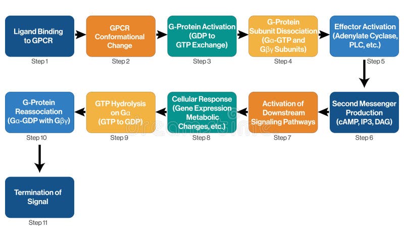
Free with trial This vector illustration presents an informative flowchart of G-Protein Coupled Receptor (GPCR) signaling processes, set on a clean white background. The visual details the key steps in GPCR signaling, from receptor activation through signal transduction pathways. Designed for clarity and educational use, this flowchart helps illustrate the intricate mechanisms of GPCR function and their impact on cellular signaling and response. G protein vectors Illustrative Flowchart of G Protein Coupled Receptor Signaling Processes with White Background. This vector illustration presents an informative flowchart of G-Protein Coupled Receptor (GPCR) signaling processes, set on a clean white background. The visual details the key steps in GPCR signaling, from receptor activation through signal transduction pathways. Designed for clarity and educational use, this flowchart helps illustrate the intricate mechanisms of GPCR function and their impact on cellular signaling and response.
Free with trial Protein rich Foods A selection of protein rich foods e g lentils chickpeas Greek yogurt isolated on white Description A range of foods high in protein essential for building and repairing tissues. G protein illustrations Protein rich Foods A selection of protein rich foods e g lentils
Free with trial This image features a black jar of Luxury Protein Powder isolated on a white background. The jar has a black lid and contains 2. 83 lbs (1287 g) of protein powder. The image is on a transparent background, making it suitable for various digital applications. G protein illustrations Luxury protein powder jar isolated on white background. This image features a black jar of Luxury Protein Powder isolated on a white background. The jar has a black lid and contains 2.83 lbs (1287 g) of protein powder. The image is on a transparent background, making it suitable for various digital applications
Free with trial The image features two black protein supplement jars placed side by side against a transparent background. The jars have white text and labels indicating they are protein supplements from the brand 'NOX GEAS'. Each jar has a black lid and contains 1. 8 lbs (816 g) of protein powder. G protein illustrations Two black protein supplement jars isolated on white background. The image features two black protein supplement jars placed side by side against a transparent background. The jars have white text and labels indicating they are protein supplements from the brand 'NOX GEAS'. Each jar has a black lid and contains 1.8 lbs (816 g) of protein powder
Free with trial A colorful and perfectly plated bowl of fresh salad. Contains organic mixed greens, various vegetables, and visible protein (e. g. , grilled chicken or chickpeas). Presented in a clean, minimalist setting, ideal for themes of healthy eating, diet, nutrition, wellness, and restaurant menus. G protein illustrations Vibrant Fresh Salad Bowl with Organic Vegetables and Protein on Minimalist Background. A colorful and perfectly plated bowl of fresh salad. Contains organic mixed greens, various vegetables, and visible protein (e.g., grilled chicken or chickpeas). Presented in a clean, minimalist setting, ideal for themes of healthy eating, diet, nutrition, wellness, and restaurant menus.
Free with trial A 3d illustrates a genetic mutation within a DNA sequence. The word "MUTATION" is highlighted in a glowing rectangular box against a background of repetitive DNA base pair letters (A, T, C, G). G protein illustrations A \'MUTATION\' word and DNA base pair letters (A, T, C, G) as a background. A 3d illustrates a genetic mutation within a DNA sequence. The word "MUTATION" is highlighted in a glowing rectangular box against a background of repetitive DNA base pair letters (A, T, C, G)
Free with trial A 3d illustrates a genetic mutation within a DNA sequence. The word "MUTATION" is highlighted in a glowing rectangular box against a background of repetitive DNA base pair letters (A, T, C, G). G protein illustrations A \'MUTATION\' word and DNA base pair letters (A, T, C, G) as a background. A 3d illustrates a genetic mutation within a DNA sequence. The word "MUTATION" is highlighted in a glowing rectangular box against a background of repetitive DNA base pair letters (A, T, C, G)
Free with trial A piece of raw meat shaped like the letter "G. G protein illustrations A piece of raw meat shaped like the letter \'G
Free with trial A whimsical 3D rendering of the letter 'G' shaped like a miniature grocery store. The interior is filled with baskets of fresh fruits and displays of baked goods. Two small shopping carts are positioned near the entrance. This playful image can be used to represent grocery shopping, healthy eating, or educational themes for children. G protein illustrations Letter G Grocery Store Concept. A whimsical 3D rendering of the letter 'G' shaped like a miniature grocery store. The interior is filled with baskets of fresh fruits and displays of baked goods. Two small shopping carts are positioned near the entrance. This playful image can be used to represent grocery shopping, healthy eating, or educational themes for children.
Free with trial Microscopic view of immunoglobulin G antibodies. G protein illustrations Microscopic view of immunoglobulin G antibodies.
Free with trial A stylized letter \'G\' made from salmon, showcasing food art and creativity. G protein illustrations A stylized letter \'G\' made from salmon, showcasing food art and creativity
Free with trial Illustration showing a central gastric D-cell releasing somatostatin to inhibit G, ECL, and parietal cells via SSTR signaling, reducing acid output. This illustration was created using AI Technology. G protein illustrations Somatostatin D-cell paracrine inhibition of G, ECL, parietal cells. Illustration showing a central gastric D-cell releasing somatostatin to inhibit G, ECL, and parietal cells via SSTR signaling, reducing acid output. This illustration was created using AI Technology
Free with trial A scientific diagram the precipitation reaction between antibodies and antigens. Blue Y-shaped antibody molecules with 'VL' labels are shown binding to red cluster-like antigen molecules. Arrows indicate the interaction and the formation of a complex. Labels identify 'Immunoglobulin G (GG)', 'Antibodies', 'Antigens', and 'Precipitation Reaction. G protein illustrations Antibody antigen precipitation reaction diagram immunoglobulin G. A scientific diagram the precipitation reaction between antibodies and antigens. Blue Y-shaped antibody molecules with 'VL' labels are shown binding to red cluster-like antigen molecules. Arrows indicate the interaction and the formation of a complex. Labels identify 'Immunoglobulin G (GG)', 'Antibodies', 'Antigens', and 'Precipitation Reaction
Free with trial An artistic arrangement of different grains and legumes, including wheat, rice, barley, and beans, meticulously placed to form the shape of the letter G. The colors range from golden yellows to deep browns, creating a visually appealing and nutritious display. G protein illustrations A vibrant display of various grains and legumes forming the letter g. An artistic arrangement of different grains and legumes, including wheat, rice, barley, and beans, meticulously placed to form the shape of the letter G. The colors range from golden yellows to deep browns, creating a visually appealing and nutritious display
Free with trial A hand is adding white powdered supplement into a blender jar containing fresh spinach leaves, cherry tomatoes, and cucumber slices. The scoop is metallic, and the jar is transparent. Nearby, a bowl of blueberries and a glass of water are on the countertop. The setting is a clean, white kitchen surface, suggesting preparation for a healthy smoothie or drink. G protein illustrations Hand using a measuring scoop to carefully drop white powdered supplement (e.g., protein, collagen). A hand is adding white powdered supplement into a blender jar containing fresh spinach leaves, cherry tomatoes, and cucumber slices. The scoop is metallic, and the jar is transparent. Nearby, a bowl of blueberries and a glass of water are on the countertop. The setting is a clean, white kitchen surface, suggesting preparation for a healthy smoothie or drink.
Free with trial This vector illustration features a comprehensive flowchart of GPCR signaling pathways, depicted against a crisp white background. The diagram illustrates the detailed process of GPCR-mediated signal transduction, including receptor activation, G-protein interactions, and downstream effects. Ideal for use in educational materials and scientific presentations, this flowchart provides a clear and accurate depiction of the complex signaling mechanisms involving G-Protein Coupled Receptors. G protein vectors Flowchart of GPCR Signaling Pathways and Mechanisms Illustrated on White Background. This vector illustration features a comprehensive flowchart of GPCR signaling pathways, depicted against a crisp white background. The diagram illustrates the detailed process of GPCR-mediated signal transduction, including receptor activation, G-protein interactions, and downstream effects. Ideal for use in educational materials and scientific presentations, this flowchart provides a clear and accurate depiction of the complex signaling mechanisms involving G-Protein Coupled Receptors.
Free with trial 3D Gaussian surface and cartoon models, chain id color scheme, PDB 7t2h, white background. G protein illustrations CryoEM structure of mu-opioid receptor (light green) - Gi protein complex bound to lofentanil (pink). 3D Gaussian surface and cartoon models, chain id color scheme, PDB 7t2h, white background
Free with trial Each molecule of DNA is a double helix formed from two complementary strands of nucleotides held together by hydrogen bonds between G-C and A-T base pairs. Duplication of the genetic information occurs by the use of one DNA strand as a template for formation of a complementary strand. G protein illustrations Double Helix Structure of the DNA Strand. Each molecule of DNA is a double helix formed from two complementary strands of nucleotides held together by hydrogen bonds between G-C and A-T base pairs. Duplication of the genetic information occurs by the use of one DNA strand as a template for formation of a complementary strand.
Free with trial Each molecule of DNA is a double helix formed from two complementary strands of nucleotides held together by hydrogen bonds between G-C and A-T base pairs. Duplication of the genetic information occurs by the use of one DNA strand as a template for formation of a complementary strand. G protein illustrations Double Helix Structure of the DNA Strand. Each molecule of DNA is a double helix formed from two complementary strands of nucleotides held together by hydrogen bonds between G-C and A-T base pairs. Duplication of the genetic information occurs by the use of one DNA strand as a template for formation of a complementary strand.
Free with trial A detailed 3D illustration depicts multiple IgG antibodies, also known as immunoglobulins G, floating in a fluid, cellular environment. The antibodies, characterized by their distinctive Y-shape, are rendered with translucent blue textures, highlighting their protein structure. Labels indicate 'IgG' and 'Antigen-binding site', emphasizing their crucial role in the immune system's defense mechanism. This visual is ideal for scientific, medical, and educational content related to immunology, biotechnology, and disease research. G protein illustrations 3D Render of IgG Antibodies in Aqueous Environment. A detailed 3D illustration depicts multiple IgG antibodies, also known as immunoglobulins G, floating in a fluid, cellular environment. The antibodies, characterized by their distinctive Y-shape, are rendered with translucent blue textures, highlighting their protein structure. Labels indicate 'IgG' and 'Antigen-binding site', emphasizing their crucial role in the immune system's defense mechanism. This visual is ideal for scientific, medical, and educational content related to immunology, biotechnology, and disease research.
Free with trial Traditional Polish curd cheese, Twar�g, served in a rustic bowl and garnished with fresh herbs. Showcases the creamy texture of this popular dairy product in Polish cuisine. G protein illustrations . Traditional Polish curd cheese, Twar�g, served in a rustic bowl and garnished with fresh herbs. Showcases the creamy texture of this popular dairy product in Polish cuisine
Free with trial A three-dimensional rendering of the letter G, creatively formed from a piece of Swiss cheese with its characteristic holes. The image is perfect for illustrating food, alphabet, or design concepts. G protein illustrations 3D Render of Letter G Shaped Swiss Cheese. A three-dimensional rendering of the letter G, creatively formed from a piece of Swiss cheese with its characteristic holes. The image is perfect for illustrating food, alphabet, or design concepts.
Free with trial A 3d illustrates a genetic mutation within a DNA sequence. The word "MUTATION" is highlighted in a glowing rectangular box against a background of repetitive DNA base pair letters (A, T, C, G). G protein illustrations A \'MUTATION\' word and DNA base pair letters (A, T, C, G) as a background. A 3d illustrates a genetic mutation within a DNA sequence. The word "MUTATION" is highlighted in a glowing rectangular box against a background of repetitive DNA base pair letters (A, T, C, G)
Free with trial A sausage shaped like the letter \'G,\' showcasing a unique food presentation. G protein illustrations A sausage shaped like the letter \'G,\' showcasing a unique food presentation
Free with trial A 3D rendering of a DNA double helix structure, showcasing its twisted ladderlike form with labeled nucleotide bases A, T, C, G on a dark blue background. G protein illustrations A 3D rendering of a DNA double helix structure, showcasing its twisted ladderlike form with labeled nucleotide bases A, T, C, G on
Free with trial Two vibrant Vietnamese spring rolls, also known as G? i cu? n or summer rolls, are beautifully presented on a clean white background. Each roll is generously filled with fresh, colorful ingredients including succulent shrimp, crisp carrots, cucumber, white radish, and leafy green herbs, all wrapped in delicate, translucent rice paper. Drizzled with a rich peanut sauce and garnished with crushed peanuts, this healthy and appetizing Asian dish is perfect for a light meal, appetizer, or a refreshing snack. G protein illustrations Fresh Vietnamese Spring Rolls with Peanut Sauce on White. Two vibrant Vietnamese spring rolls, also known as G?i cu?n or summer rolls, are beautifully presented on a clean white background. Each roll is generously filled with fresh, colorful ingredients including succulent shrimp, crisp carrots, cucumber, white radish, and leafy green herbs, all wrapped in delicate, translucent rice paper. Drizzled with a rich peanut sauce and garnished with crushed peanuts, this healthy and appetizing Asian dish is perfect for a light meal, appetizer, or a refreshing snack.
Free with trial Top-down image of capital letters M and G formed from magnesium-rich foods on white background, including seeds, nuts, avocado, banana, spinach, black beans and salmon. G protein illustrations Top-down image of capital letters M and G formed from magnesium-rich foods on white background
Free with trial Top-down image of capital letters M and G formed from magnesium-rich foods on white background, including seeds, almonds, avocado, chocolate, spinach, black beans and salmon. G protein illustrations Top-down image of capital letters M and G formed from magnesium-rich foods on white background, including seeds, almonds, avocado
Free with trial A bowl of sweet laddu, also known as sweets or traditional Indian confectionery made from flour and g embracing the golden hues of Diwali. G protein illustrations A bowl of sweet laddu, also known as sweets or traditional Indian confectionery made from flour and g embracing the golden hues of
Free with trial A bowl of sweet laddu, also known as sweets or traditional Indian confectionery made from flour and g embracing the golden hues of Diwali. G protein illustrations A bowl of sweet laddu, also known as sweets or traditional Indian confectionery made from flour and g embracing the golden hues of
Free with trial Receptor word block on white background. G protein illustrations Receptor word block on white
Free with trial Nutrition facts label features a 100 g serving size. Key nutrients include 250 kcal from calories, with 10 kcal from fat. Cholesterol is 50 mg (28% DV), sodium 150 mg (15% DV), and total carbohydrates 10 g (3% DV) with 5 g dietary fiber and 3 g sugars. Protein provides 16% DV. Vitamins and minerals include Vitamin A (1% DV), Vitamin C (3% DV), calcium (2% DV), and iron (2% DV). Total fat, saturated fat, trans fat, and exact protein amount are unspecified. G protein vectors Nutrition Facts Food Label. Nutrition facts label features a 100 g serving size. Key nutrients include 250 kcal from calories, with 10 kcal from fat. Cholesterol is 50 mg (28% DV), sodium 150 mg (15% DV), and total carbohydrates 10 g (3% DV) with 5 g dietary fiber and 3 g sugars. Protein provides 16% DV. Vitamins and minerals include Vitamin A (1% DV), Vitamin C (3% DV), calcium (2% DV), and iron (2% DV). Total fat, saturated fat, trans fat, and exact protein amount are unspecified.
Free with trial Nutritional label for coconut sugar shows values per teaspoon serving: 15 kcal, 0 g total fat, 0 mg sodium, 4 g total carbohydrates (all sugars), 0 g protein, and 10 mg potassium. G protein illustrations Coconut Sugar Nutritional Label. Nutritional label for coconut sugar shows values per teaspoon serving: 15 kcal, 0 g total fat, 0 mg sodium, 4 g total carbohydrates (all sugars), 0 g protein, and 10 mg potassium.
Free with trial A tantalizing close-up of a steak sizzling on a grill, seasoned generously with spices. The background features vibrant flames, enhancing the cooking process. The detail captures the char and smoky essence of the food. The image conveys the essence of outdoor grilling and delicious, smoky flavors, g. G protein illustrations Steak on Fire. A tantalizing close-up of a steak sizzling on a grill, seasoned generously with spices. The background features vibrant flames, enhancing the cooking process. The detail captures the char and smoky essence of the food. The image conveys the essence of outdoor grilling and delicious, smoky flavors, g
Free with trial Chicken is a dietary, protein-rich (about 20-25 g per 100 g) product that is easily digestible and contains B vitamins, phosphorus, selenium and zinc. Low-calorie white meat (breast) is ideal for dieting, while dark meat (thigh) contains more fat and iron. Regular consumption supports muscles, heart and immunity. G protein illustrations Fresh raw chicken meat isolated on white background, macro. Chicken is a dietary, protein-rich (about 20-25 g per 100 g) product that is easily digestible and contains B vitamins, phosphorus, selenium and zinc. Low-calorie white meat (breast) is ideal for dieting, while dark meat (thigh) contains more fat and iron. Regular consumption supports muscles, heart and immunity.
Free with trial A close-up, mouth-watering view of flaky, layered flatbread served alongside a rich, vibrant curry. The bread's texture is highlighted by its numerous thin layers, suggesting a delightful chewiness. The curry, featuring tender chunks of meat in a deep red sauce, is presented in a rustic clay bowl, g. G protein illustrations Layered flatbread with curry. A close-up, mouth-watering view of flaky, layered flatbread served alongside a rich, vibrant curry. The bread's texture is highlighted by its numerous thin layers, suggesting a delightful chewiness. The curry, featuring tender chunks of meat in a deep red sauce, is presented in a rustic clay bowl, g