Free with trial Set of human cells, eps8, gradient and mesh printing compatible. Goblet cells vectors Human cell collection. Set of human cells, eps8, gradient and mesh printing compatible
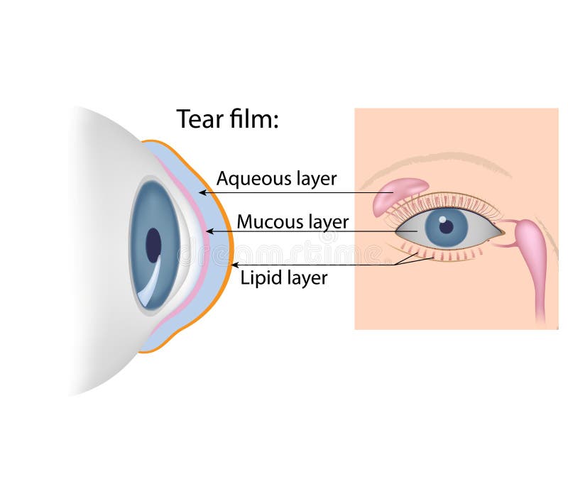
Free with trial The lacrimal gland, meibomian glands and conjunctival goblet cells secretion. Goblet cells vectors Tear film formation. The lacrimal gland, meibomian glands and conjunctival goblet cells secretion
Free with trial Anatomy of human cells (useful for education in schools and clinics ) - vector illustration. Goblet cells illustrations Anatomy of human cells
Free with trial The lacrimal gland, meibomian glands and conjunctival goblet cells secretion, anterior and lateral view. Goblet cells vectors Tears chemical composition. The lacrimal gland, meibomian glands and conjunctival goblet cells secretion, anterior and lateral view
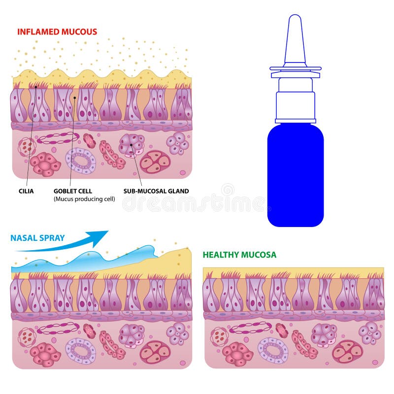
Free with trial Inflamed and normal nasal mucosa cells and micro cilia vector scheme with nasal spray effect and bottle. Goblet cells vectors Nasal mucosa cells and micro cilia vector scheme
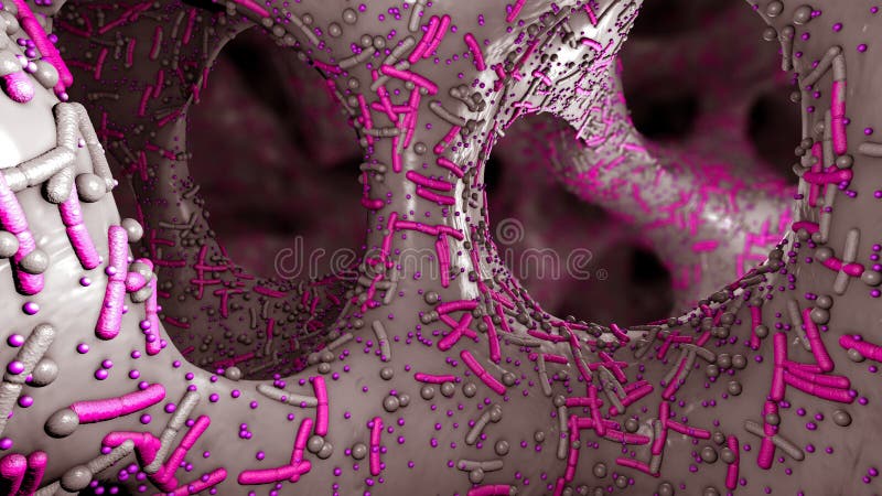
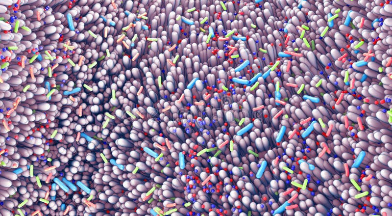
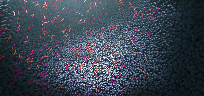
Free with trial Different germs in the human intestines called microbiome - 3d illustration. Goblet cells illustrations Germs in the human intestines called microbiome
Free with trial Nasal mucosa cells. Nasal secretions. Ciliated, basal and goblet cells. Olfactory epithelium. Cells act as a low resistance filter. Vector illustration. Goblet cells vectors Nasal mucosa cells. Nasal secretions. Ciliated, basal and goblet cells
Free with trial Wall of small intestine with villi and epithelial cells Enterocyte, Goblet and Paneth cell. Goblet cells vectors Small intestine with villi and epithelial cells
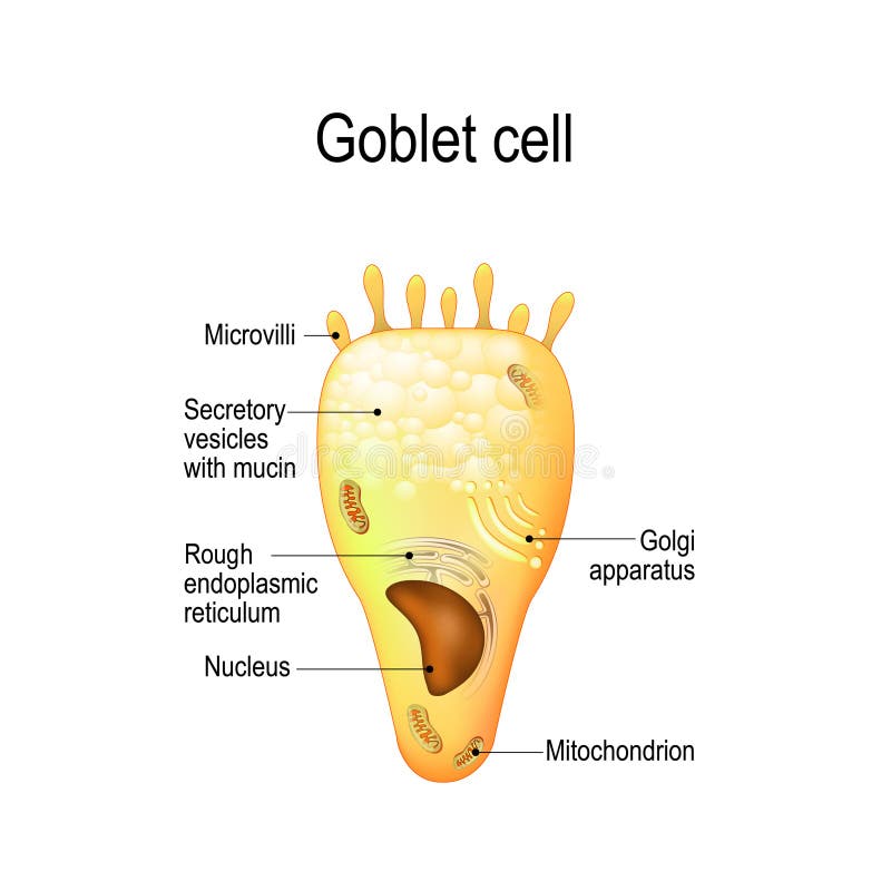
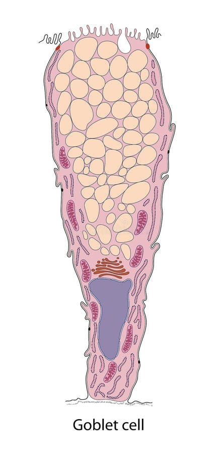
Free with trial Goblet cell. simple columnar epithelial cell for secrete mucus. They are found inside the trachea, bronchi, small and large intestine, and conjunctiva in the eyes. Structure cell nucleus and other organelles. Goblet cells vectors Goblet cell. Structure cell nucleus and other organelles. Goblet cell. simple columnar epithelial cell for secrete mucus. They are found inside the trachea, bronchi, small and large intestine, and conjunctiva in the eyes. Structure cell nucleus and other organelles.
Free with trial The Airway Epithelium. Human airway epithelial cells respond to environmental differential. Goblet cells vectors The Airway Epithelium. Health care illustration. The Airway Epithelium. Human airway epithelial cells respond to environmental differential
Free with trial Intestinal villi in the surface area of intestinal walls. Folds, villus, microvilli, epithelial and goblet cells. Small intestine anatomical poster. Digestive system medical flat vector illustration. Goblet cells vectors Intestinal villi anatomy. Intestinal villi in the surface area of intestinal walls. Folds, villus, microvilli, epithelial and goblet cells. Small intestine anatomical poster. Digestive system medical flat vector illustration.
Free with trial Nasal mucosa anatomy. Nasal mucous membrane lining the respiratory tract. Ciliated, basal and goblet cells. Flat vector illustration. Goblet cells vectors Nasal mucosa anatomy. Nasal mucous membrane lining the respiratory tract
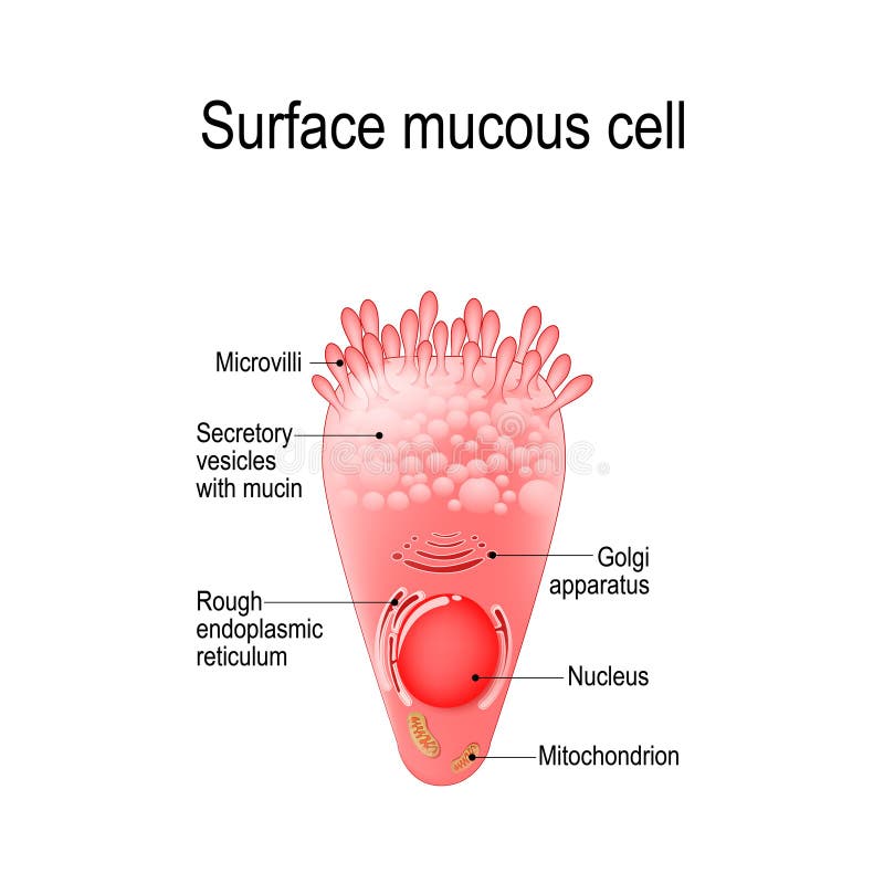
Free with trial Surface mucous cell is a foveolar mucus-producing cell that covering the inside of the stomach. Structure cell: golgi apparatus, secretory vesicle, mucin, nucleus, mitochondrion, microvilli. Goblet cells vectors Surface mucous cell
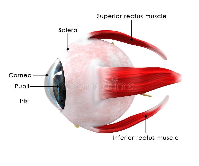
Free with trial The human eye is an organ that reacts to light and has several purposes. As a sense organ, the mammalian eye allows vision. Rod and cone cells in the retina allow conscious light perception and vision including color differentiation and the perception of depth. Goblet cells illustrations Human Eye
Free with trial The human eye is an organ that reacts to light and has several purposes. As a sense organ, the mammalian eye allows vision. Rod and cone cells in the retina allow conscious light perception and vision including color differentiation and the perception of depth. Goblet cells illustrations Human eye
Free with trial 3d illustration of microscopic closeup showing viruses and intestine villus into digestive tract. Goblet cells illustrations Microscopic closeup showing viruses and intestine villus into digestive tract
Free with trial Different germs in the human intestines called microbiome - 3d illustration. Goblet cells illustrations Germs in the human intestines called microbiome
Free with trial Bacteria as part of the intestinal microbiome in the digestive tract - 3d illustration. Goblet cells illustrations Bacteria as part of the intestinal microbiome in the digestive tract
Free with trial 3d illustration of microscopic closeup of intestine villus. Goblet cells illustrations 3d illustration of microscopic closeup of intestine villus
Free with trial 3d illustration of microscopic closeup of intestine villus. Goblet cells illustrations 3d illustration of microscopic closeup of intestine villus
Free with trial Different germs in the human intestines called microbiota - 3d illustration. Goblet cells illustrations Different germs in the human intestines called microbiota
Free with trial Respiratory Epithelium Cell Structure Illustration. Ciliated, Basal, and Goblet Cells Diagram. Mucociliary Epithelium Cross Section Illustration. Goblet cells vectors Respiratory Epithelium Cell Structure Illustration. Ciliated, Basal, and Goblet Cells Diagram. Mucociliary Epithelium
Free with trial Small intestine anatomy. Layers of the small bowel Mucosa, Submucosa, muscularis externa, serosa. Cross section of Intestinal villi. Close-up of Stem cell, Goblet and Paneth cells, Enterocyte. Detailed Vector poster. Goblet cells vectors Small intestine anatomy. Layers and cells. small intestine anatomy. Layers of the small bowel Mucosa, Submucosa, muscularis externa, serosa. Cross section of Intestinal villi. Close-up of Stem cell, Goblet and Paneth cells, Enterocyte. Detailed Vector poster
Free with trial Epithelial Tissue with Goblet Cells. Goblet Cell Structure Illustration. Mucus Secreting. Human Goblet Cell Anatomy Educational Chart. Goblet cells vectors Epithelial Tissue with Goblet Cells. Goblet Cell Structure Illustration. Mucus Secreting. Human Goblet Cell Anatomy
Free with trial Goblet cells are a type of intestinal mucosal epithelial cell, which serves as the primary site for nutrient digestion and mucosal absorption. The primary function of goblet cells is to synthesize and secrete mucus. As the primary secretory cell in the superficial epithelium of large airways, goblet cells secrete. Goblet cells illustrations Diagram of goblet cell. Cells are located in the gastric glands into stomach. Goblet cells are a type of intestinal mucosal epithelial cell, which serves as the primary site for nutrient digestion and mucosal absorption. The primary function of goblet cells is to synthesize and secrete mucus. As the primary secretory cell in the superficial epithelium of large airways, goblet cells secrete
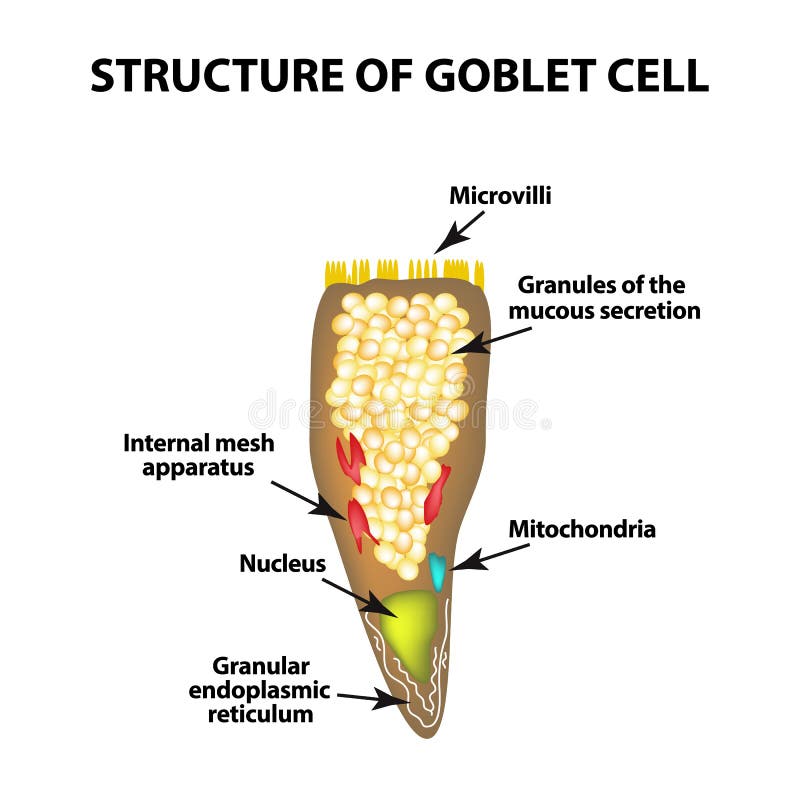
Free with trial Structure Goblet cells of the intestine. Infographics. Vector illustration on isolated background. Goblet cells vectors Structure Goblet cells of the intestine. Infographics. Vector illustration on isolated background
Free with trial Structure Goblet cells of the intestine. Infographics. Vector illustration on isolated background. Goblet cells vectors Structure Goblet cells of the intestine. Infographics. Vector illustration on isolated background
Free with trial Intestinal villi in the surface of intestinal. Epithelial and goblet cells. Healthy digestive system, with microorganisms maintenaning intestinal cells. Flat vector illustration. Goblet cells illustrations Intestinal villi in the surface of intestinal. Epithelial and goblet cells.
Free with trial Intestinal epithelium showing functions of enterocytes, dendritic cell, goblet cell, neuroendocrine cell, stem cell, mucosa, and mucus. Source: Kong, S. , Zhang, Y. H. , & Zhang, W. 2018. Regulation of intestinal epithelial cells properties and functions by amino acids. BioMed research international, 2018. Goblet cells illustrations Intestinal epithelium showing functions of cells. Intestinal epithelium showing functions of enterocytes, dendritic cell, goblet cell, neuroendocrine cell, stem cell, mucosa, and mucus. Source: Kong, S., Zhang, Y. H., & Zhang, W. 2018. Regulation of intestinal epithelial cells properties and functions by amino acids. BioMed research international, 2018.
Free with trial Olfactory Bulb Anatomy. Epithelium receptor cells. Nasal mucosa cells. Goblet cells vectors Olfactory Bulb Anatomy. Epithelium receptor cells. Nasal mucosa cells
Free with trial Histamine. Immune response and Allergic reaction. Histamine-releasing cells produce of Histamine then Stomach secretes gastric acid, smooth muscle is reduced and in the bronchi there are difficulties with breathing, Goblet cells secrete mucus, and Skin becomes redness and itching. Vector poster. Isometric flat illustration. Goblet cells vectors Histamine. Immune response and Allergic reaction
Free with trial Intestinal epithelium showing enterocytes, dendritic cell, goblet cell, neuroendocrine cell, stem cell, mucosa, and mucus. Goblet cells illustrations Intestinal epithelium showing enterocytes and other cells. Intestinal epithelium showing enterocytes, dendritic cell, goblet cell, neuroendocrine cell, stem cell, mucosa, and mucus.
Free with trial Goblet cells are specialized epithelial cells found in the lining of various organs, particularly in the respiratory and digestive tracts. They are named for their goblet-like shape, with a wide base and a narrower opening at the top. Goblet cells are responsible for secreting mucins, which are glycoproteins that form mucous. Goblet cells vectors Goblet cell
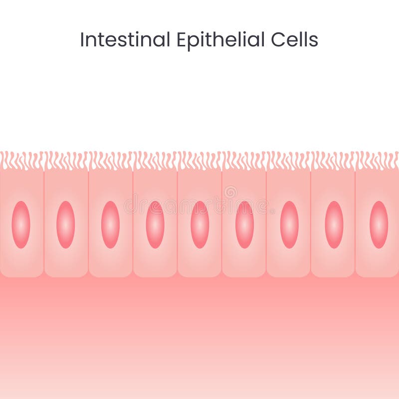
Free with trial Vector illustration science background of the epithelial cells that line the large and small intestines. Goblet cells vectors Intestinal Epithelial Cells background. vector illustration science background of the epithelial cells that line the large and small intestines
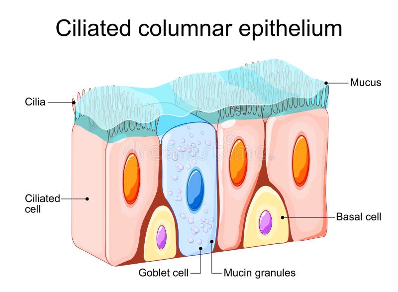
Free with trial Nasal epithelium. Ciliated columnar epithelium. epithelial cells forms the lining of the stomach and intestines, duodenum, fallopian tubes, uterus, central canal of the spinal cord, nose, ears and the taste buds. Ciliated cells. Respiratory defense mechanisms. Vector poster. Isometric Flat illustration. Goblet cells vectors Nasal epithelium. Ciliated columnar epithelium
Free with trial Ciliated columnar epithelium. Epithelial cells forms the lining of the stomach and intestines, duodenum, fallopian tubes, uterus, central canal of the spinal cord, nose, ears and the taste buds. Nasal epithelium. Ciliated cells. Respiratory defense mechanisms. Vector illustration. Medical poster. Schematic diagram. Goblet cells vectors Ciliated columnar epithelium. Nasal epithelium. Ciliated columnar epithelium. Epithelial cells forms the lining of the stomach and intestines, duodenum, fallopian tubes, uterus, central canal of the spinal cord, nose, ears and the taste buds. Nasal epithelium. Ciliated cells. Respiratory defense mechanisms. Vector illustration. Medical poster. Schematic diagram
Free with trial Pseudostratified epithelium is a type of epithelium that, though comprising only a single layer of cells. Goblet cells illustrations Pseudostratified epithelium is a type of epithelium that
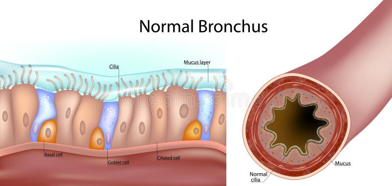
Free with trial Structure of a normal human bronchus. Cross-section of the airway and a close-up of the pseudostratified ciliated epithelium with ciliated cells, goblet cells, basal cells, and the mucus layer. Goblet cells vectors Structure of a normal human bronchus. Cross-section of the airway and a close-up of the pseudostratified ciliated epithelium with
Free with trial Intestinal villi in the surface area of intestinal walls. Folds, villus, microvilli, epithelial and goblet cells. Small intestine anatomical poster. Digestive system medical flat vector illustration. Goblet cells vectors Intestinal villi anatomy. Intestinal villi in the surface area of intestinal walls. Folds, villus, microvilli, epithelial and goblet cells. Small intestine anatomical poster. Digestive system medical flat vector illustration.
Free with trial Intestinal epithelial cells with capillary network. Enterocyte and Goblet cell. Vector illustration. Goblet cells vectors Intestinal villi. Microvilli. Intestinal epithelium. Villi absorb nutrients from the food. Intestinal epithelial cells with capillary network. Enterocyte and Goblet cell. Vector illustration.
Free with trial Nasal epithelium anatomical poster. Human respiratory system concept. Mucus, goblet, ciliated and basal cells. Smell organs respiratory system. Medical flat vector illustration for clinic or education. Goblet cells vectors Nasal epithelium concept. Nasal epithelium anatomical poster. Human respiratory system concept. Mucus, goblet, ciliated and basal cells. Smell organs respiratory system. Medical flat vector illustration for clinic or education
Free with trial Intestinal villi in the surface area of intestinal walls. Folds, villus, microvilli, epithelial and goblet cells. Small intestine anatomical poster. Digestive system medical flat vector illustration. Goblet cells vectors Intestinal villi anatomy. Intestinal villi in the surface area of intestinal walls. Folds, villus, microvilli, epithelial and goblet cells. Small intestine anatomical poster. Digestive system medical flat vector illustration.
Free with trial Single layer of cells. Each cell is in contact with the basement membrane. Found in respiratory tract, prostate, endometrium. Vector illustration. Goblet cells vectors Pseudostratified epithelium. Epithelial tissue types. Single layer of cells. Each cell is in contact with the basement membrane. Found in respiratory tract, prostate, endometrium. Vector illustration
Free with trial Intestinal villi in the surface area of intestinal walls. Folds, villus, microvilli, epithelial and goblet cells. Small intestine anatomical poster. Digestive system medical flat vector illustration. Goblet cells vectors Intestinal villi anatomy. Intestinal villi in the surface area of intestinal walls. Folds, villus, microvilli, epithelial and goblet cells. Small intestine anatomical poster. Digestive system medical flat vector illustration.
Free with trial Nasal epithelium anatomical poster. Human respiratory system concept. Mucus, goblet, ciliated and basal cells. Smell organs respiratory system. Medical flat vector illustration for clinic or education. Goblet cells vectors Nasal epithelium concept. Nasal epithelium anatomical poster. Human respiratory system concept. Mucus, goblet, ciliated and basal cells. Smell organs respiratory system. Medical flat vector illustration for clinic or education
Free with trial Nasal epithelium anatomical poster. Human respiratory system concept. Mucus, goblet, ciliated and basal cells. Smell organs respiratory system. Medical flat vector illustration for clinic or education. Goblet cells vectors Nasal epithelium concept. Nasal epithelium anatomical poster. Human respiratory system concept. Mucus, goblet, ciliated and basal cells. Smell organs respiratory system. Medical flat vector illustration for clinic or education
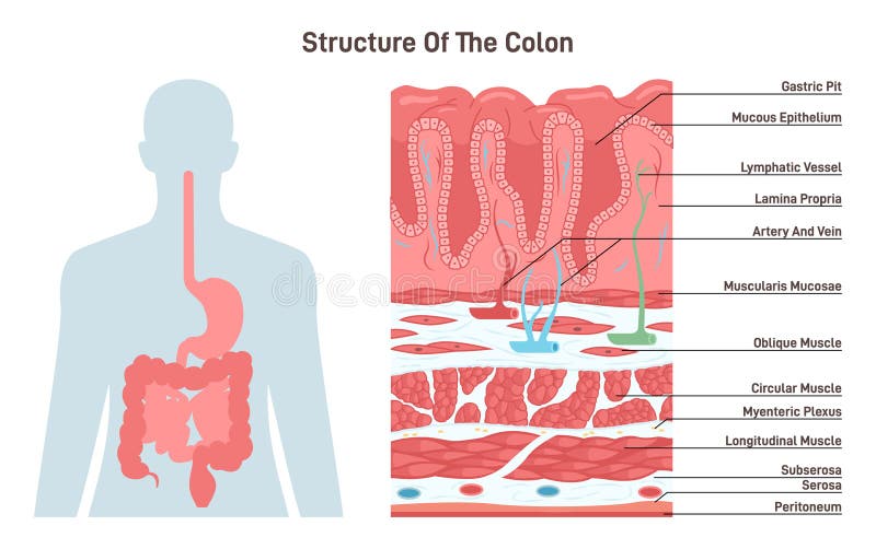
Free with trial Structure of the colon. Human digestive system anatomy. Intestinal villi in the surface of intestinal. Epithelial and goblet cells. Healthy digestive system. Flat vector illustration. Goblet cells vectors Structure of the colon. Human digestive system anatomy. Intestinal villi
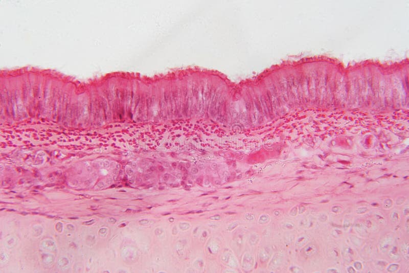
Free with trial The conducting passageways of the respiratory system nasal cavity, trachea, bronchi and bronchioles are lined by pseudostratified columnar epithelial tissue, which is ciliated and which includes mucus-secreting goblet cells. ... This epithelium is exceedingly thin to facilitate diffusion of oxygen and CO2. Goblet cells illustrations Pseudostratified columnar epithelium of nasal mucosa. The conducting passageways of the respiratory system nasal cavity, trachea, bronchi and bronchioles are lined by pseudostratified columnar epithelial tissue, which is ciliated and which includes mucus-secreting goblet cells. ... This epithelium is exceedingly thin to facilitate diffusion of oxygen and CO2
Free with trial The esophagus or oesophagus is an organ in vertebrates through which food passes, aided by peristaltic contractions, from the pharynx to the stomach. The esophagus is a fibromuscular tube, that travels behind the trachea and heart. During swallowing, the epiglottis tilts backwards to prevent food from going down the larynx and lungs. The trachea is the long tube that connects your larynx to your bronchi. Bronchi send air to your lungs. Trachea is a key part of respiratory system. The trachea is made of rings of cartilage. It is lined with cells that produce mucus. Goblet cells illustrations Anatomy of Trachea and Esophagus. The esophagus or oesophagus is an organ in vertebrates through which food passes, aided by peristaltic contractions, from the pharynx to the stomach. The esophagus is a fibromuscular tube, that travels behind the trachea and heart. During swallowing, the epiglottis tilts backwards to prevent food from going down the larynx and lungs.The trachea is the long tube that connects your larynx to your bronchi. Bronchi send air to your lungs. Trachea is a key part of respiratory system. The trachea is made of rings of cartilage. It is lined with cells that produce mucus.
Free with trial The esophagus or oesophagus is an organ in vertebrates through which food passes, aided by peristaltic contractions, from the pharynx to the stomach. The esophagus is a fibromuscular tube, that travels behind the trachea and heart. During swallowing, the epiglottis tilts backwards to prevent food from going down the larynx and lungs. The trachea is the long tube that connects your larynx to your bronchi. Bronchi send air to your lungs. Trachea is a key part of respiratory system. The trachea is made of rings of cartilage. It is lined with cells that produce mucus. Goblet cells illustrations Trachea and Esophagus cross section. The esophagus or oesophagus is an organ in vertebrates through which food passes, aided by peristaltic contractions, from the pharynx to the stomach. The esophagus is a fibromuscular tube, that travels behind the trachea and heart. During swallowing, the epiglottis tilts backwards to prevent food from going down the larynx and lungs.The trachea is the long tube that connects your larynx to your bronchi. Bronchi send air to your lungs. Trachea is a key part of respiratory system. The trachea is made of rings of cartilage. It is lined with cells that produce mucus
Free with trial Ciliated columnar epithelial cells are found mainly in the tracheal and bronchial regions of the pulmonary system and also in the fallopian tubes of the female reproductive system. Goblet cells illustrations Illustration of Pseudostratified Columnar Epithelia. Ciliated columnar epithelial cells are found mainly in the tracheal and bronchial regions of the pulmonary system and also in the fallopian tubes of the female reproductive system.
Free with trial Medical illustration shows a comparison between normal ciliated respiratory epithelium and damaged epithelium with impaired mucociliary clearance. Healthy Versus Impaired Ciliated Epithelium. Goblet cells vectors Medical illustration shows a comparison between normal ciliated respiratory epithelium and damaged epithelium with impaired
Free with trial Bronchus. Medical illustration depicting Respiratory cilia and mucus. Pseudostratified columnar epithelium. Structure Ciliated Epithelium Infographics. Goblet cells vectors Bronchus. Medical illustration depicting Respiratory cilia and mucus. Pseudostratified columnar epithelium. Structure
Free with trial Small intestine anatomical poster. Surface area of intestinal walls. Intestinal villi, cross section, fold, villus and epithelial cells. Digestive system medical flat vector illustration in human body. Goblet cells vectors Small intestine anatomy. Small intestine anatomical poster. Surface area of intestinal walls. Intestinal villi, cross section, fold, villus and epithelial cells. Digestive system medical flat vector illustration in human body
Free with trial 3d illustration of microscopic closeup of intestine villus. Goblet cells illustrations 3d illustration of microscopic closeup of intestine villus
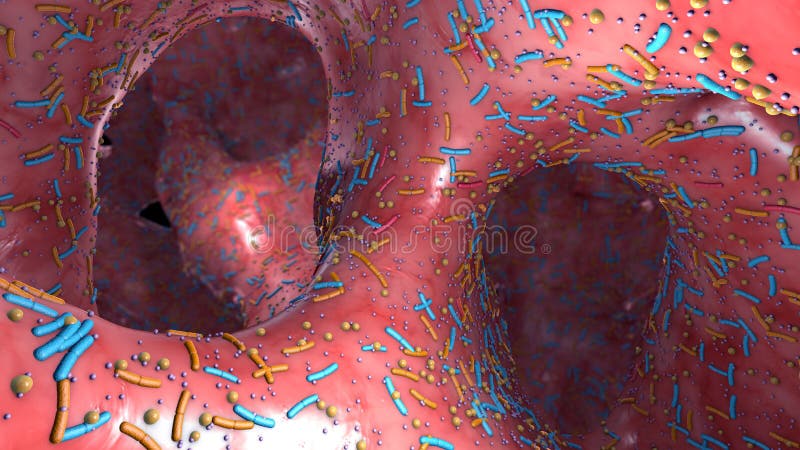
Free with trial 3d illustration of the inner side of the colon or intestinal tract. Goblet cells illustrations Inner side of the colon or intestinal tract
Free with trial 3d illustration of the inner side of the colon or intestinal tract. Goblet cells illustrations Inner side of the colon or intestinal tract
Free with trial 3d illustration of microscopic closeup of intestine villus. Goblet cells illustrations Microscopic closeup of intestine villus
Free with trial Different germs in the human intestines called microbiota - 3d illustration. Goblet cells illustrations Different germs in the human intestines called microbiota
Free with trial Different germs in the human intestines called microbiota - 3d illustration. Goblet cells illustrations Different germs in the human intestines called microbiota
Free with trial 3d illustration of microscopic closeup of intestine villus. Goblet cells illustrations 3d illustration of microscopic closeup of intestine villus
Free with trial Bacteria as part of the intestinal microbiome in the digestive tract - 3d illustration. Goblet cells illustrations Bacteria as part of the intestinal microbiome in the digestive tract
Free with trial Different germs in the human intestines called microbiome - 3d illustration. Goblet cells illustrations Germs in the human intestines called microbiome
Free with trial 3d illustration of a inflammation inside the colon or intestinal tract called intestine inflammation. Goblet cells illustrations 3d illustration of a bowel disease an inflammation inside the colon or intestinal tract. 3d illustration of a inflammation inside the colon or intestinal tract called intestine inflammation
Free with trial The nasal mucosa lines the nasal cavity. It is part of the respiratory mucosa, the mucous membrane lining the respiratory tract. The nasal mucosa is intimately adherent to the periosteum or perichondrium of the nasal conchae. Goblet cells illustrations Nasal mucosa lines (respiratory epihtelium) - closeup view 3d illustration. The nasal mucosa lines the nasal cavity. It is part of the respiratory mucosa, the mucous membrane lining the respiratory tract. The nasal mucosa is intimately adherent to the periosteum or perichondrium of the nasal conchae.
Free with trial Different germs in the human intestines called microbiome - 3d illustration. Goblet cells illustrations Germs in the human intestines called microbiome
Free with trial Different germs in the human intestines called microbiome - 3d illustration. Goblet cells illustrations Germs in the human intestines called microbiome
Free with trial Bacteria as part of the intestinal microbiome in the digestive tract - 3d illustration. Goblet cells illustrations Bacteria as part of the intestinal micro biota in the digestive tract. Bacteria as part of the intestinal microbiome in the digestive tract - 3d illustration
Free with trial Detailed anatomical illustration of the colon with labeled sections of the large intestine and a magnified inset highlighting goblet cells and crypts. Goblet cells illustrations Colonic Mecala Diagram Showing Large Intestine and Goblet Cells. Detailed anatomical illustration of the colon with labeled sections of the large intestine and a. Detailed anatomical illustration of the colon with labeled sections of the large intestine and a magnified inset highlighting goblet cells and crypts
Free with trial A detailed, high-resolution microscopic view of the intestinal lining, showcasing villi and goblet cells. Goblet cells illustrations Microscopic View of Intestinal Villi and Goblet Cells. A detailed, high-resolution microscopic view of the intestinal lining, showcasing villi and goblet cells
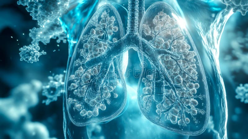
Free with trial Discover a stunning digital artwork depicting bioengineered goblet cells within human lungs, highlighting a sci-fi concept of advanced biological technology in healthcare. Goblet cells illustrations Bioengineered Goblet Cells Inside Lungs in Sci-Fi Concept Artwork. Discover a stunning digital artwork depicting bioengineered goblet cells within human lungs, highlighting a sci-fi concept of advanced biological technology in healthcare
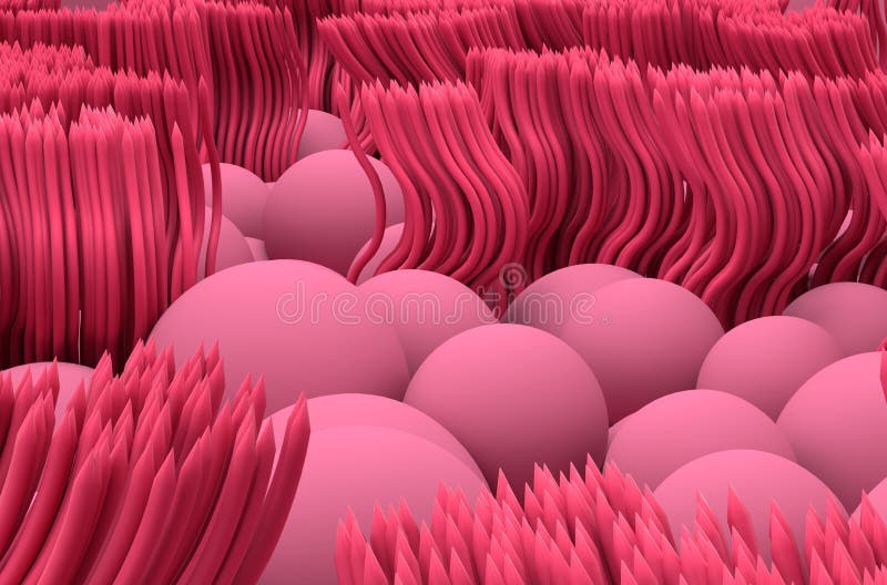
Free with trial This vibrant 3D illustration showcases goblet cells actively secreting mucus, highlighting their crucial role in human anatomy and biology. Perfect for educational use. Goblet cells illustrations 3D Medical Illustration of Goblet Cells Secreting Mucus in Detail. This vibrant 3D illustration showcases goblet cells actively secreting mucus, highlighting their crucial role in human anatomy and biology. Perfect for educational use
Free with trial Detailed microscopic image showcasing the structure of intestinal villi and crypts. The image highlights the abundance of goblet cells filled with mucin, epithelial lining, and a prominent blood vessel within the submucosa, illustrating key digestive system histology. Goblet cells illustrations Microscopic View of Intestinal Villi and Crypts with Goblet Cells and Blood Vessels. Detailed microscopic image showcasing the structure of intestinal villi and crypts. The image highlights the abundance of goblet cells filled with mucin, epithelial lining, and a prominent blood vessel within the submucosa, illustrating key digestive system histology.
Free with trial This stunning 3D medical illustration showcases goblet cells actively secreting mucus. Observe the intricate details and vibrant textures in this cellular environment. Goblet cells illustrations 3D Medical Illustration of Goblet Cells Secreting Mucus Extracellularly. This stunning 3D medical illustration showcases goblet cells actively secreting mucus. Observe the intricate details and vibrant textures in this cellular environment
Free with trial Delve into the intricate world of human nasal mucosa under a microscope. This captivating image reveals the diverse cellular components at play, crucial for maintaining respiratory health. The image, captured at night, highlights the delicate structure of ciliated cells, responsible for sweeping away debris and pathogens. Notice the interspersed goblet cells, secreting mucus to trap and remove. Goblet cells illustrations Nighttime Nasal Mucosa A Microscopic Exploration of Ciliated Goblet and Basal Cells. Delve into the intricate world of human nasal mucosa under a microscope. This captivating image reveals the diverse cellular components at play, crucial for maintaining respiratory health. The image, captured at night, highlights the delicate structure of ciliated cells, responsible for sweeping away debris and pathogens. Notice the interspersed goblet cells, secreting mucus to trap and remove
Free with trial Explore this vibrant 3D medical illustration showcasing goblet cells actively secreting mucus, perfect for educational and scientific purposes in biology and health fields. Goblet cells illustrations Detailed 3D Medical Illustration of Goblet Cells Secreting Mucus. Explore this vibrant 3D medical illustration showcasing goblet cells actively secreting mucus, perfect for educational and scientific purposes in biology and health fields
Free with trial Futuristic visualization of bioengineered goblet cells within a lung structure, blending science and fiction for a captivating digital art piece. Goblet cells illustrations Concept of Bioengineered Goblet Cells in Sci-Fi Style Visualization. Futuristic visualization of bioengineered goblet cells within a lung structure, blending science and fiction for a captivating digital art piece
Free with trial Large intestine anatomy. Layers of the colon Muscularis propria, Submucosa, Mucosa, Serosa. Colonic crypt. Cross section of large bowel. Part of intestinal glands. Close-up of Stem cell, Goblet cells, Enterocyte. Detailed Vector poster. Goblet cells vectors Large intestine anatomy. Colonic crypt. Stem cell, Goblet cell, Enterocyte. Large intestine anatomy. Layers of the colon Muscularis propria, Submucosa, Mucosa, Serosa. Colonic crypt. Cross section of large bowel. Part of intestinal glands. Close-up of Stem cell, Goblet cells, Enterocyte. Detailed Vector poster