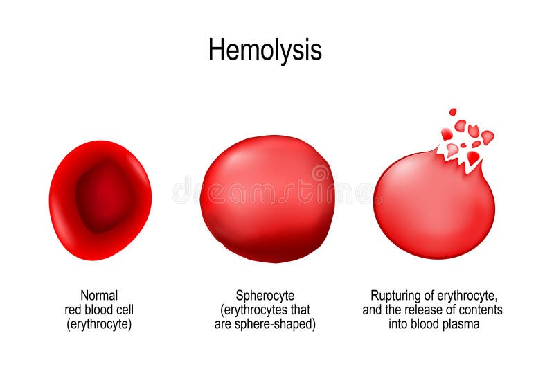
Free with trial Hemolysis. Normal red blood cell, spherocyte, and rupturing of erythrocyte release of contents into blood plasma. Vector illustration for medical use. Hemolytic anemia vectors Hemolysis. Normal red blood cell, spherocyte, and rupturing of e
Free with trial Medical illustration of the symptoms of enlarged spleen. Hemolytic anemia vectors Enlarged spleen
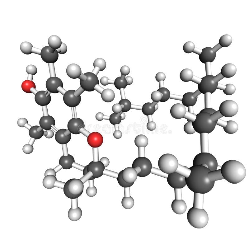
Free with trial Ball and stick model of vitamin E, also known as alpha-tocopherol. Atoms are coloured according to convention (carbon-gray; oxygen-red; hydrogen-white). Hemolytic anemia illustrations Vitamin E molecule. Ball and stick model of vitamin E, also known as alpha-tocopherol. Atoms are coloured according to convention (carbon-gray; oxygen-red; hydrogen-white).
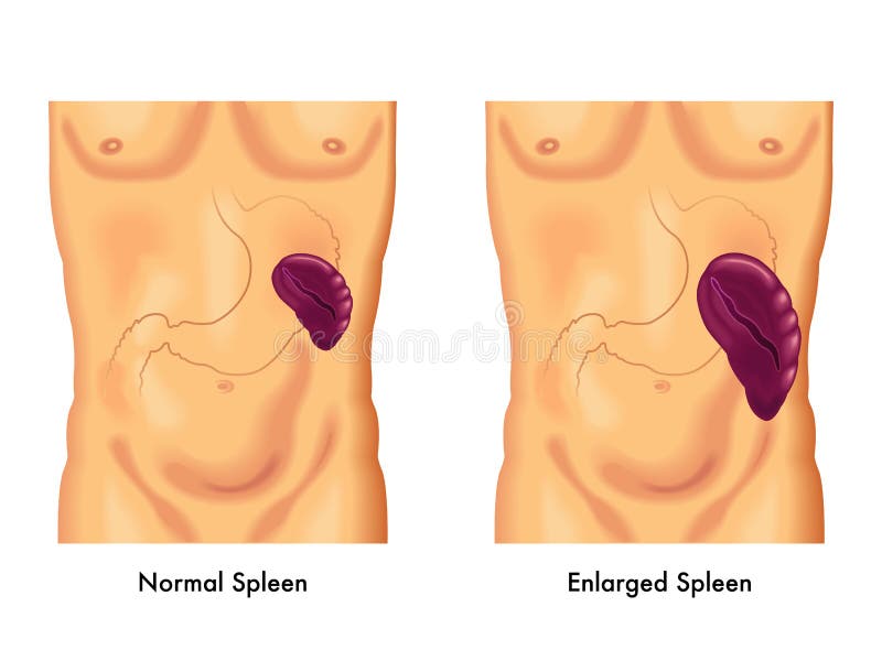
Free with trial Splenomegaly is an enlargement of the spleen. Hemolytic anemia vectors Splenomegaly or enlarged spleen. Splenomegaly is an enlargement of the spleen
Free with trial This captivating medical illustration provides a microscopic view of the profound impact of anemia on red blood cell count and function. The image showcases the cellular level of red blood cell deficiency, highlighting the critical role these cells play in oxygen transport throughout the body. Notice the stark contrast between healthy, robust erythrocytes and the deficient, smaller, and. Hemolytic anemia illustrations Unveiling the Cellular Depths of Anemia A Microscopic Exploration of Red Blood Cell Deficiency Featuring Detailed. This captivating medical illustration provides a microscopic view of the profound impact of anemia on red blood cell count and function. The image showcases the cellular level of red blood cell deficiency, highlighting the critical role these cells play in oxygen transport throughout the body. Notice the stark contrast between healthy, robust erythrocytes and the deficient, smaller, and
Free with trial Glucose-6-phosphate dehydrogenase (G6PD) protein. Enzyme of the pentose phosphate pathway that generates co-enzyme NADPH. Deficiency causes hemolytic anemia. 3D illustration. Cartoon representation with secondary structure coloring (green sheets, red helices. Hemolytic anemia illustrations Glucose-6-phosphate dehydrogenase (G6PD) protein. Enzyme of the pentose phosphate pathway that generates co-enzyme NADPH.
Free with trial Fanconi anemia color icon vector. fanconi anemia sign. isolated symbol illustration. Hemolytic anemia vectors Fanconi anemia color icon vector illustration. fanconi anemia color icon vector. fanconi anemia sign. isolated symbol illustration
Free with trial Sickle cell disease. Normal Red blood cell and Sickle-shaped RBCs. Close-up of Amino acid chain and Nucleotide sequence in a DNA strand. Vector illustration. Hemolytic anemia vectors Sickle cell disease. Anemia. Red blood cell. Sickle cell disease. Normal Red blood cell and Sickle-shaped RBCs. Close-up of Amino acid chain and Nucleotide sequence in a DNA strand. Vector illustration
Free with trial Illustration of a young child with Erythema Infectiosum (Fifth Disease), showing two characteristic rashes. The "slapped-cheek" rash appears prominently on the cheeks while a "lacy body rash" is visible on the chest and arms. The child has short black hair and a neutral expression. Arrows and labels highlight the locations of the rashes for educational purposes. Hemolytic anemia vectors Fifth Disease (Erythema Infectiosum) Illustration. Illustration of a young child with Erythema Infectiosum (Fifth Disease), showing two characteristic rashes. The "slapped-cheek" rash appears prominently on the cheeks while a "lacy body rash" is visible on the chest and arms. The child has short black hair and a neutral expression. Arrows and labels highlight the locations of the rashes for educational purposes.
Free with trial Shortness of breath line icon vector. shortness of breath sign. isolated contour symbol black illustration. Hemolytic anemia vectors Shortness of breath line icon vector illustration. shortness of breath line icon vector. shortness of breath sign. isolated contour symbol black illustration
Free with trial A scientific vector illustration graphic of the erythrocyte glutathione enzymatic pathway. Hemolytic anemia vectors Erythrocyte glutathione enzymatic pathway vector illustration graphic. a scientific vector illustration graphic of the erythrocyte glutathione enzymatic pathway
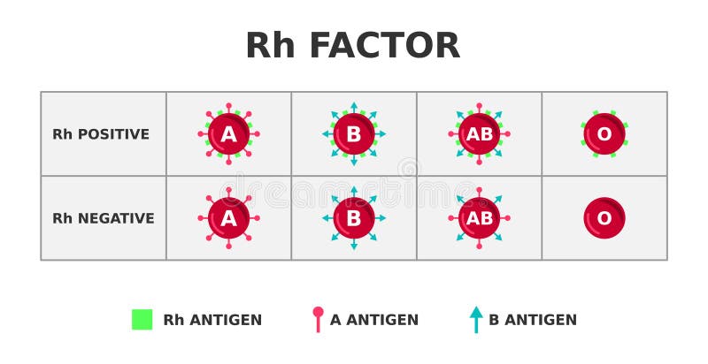
Free with trial Importnace in blood transfusion. 85% of people are Rh-positive. Vector illustration. Hemolytic anemia vectors Rh factor blood group system. Rh positive on Rh negative. Rhesus D antigen on the surface of red blood cells. Importnace in blood transfusion. 85% of people are Rh-positive. Vector illustration.
Free with trial Ferritin is the main iron storage protein in humans. Serum ferritin correlates with amount of iron present in the body. Cartoon model. transferrin 2d graphic. Hemolytic anemia illustrations Ferritin is the main iron storage protein in humans. Serum ferritin correlates with amount of iron present in the body. Cartoon
Free with trial A mild, inherited liver condition in which the liver doesn't process bilirubin properly, leading to occasional yellowing of the skin and eyes (jaundice). Hemolytic anemia vectors Gilbert\'s Syndrome (Jaundice) Flashcard Illustration. A mild, inherited liver condition in which the liver doesn't process bilirubin properly, leading to occasional yellowing of the skin and eyes (jaundice).
Free with trial Illustration comparing a healthy spleen and an enlarged spleen (splenomegaly). The left side shows a normal spleen with splenic vein and artery highlighted in blue and red. The right side depicts an enlarged spleen, emphasizing increased size. The spleens are labeled with connecting blood vessels to illustrate anatomical differences. The image explains the condition of splenomegaly through visual contrast. Hemolytic anemia vectors Enlarged Spleen (Splenomegaly) spleen flashcard illustration. Illustration comparing a healthy spleen and an enlarged spleen (splenomegaly). The left side shows a normal spleen with splenic vein and artery highlighted in blue and red. The right side depicts an enlarged spleen, emphasizing increased size. The spleens are labeled with connecting blood vessels to illustrate anatomical differences. The image explains the condition of splenomegaly through visual contrast.
Free with trial Fatigue person patient color icon vector. fatigue person patient sign. isolated symbol illustration. Hemolytic anemia vectors Fatigue person patient color icon vector illustration. fatigue person patient color icon vector. fatigue person patient sign. isolated symbol illustration
Free with trial Iron supplements package line icon vector. iron supplements package sign. isolated contour symbol black illustration. Hemolytic anemia vectors Iron supplements package line icon vector illustration. iron supplements package line icon vector. iron supplements package sign. isolated contour symbol black illustration
Free with trial Hemolytic anemia icons set. Low number of red blood cells due to too much hemolysis in the body. Medical pictograms. Symptoms signs. Editable stroke. Vector illustration isolated on a white background. Hemolytic anemia vectors Hemolytic anemia icons set. Low number of red blood cell
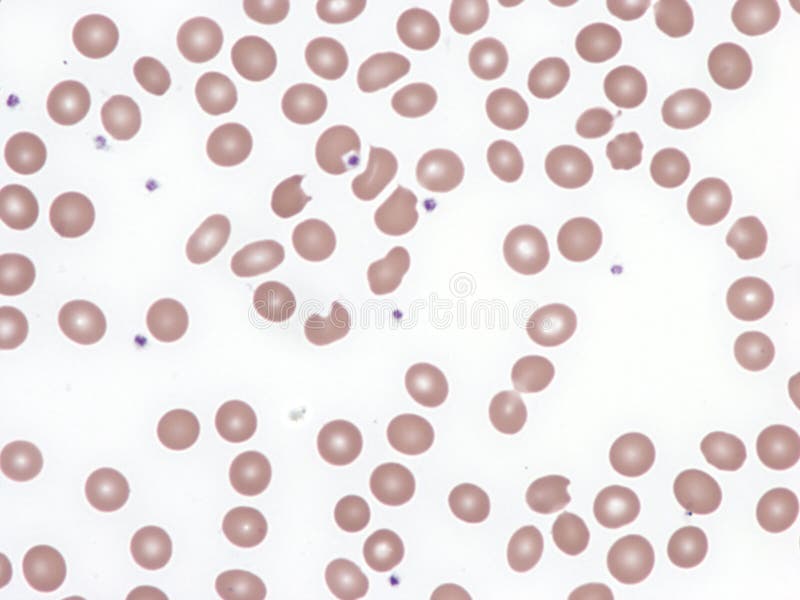
Free with trial Autoimmune hemolytic anemia. It occurs when a person's immune system produces antibodies directed against their own red blood cells Note the numerous spherocytes. Peripheral blood. Wright x1000. Hemolytic anemia illustrations Autoimmune hemolytic anemia.
Free with trial Hemolytic anemia icons set. Low number of red blood cells due to too much hemolysis in the body. Medical pictograms. Symptoms signs. Editable stroke. Vector illustration isolated on a white background. Hemolytic anemia vectors Hemolytic anemia icons set. Vector illustration isolated on a white background. Hemolytic anemia icons set. Low number of red blood cells due to too much hemolysis in the body. Medical pictograms. Symptoms signs. Editable stroke. Vector illustration isolated on a white background
Free with trial Hemolytic anemia banner. Low number of red blood cells due to too much hemolysis in the body. Medical poster with linear icons. Editable stroke. Vector illustration isolated on a white background. Hemolytic anemia vectors Hemolytic anemia banner. Editable stroke. Vector illustration. Hemolytic anemia banner. Low number of red blood cells due to too much hemolysis in the body. Medical poster with linear icons. Editable stroke. Vector illustration isolated on a white background
Free with trial Hemolytic anemia infographic. Low number of red blood cells due to too much hemolysis in the body. Medical schema with linear icons. Editable stroke. Vector illustration isolated on a white background. Hemolytic anemia vectors Hemolytic anemia infographic. Vector illustration isolated on a white background. Hemolytic anemia infographic. Low number of red blood cells due to too much hemolysis in the body. Medical schema with linear icons. Editable stroke. Vector illustration isolated on a white background
Free with trial Hemolytic anemia infographic. Low number of red blood cells due to too much hemolysis in the body. Medical schema with linear icons. Editable stroke. Vector illustration isolated on a white background. Hemolytic anemia vectors Hemolytic anemia infographic. Vector illustration isolated on a white background. Hemolytic anemia infographic. Low number of red blood cells due to too much hemolysis in the body. Medical schema with linear icons. Editable stroke. Vector illustration isolated on a white background
Free with trial Hemolytic anemia is a sub-type of anemia, a common blood disorder that occurs when the body has fewer red blood cells than normal. In hemolytic anemias, the low red blood cell count is caused by the destruction � rather than the underproduction � of red blood cells. Hemolytic anemia illustrations Hemolytic Anemia (HA) cells in the blood flow - closeup view 3d illustration. Hemolytic anemia is a sub-type of anemia, a common blood disorder that occurs when the body has fewer red blood cells than normal. In hemolytic anemias, the low red blood cell count is caused by the destruction � rather than the underproduction � of red blood cells.
Free with trial Hemolytic anemia is a sub-type of anemia, a common blood disorder that occurs when the body has fewer red blood cells than normal. In hemolytic anemias, the low red blood cell count is caused by the destruction � rather than the underproduction � of red blood cells. Hemolytic anemia illustrations Hemolytic Anemia (HA) cells in the blood flow - isometric view 3d illustration. Hemolytic anemia is a sub-type of anemia, a common blood disorder that occurs when the body has fewer red blood cells than normal. In hemolytic anemias, the low red blood cell count is caused by the destruction � rather than the underproduction � of red blood cells.
Free with trial Autoimmune hemolytic anemia. Note the numerous spherocytes. Peripheral blood. Wright x200. Hemolytic anemia illustrations Autoimmune hemolytic anemia.
Free with trial Autoimmune hemolytic anemia ill doodle icon sketch vector. autoimmune hemolytic anemia ill sign. isolated symbol illustration. Hemolytic anemia illustrations Autoimmune hemolytic anemia ill icon doodle illustration. autoimmune hemolytic anemia ill doodle icon sketch vector. autoimmune hemolytic anemia ill sign. isolated symbol illustration
Free with trial Anemia Patient Health Problem doodle icons set vector. Fanconi Autoimmune Hemolytic Anemia Disease, Dizziness Or Lightheadedness, Shortness Of Breath Irregular Heartbeats sketch hand drawn. Hemolytic anemia vectors Anemia Patient Health Problem Icons hand drawn. Anemia Patient Health Problem doodle icons set vector. Fanconi Autoimmune Hemolytic Anemia Disease, Dizziness Or Lightheadedness, Shortness Of Breath Irregular Heartbeats sketch hand drawn
Free with trial Anemia Patient Health Problem isometric icons set. Fanconi And Autoimmune Hemolytic Anemia Disease, Dizziness Or Lightheadedness, Shortness Of Breath And Irregular Heartbeats Line. Color. Hemolytic anemia vectors Anemia Patient Health Problem isometric icons set vector. Anemia Patient Health Problem isometric icons set. Fanconi And Autoimmune Hemolytic Anemia Disease, Dizziness Or Lightheadedness, Shortness Of Breath And Irregular Heartbeats Line. Color .
Free with trial Autoimmune hemolytic anemia (AIHA) is a complex blood disorder characterized by the premature destruction of red blood cells. The body's immune system mistakenly identifies its own red blood cells as foreign invaders, triggering an attack that leads to anemia. This condition can manifest in various ways, impacting overall health and well-being. Symptoms of AIHA can range from mild fatigue and. Hemolytic anemia illustrations Understanding Autoimmune Hemolytic Anemia A Comprehensive Overview of AIHA and its Impact on Blood Health. Autoimmune hemolytic anemia (AIHA) is a complex blood disorder characterized by the premature destruction of red blood cells. The body's immune system mistakenly identifies its own red blood cells as foreign invaders, triggering an attack that leads to anemia. This condition can manifest in various ways, impacting overall health and well-being. Symptoms of AIHA can range from mild fatigue and
Free with trial Hemolytic uremic syndrome on white paper background. Hemolytic anemia illustrations Hemolytic uremic syndrome on white paper
Free with trial Bite cells, in a case of glucose-6-phosphate deficiency, a type of hemolytic anemia. Peripheral blood. Wright x1000. Hemolytic anemia illustrations Bite cells. Bite cells, in a case of glucose-6-phosphate deficiency, a type of hemolytic anemia. Peripheral blood. Wright x1000
Free with trial Fanconi anemia doodle icon sketch vector. fanconi anemia sign. isolated symbol illustration. Hemolytic anemia vectors Fanconi anemia icon doodle illustration. fanconi anemia doodle icon sketch vector. fanconi anemia sign. isolated symbol illustration
Free with trial Sideroblastic anemia sick doodle icon sketch vector. sideroblastic anemia sick sign. isolated symbol illustration. Hemolytic anemia vectors Sideroblastic anemia sick icon doodle illustration. sideroblastic anemia sick doodle icon sketch vector. sideroblastic anemia sick sign. isolated symbol illustration
Free with trial Megaloblastic anemia researching doodle icon sketch vector. megaloblastic anemia researching sign. isolated symbol illustration. Hemolytic anemia vectors Megaloblastic anemia researching icon doodle illustration. megaloblastic anemia researching doodle icon sketch vector. megaloblastic anemia researching sign. isolated symbol illustration
Free with trial Aplastic anemia disease doodle icon sketch vector. aplastic anemia disease sign. isolated symbol illustration. Hemolytic anemia illustrations Aplastic anemia disease icon doodle illustration. aplastic anemia disease doodle icon sketch vector. aplastic anemia disease sign. isolated symbol illustration
Free with trial Diamond-blackfan anemia health problem doodle icon sketch vector. diamond-blackfan anemia health problem sign. isolated symbol illustration. Hemolytic anemia illustrations Diamond-blackfan anemia health problem icon doodle illustration. diamond-blackfan anemia health problem doodle icon sketch vector. diamond-blackfan anemia health problem sign. isolated symbol illustration
Free with trial Congenital dyserythropoietic anemia disease doodle icon sketch vector. congenital dyserythropoietic anemia disease sign. isolated symbol illustration. Hemolytic anemia vectors Congenital dyserythropoietic anemia disease icon doodle illustration. congenital dyserythropoietic anemia disease doodle icon sketch vector. congenital dyserythropoietic anemia disease sign. isolated symbol illustration
Free with trial Medical concept of schistocytes in blood smear, indicating hemolytic syndromes and dic, Generated by AI. Hemolytic anemia illustrations Medical concept of schistocytes in blood smear, indicating hemolytic syndromes and dic
Free with trial 3D medical illustration of schistocytes: fragmented, irregular red blood cells. These jagged "helmet" shapes signify microangiopathic hemolytic anemia or physical blood cell damage. Hemolytic anemia illustrations 3D medical illustration of schistocytes
Free with trial 3D medical illustration of schistocytes: fragmented, irregular red blood cells. These jagged "helmet" shapes signify microangiopathic hemolytic anemia or physical blood cell damage. Hemolytic anemia illustrations 3D medical illustration of schistocytes
Free with trial 3D medical illustration of schistocytes: fragmented, irregular red blood cells. These jagged "helmet" shapes signify microangiopathic hemolytic anemia or physical blood cell damage. Hemolytic anemia illustrations 3D medical illustration of schistocytes. Fragmented, irregular red blood cells. These jagged 'helmet' shapes signify microangiopathic hemolytic anemia or. 3D medical illustration of schistocytes: fragmented, irregular red blood cells. These jagged "helmet" shapes signify microangiopathic hemolytic anemia or physical blood cell damage.
Free with trial Anemia. Vector Icons about blood disorders. Red Drop shapes with blood level on dark background. Hemolytic anemia vectors Red Drop shapes with blood level
Free with trial Glucose-6-phosphate dehydrogenase deficiency, also known as favism, is the most common enzyme deficiency anemia worldwide. It is an X-linked recessive disorder, an inborn error of metabolism that predisposes to red blood cell breakdown. Note blister cells, bite cells and a nucleated red cell. Peripheral blood, Wright x1000. Hemolytic anemia illustrations Glucose-6-phosphate dehydrogenase deficiency. Peripheral blood. Glucose-6-phosphate dehydrogenase deficiency, also known as favism, is the most common enzyme deficiency anemia worldwide. It is an X-linked recessive disorder, an inborn error of metabolism that predisposes to red blood cell breakdown. Note blister cells, bite cells and a nucleated red cell. Peripheral blood, Wright x1000.
Free with trial Direct and Indirect Coombs Test Diagram for Diagnosis of Anemia. Hemolytic anemia vectors Direct and Indirect Coombs Test
Free with trial Glucose-6-phosphate dehydrogenase deficiency, also known as favism, is the most common enzyme deficiency anemia worldwide. It is an X-linked recessive disorder, an inborn error of metabolism that predisposes to red blood cell breakdown. Note blister cells (hemighost cells) are abnormal erythrocytes with a peripheral clearing due to hemoglobin damage. Peripheral blood, Wright x1000. Hemolytic anemia illustrations Glucose-6-phosphate dehydrogenase deficiency. Blister cells. Glucose-6-phosphate dehydrogenase deficiency, also known as favism, is the most common enzyme deficiency anemia worldwide. It is an X-linked recessive disorder, an inborn error of metabolism that predisposes to red blood cell breakdown. Note blister cells (hemighost cells) are abnormal erythrocytes with a peripheral clearing due to hemoglobin damage. Peripheral blood, Wright x1000.
Free with trial Glucose-6-phosphate dehydrogenase deficiency, also known as favism, is the most common enzyme deficiency anemia worldwide. It is an X-linked recessive disorder, an inborn error of metabolism that predisposes to red blood cell breakdown. Note blister cells (hemighost cells) are abnormal erythrocytes with a peripheral clearing due to hemoglobin damage. Peripheral blood, Wright x1000. Hemolytic anemia illustrations Glucose-6-phosphate dehydrogenase deficiency. Blister cells. Glucose-6-phosphate dehydrogenase deficiency, also known as favism, is the most common enzyme deficiency anemia worldwide. It is an X-linked recessive disorder, an inborn error of metabolism that predisposes to red blood cell breakdown. Note blister cells (hemighost cells) are abnormal erythrocytes with a peripheral clearing due to hemoglobin damage. Peripheral blood, Wright x1000.
Free with trial Glucose-6-phosphate dehydrogenase deficiency, also known as favism, is the most common enzyme deficiency anemia worldwide. It is an X-linked recessive disorder, an inborn error of metabolism that predisposes to red blood cell breakdown. Note blister cells (hemighost cells) are abnormal erythrocytes with a peripheral clearing due to hemoglobin damage. Peripheral blood, Wright x1000. Hemolytic anemia illustrations Glucose-6-phosphate dehydrogenase deficiency. Blister cells. Glucose-6-phosphate dehydrogenase deficiency, also known as favism, is the most common enzyme deficiency anemia worldwide. It is an X-linked recessive disorder, an inborn error of metabolism that predisposes to red blood cell breakdown. Note blister cells (hemighost cells) are abnormal erythrocytes with a peripheral clearing due to hemoglobin damage. Peripheral blood, Wright x1000.
Free with trial Glucose-6-phosphate dehydrogenase deficiency, also known as favism, is the most common enzyme deficiency anemia worldwide. It is an X-linked recessive disorder, an inborn error of metabolism that predisposes to red blood cell breakdown. Note blister cells (hemighost cells) are abnormal erythrocytes with a peripheral clearing due to hemoglobin damage. Peripheral blood, Wright x1000. Hemolytic anemia illustrations Glucose-6-phosphate dehydrogenase deficiency. Blister cells. Glucose-6-phosphate dehydrogenase deficiency, also known as favism, is the most common enzyme deficiency anemia worldwide. It is an X-linked recessive disorder, an inborn error of metabolism that predisposes to red blood cell breakdown. Note blister cells (hemighost cells) are abnormal erythrocytes with a peripheral clearing due to hemoglobin damage. Peripheral blood, Wright x1000.
Free with trial Glucose-6-phosphate dehydrogenase deficiency, also known as favism, is the most common enzyme deficiency anemia worldwide. It is an X-linked recessive disorder, an inborn error of metabolism that predisposes to red blood cell breakdown. Note blister cells (hemighost cells) are abnormal erythrocytes with a peripheral clearing due to hemoglobin damage. Peripheral blood, Wright x1000. Hemolytic anemia illustrations Glucose-6-phosphate dehydrogenase deficiency. Blister cells. Glucose-6-phosphate dehydrogenase deficiency, also known as favism, is the most common enzyme deficiency anemia worldwide. It is an X-linked recessive disorder, an inborn error of metabolism that predisposes to red blood cell breakdown. Note blister cells (hemighost cells) are abnormal erythrocytes with a peripheral clearing due to hemoglobin damage. Peripheral blood, Wright x1000.
Free with trial TTP pathophysiology illustrated, ADAMTS13 deficiency leads to ultra-large vWF, platelet-rich microthrombi in a blood vessel, key items are platelets, RBCs, microthrombus. Outline diagram. Hemolytic anemia vectors TTP pathophysiology illustrated, ADAMTS13 deficiency leads to ultra-large vWF
Free with trial Red blood cells inside blood drop. Erythrocyte analysis, blood sample diagnostics, hematology testing, medical laboratory research, health monitoring concept. Low poly digital wireframe style. Vector. Hemolytic anemia vectors Red blood cells inside blood drop. Erythrocyte analysis, blood sample diagnostics, hematology testing, medical
Free with trial Direct and Indirect Coombs Test Vector Diagram. Hemolytic anemia vectors Coombs Test Vector Diagram
Free with trial Hemolysis of Red blood cells. Hypertonic, Hypotonic, and Isotonic Erythrocytes. Vector illustration. Hemolytic anemia illustrations Hemolysis of Red blood cells
Free with trial Drop of blood from puzzles in hands, as metaphor for donation and fight against cancer caused by leukocyte disease. People donate blood to help sick patients in need of plasma transfusions. Hemolytic anemia vectors Drop of blood from puzzles in hands, as metaphor for donation and fight against cancer
Free with trial Jaundice icons in outline style. Yellowing of skin and eyes, caused by bilirubin buildup. Hyperbilirubinemia linear pictogram. Medical concept. Vector illustration isolated on a white background. Hemolytic anemia vectors Jaundice icons set in outline style. Vector illustration. Jaundice icons in outline style. Yellowing of skin and eyes, caused by bilirubin buildup. Hyperbilirubinemia linear pictogram. Medical concept. Vector illustration isolated on a white background
Free with trial Enlarged spleen line icon. Medical symptom outline sign. Splenomegaly pictogram. Simple symbol in black and red color. Editable stroke. Vector illustration isolated on a white background. Hemolytic anemia vectors Enlarged spleen line icon. Medical symptom outline sign.
Free with trial Enlarged spleen line icon. Medical symptom outline sign. Splenomegaly pictogram. Simple symbol in black and red color. Editable stroke. Vector illustration isolated on a white background. Hemolytic anemia vectors Enlarged spleen line icon. Medical symptom outline sign
Free with trial A viral infection caused by Parvovirus B19 that commonly affects children and causes Fifth disease (erythema infectiosum). Hemolytic anemia vectors Parvovirus Infection (Parvovirus B19) Flashcard. A viral infection caused by Parvovirus B19 that commonly affects children and causes Fifth disease (erythema infectiosum).
Free with trial Erythema infectiosum, fifth disease, or slapped cheek syndrome is one of several possible manifestations of infection by parvovirus B19. Fifth disease typically presents as a rash and is more common in children. While parvovirus B19 can affect humans of all ages, only two out of ten individuals will present with physical symptoms. Hemolytic anemia illustrations Monoclonal antibody treatment in B19 Parvovirus - closeup view 3d illustration. Erythema infectiosum, fifth disease, or slapped cheek syndrome is one of several possible manifestations of infection by parvovirus B19. Fifth disease typically presents as a rash and is more common in children. While parvovirus B19 can affect humans of all ages, only two out of ten individuals will present with physical symptoms.
Free with trial Erythema infectiosum, fifth disease, or slapped cheek syndrome is one of several possible manifestations of infection by parvovirus B19. Fifth disease typically presents as a rash and is more common in children. While parvovirus B19 can affect humans of all ages, only two out of ten individuals will present with physical symptoms. Hemolytic anemia illustrations Monoclonal antibody treatment in B19 Parvovirus - isometric view 3d illustration. Erythema infectiosum, fifth disease, or slapped cheek syndrome is one of several possible manifestations of infection by parvovirus B19. Fifth disease typically presents as a rash and is more common in children. While parvovirus B19 can affect humans of all ages, only two out of ten individuals will present with physical symptoms.
Free with trial Erythema infectiosum, fifth disease, or slapped cheek syndrome is one of several possible manifestations of infection by parvovirus B19. Fifth disease typically presents as a rash and is more common in children. While parvovirus B19 can affect humans of all ages, only two out of ten individuals will present with physical symptoms. Hemolytic anemia illustrations Parvovirus B19 in erythema infectiosum - isometric view 3d illustration. Erythema infectiosum, fifth disease, or slapped cheek syndrome is one of several possible manifestations of infection by parvovirus B19. Fifth disease typically presents as a rash and is more common in children. While parvovirus B19 can affect humans of all ages, only two out of ten individuals will present with physical symptoms.
Free with trial Jaundice icon in outline style. Yellowing of skin and eyes, caused by bilirubin buildup. Hyperbilirubinemia linear pictogram. Medical concept. Vector illustration isolated on a white background. Hemolytic anemia vectors Jaundice icon in outline style. Yellowing of skin and eyes
Free with trial 3d rendering of antibody molecules scattered around red blood cells or erythrocytes. Hemolytic anemia illustrations Antibody molecules surrounding red blood cells. 3d rendering of antibody molecules scattered around red blood cells or erythrocytes.
Free with trial 3d rendering of antibody molecules scattered around red blood cells or erythrocytes. Hemolytic anemia illustrations Antibody molecules surrounding red blood cells. 3d rendering of antibody molecules scattered around red blood cells or erythrocytes.
Free with trial 3d rendering of antibody molecules scattered around red blood cells or erythrocytes. Hemolytic anemia illustrations Antibody molecules surrounding red blood cells. 3d rendering of antibody molecules scattered around red blood cells or erythrocytes.
Free with trial 3d rendering of antibody molecules scattered around red blood cells or erythrocytes. Hemolytic anemia illustrations Antibody molecules surrounding red blood cells. 3d rendering of antibody molecules scattered around red blood cells or erythrocytes.
Free with trial 3d rendering of antibody molecules scattered around red blood cells or erythrocytes. Hemolytic anemia illustrations Antibody molecules surrounding red blood cells. 3d rendering of antibody molecules scattered around red blood cells or erythrocytes.
Free with trial 3d rendering of Hemolysis, is the process of red blood cells breaking down and releasing their contents into surrounding fluid. Hemolytic anemia illustrations 3d rendering of Hemolysis red blood cells. 3d rendering of Hemolysis, is the process of red blood cells breaking down and releasing their contents into surrounding fluid
Free with trial 3d rendering of Hemolysis, is the process of red blood cells breaking down and releasing their contents into surrounding fluid. Hemolytic anemia illustrations 3d rendering of Hemolysis red blood cells. 3d rendering of Hemolysis, is the process of red blood cells breaking down and releasing their contents into surrounding fluid