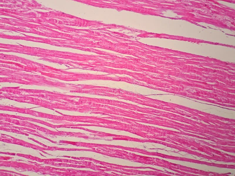
Free with trial Histology of human cardiac muscle under microscope view. Histology illustrations Histology of human cardiac muscle
Free with trial Tissue processing steps diagram illustrates the histology workflow from fixation to mounting, key objects, tissue sample, reagent jars, microscope. Outline diagram. Histology vectors Tissue processing steps diagram illustrates the histology workflow from fixation ... Tissue processing steps diagram illustrates the histology workflow from fixation to mounting, key objects, tissue sample, reagent jars, microscope. Outline diagram
Free with trial Liver diseases. Stages of liver damage from healthy liver to fatty, fibrosis, and cirrhosis. Close-up of histology. Histology vectors Liver diseases. Stages of liver damage
Free with trial The trichrome stain perpetuates visualization of collagen (blue), muscle (red, none shown) and nuclei (purple). The large portion of reddish purple color here is the top layer of skin composed of keratin. Histology illustrations Histology of Normal Skin Stained with Trichrome. The trichrome stain perpetuates visualization of collagen (blue), muscle (red, none shown) and nuclei (purple). The large portion of reddish purple color here is the top layer of skin composed of keratin.
Free with trial This is a horse patella that shows articular cartilage. There is dense bone (light pink), the articular cartilage (reddish pink) and nuclei (purple). Histology illustrations Histology Bone stained H&E. This is a horse patella that shows articular cartilage. There is dense bone (light pink), the articular cartilage (reddish pink) and nuclei (purple).
Free with trial Anatomy vs histology comparison shows the stomach in gross view and a close-up of its wall layers, key objects, stomach, magnifying glass, labeled mucosa to serosa. Outline diagram. Histology vectors Anatomy vs histology comparison shows the stomach in gross view ... Anatomy vs histology comparison shows the stomach in gross view and a close-up of its wall layers, key objects, stomach, magnifying glass, labeled mucosa to serosa. Outline diagram
Free with trial Blood cells under microscope view for histology education. Human physiology. Histology illustrations Blood cells under microscope view for histology education
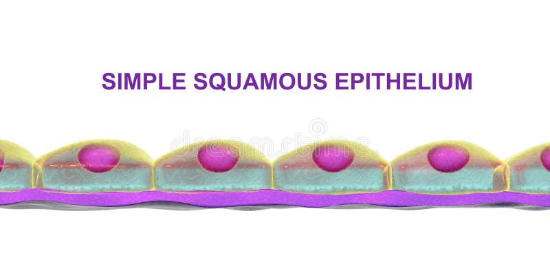
Free with trial Simple squamous epithelium, 3D illustration. Histology background. It is found in alveoli, forms lining of blood vessels, heart cavities. Histology illustrations Simple squamous epithelium
Free with trial Simple columnar epithelium, 3D illustration. Histology background. Columnar epithelium is found in digestive system, uterus. Histology illustrations Simple columnar epithelium
Free with trial Histology vs Pathology shows colon tissue comparison, normal crypts with goblet cells versus adenocarcinoma tumor mass. Main objects, colon, crypts, tumor cells. Outline diagram. Histology vectors Histology vs Pathology shows colon tissue comparison, normal crypts with ... Histology vs Pathology shows colon tissue comparison, normal crypts with goblet cells versus adenocarcinoma tumor mass. Main objects, colon, crypts, tumor cells. Outline diagram
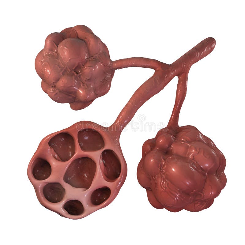
Free with trial Anatomy of alveoli in lungs, illustration. Microstructure of respiratory system, science, histology, organ, tract, medical, body, respiration, white, anatomical, normal, health, medicine, duct, education, scientific, educational, object, alveolus, isolated, air, breath, human, pulmonary, breathing, background, 3d. Histology illustrations Anatomy of alveoli in lungs
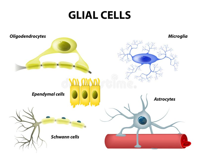
Free with trial Types of neuroglia. Classification of glial cells: microglia, astrocytes, oligodendrocytes and Schwann cells, Ependymal cells. Histology vectors Supporting Cells. Neuroglia or Glial cells. Types of neuroglia. Classification of glial cells: microglia, astrocytes, oligodendrocytes and Schwann cells, Ependymal cells
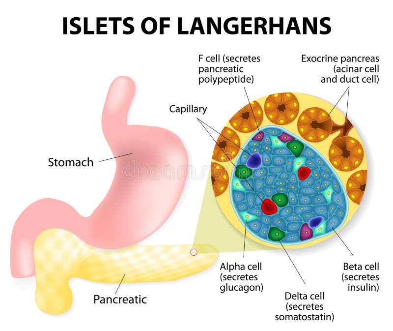
Free with trial The islets of Langerhans are responsible for the endocrine function of the pancreas. Each islet contains beta, alpha, and delta cells that are responsible for the secretion of a hormones. Histology vectors Islets of Langerhans
Free with trial Histology Word Cloud. A visual synthesis of microscopic anatomy, tissue organization, and histopathology terms essential for medical research, cellular biology, and disease diagnosis. Histology illustrations Histology Word Cloud. Core Concepts in Microscopic Anatomy and Tissue Science. Histology Word Cloud. A visual synthesis of microscopic anatomy, tissue organization, and histopathology terms essential for medical research, cellular biology, and disease diagnosis
Free with trial Types of Muscle Tissue of Human Body Diagram including cardiac skeletal smooth. Examples of heart digestive system. Along with involuntary voluntary control for medical science education. Histology vectors Types of Muscle Tissue of Human Body Diagram
Free with trial Parkinsons disease and Alzheimers disease. large black sphere inside the body of a neuron, a Lewy Body confirms the diagnosis of parkinsonism. Histology vectors Lewy body. Parkinsons disease and Alzheimers disease. Parkinsons disease and Alzheimers disease. large black sphere inside the body of a neuron, a Lewy Body confirms the diagnosis of parkinsonism.
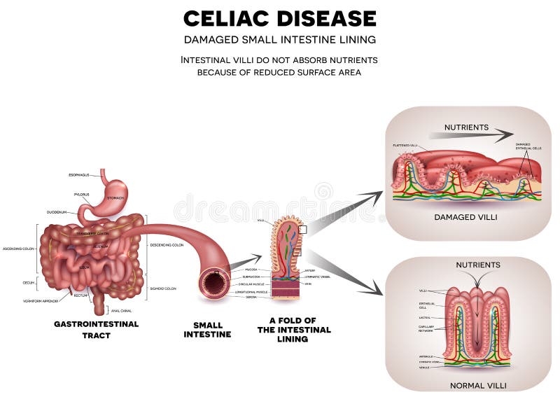
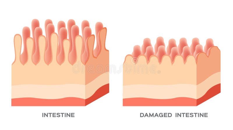
Free with trial Gastrointestinal tract anatomy and Celiac disease affected small intestine villi. Unhealthy villi with damaged cells and healthy villi. Intestinal villi do not absorb nutrients because of reduced surface area. Histology vectors Celiac disease
Free with trial Surgical Pathology Word Cloud. A Visual Guide to Diagnostic Terms, Histology Techniques, and Molecular Testing Used by Pathologists to Identify Disease in Tissue Specimens. Histology illustrations Surgical Pathology Word Cloud. A Visual Guide to Diagnostic Terms, Histology Techniques, and Molecular Testing Used by. Pathologists to Identify Disease in
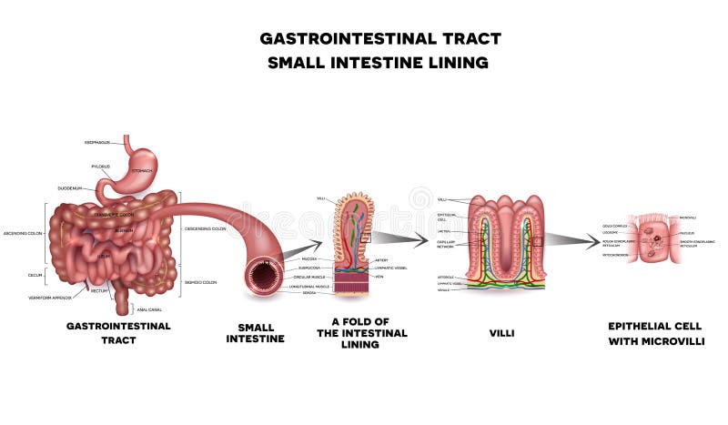
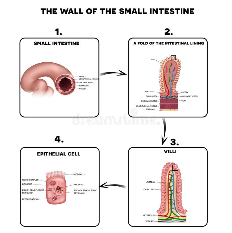
Free with trial Gastrointestinal system small intestine detailed wall anatomy. Small intestine villi and epithelial cell with microvilli detailed illustration. Histology vectors Gastrointestinal system small intestine anatomy. Gastrointestinal system small intestine detailed wall anatomy. Small intestine villi and epithelial cell with microvilli detailed illustration.
Free with trial Set of human cells, eps8, gradient and mesh printing compatible. Histology vectors Human cell collection. Set of human cells, eps8, gradient and mesh printing compatible
Free with trial Lipoma are adipose tumors located in the subcutaneous tissues. Histology vectors Lipoma
Free with trial Intestinal villi anatomy, small intestine lining, villi and epithelial cells with microvilli detailed illustration. Histology vectors Intestinal villi anatomy
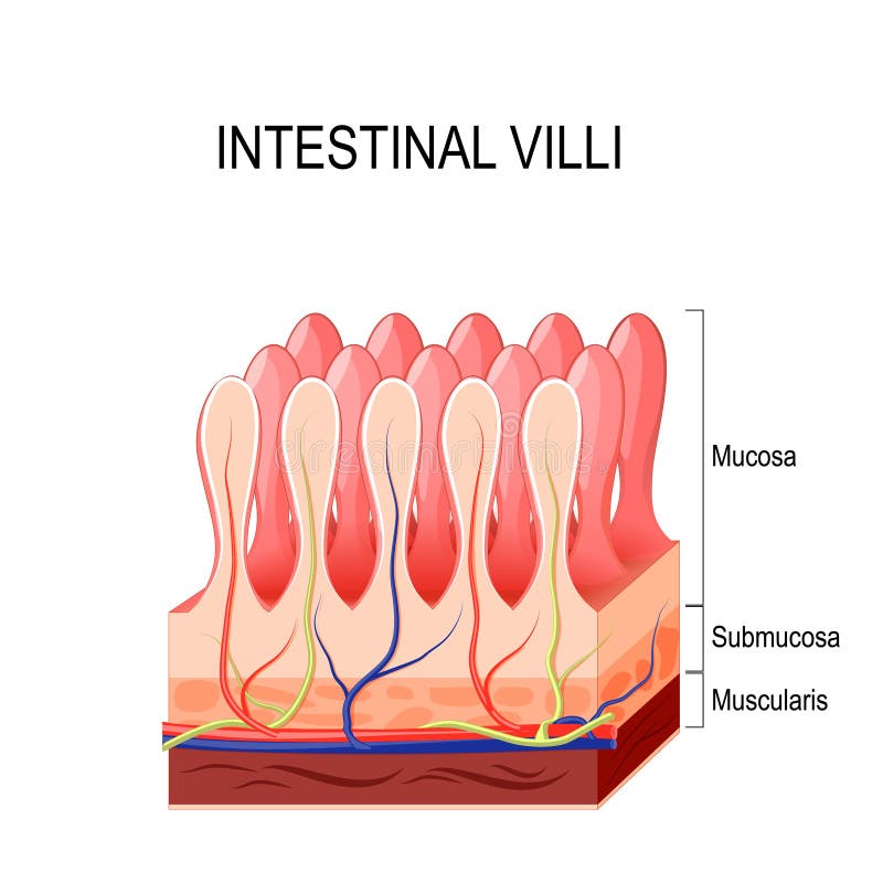
Free with trial Anatomy of the wall of the small intestine villi into the lumen of the intestine increasing its surface area for secretion of enzymes, water, mucus, and for absorption of nutrients. Vector diagram. Histology vectors Intestinal villi. Vector diagram. Anatomy of the wall of the small intestine villi into the lumen of the intestine increasing its surface area for secretion of enzymes, water, mucus, and for absorption of nutrients. Vector diagram
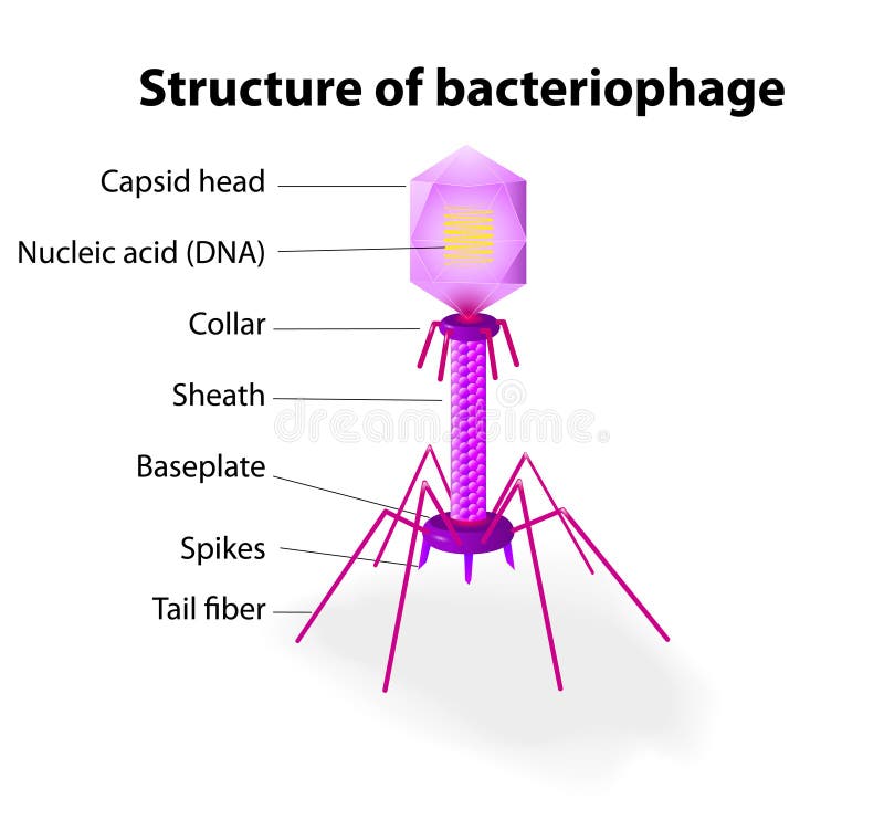
Free with trial Virus. Vector diagram of a typical tailed bacteriophage structure. Histology vectors Structure of virus Bacteriophage. Virus. Vector diagram of a typical tailed bacteriophage structure
Free with trial Classification of skin burns with cross section of skin layers, eps8. Histology vectors Skin burns
Free with trial This high-magnification photomicrograph showcases the intricate histology of human gastric glands, expertly stained with Hematoxylin and Eosin (H&E). The image reveals numerous circular and oval glandular structures, each lined by epithelial cells displaying distinct purple nuclei and pink cytoplasm, surrounding a central lumen. The surrounding connective tissue is also visible in varying shades of pink. This detailed view is ideal for medical education, scientific research, and understanding the normal morphology of gastric tissue within the digestive system. Histology illustrations Human Gastric Glands Histology, H&E Stain Micrograph. This high-magnification photomicrograph showcases the intricate histology of human gastric glands, expertly stained with Hematoxylin and Eosin (H&E). The image reveals numerous circular and oval glandular structures, each lined by epithelial cells displaying distinct purple nuclei and pink cytoplasm, surrounding a central lumen. The surrounding connective tissue is also visible in varying shades of pink. This detailed view is ideal for medical education, scientific research, and understanding the normal morphology of gastric tissue within the digestive system.
Free with trial Stages of Skin cancer, eps8, gradient and mesh printing compatible. Histology vectors Skin cancer
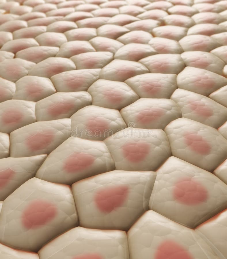
Free with trial Skin tissue with visible cells - CG illustration. Histology illustrations Skin tissue
Free with trial Celiac disease affected small intestine villi. Unhealthy villi with damaged cells and healthy villi. Intestinal villi do not absorb nutrients because of reduced surface area. Histology vectors Celiac disease
Free with trial This image depicts a microscopic histology slide showcasing cellular tissue stained in shades of pink and purple. The slide is set against a transparent background, making it suitable for various scientific and educational applications. The intricate arrangement of cells and tissue structures is clearly visible, highlighting the detailed cellular composition. Histology illustrations Microscopic histology slide showing cellular tissue with transparent background. This image depicts a microscopic histology slide showcasing cellular tissue stained in shades of pink and purple. The slide is set against a transparent background, making it suitable for various scientific and educational applications. The intricate arrangement of cells and tissue structures is clearly visible, highlighting the detailed cellular composition
Free with trial Celiac disease affected small intestine villi on a white background. Healthy villi and unhealthy villi with damaged cells. Histology vectors Celiac disease
Free with trial Healthy small intestine and stomach, bright anatomy illustration, white background. Histology vectors Small intestine and stomach anatomy. Healthy small intestine and stomach, bright anatomy illustration, white background
Free with trial A modern benchtop laboratory microtome or precision imaging instrument featuring a vertical column, adjustable arm, and a control panel with numeric keypad and display. The device is designed for precise sample sectioning, histology preparation, and micro-imaging in research, education, and clinical labs. Ideal for stock photos illustrating lab automation, scientific workflows, and technology-driven biology workflows in professional settings. Histology vectors Laboratory Microtome Precision Benchtop Imaging Instrument for Histology and Research. A modern benchtop laboratory microtome or precision imaging instrument featuring a vertical column, adjustable arm, and a control panel with numeric keypad and display. The device is designed for precise sample sectioning, histology preparation, and micro-imaging in research, education, and clinical labs. Ideal for stock photos illustrating lab automation, scientific workflows, and technology-driven biology workflows in professional settings.
Free with trial Progression of pressure sores (bed sores), eps8. Histology vectors Stages of pressure sores. Progression of pressure sores (bed sores), eps8
Free with trial Alveoli changes in different lung diseases, eps8, gradient and mesh printing compatible. Histology vectors Alveoli in lung diseases. Alveoli changes in different lung diseases, eps8, gradient and mesh printing compatible
Free with trial Biology anatomy background. Illustration of the colon villi. Histology illustrations Colon villi illustration. Biology anatomy background. Illustration of the colon villi.
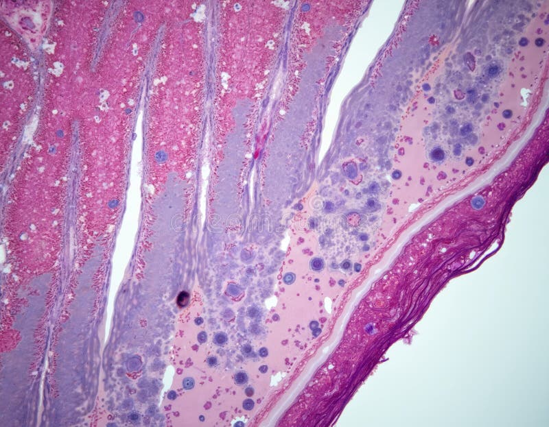
Free with trial Tissue showing microscopic view of parasitic organisms within host tissue with pink staining keywords: microscopic, histology, tissue, cells. Histology illustrations Microscopic view of parasitic organisms within host tissue with pink staining Keywords: microscopic, histology
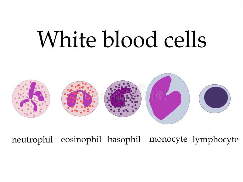
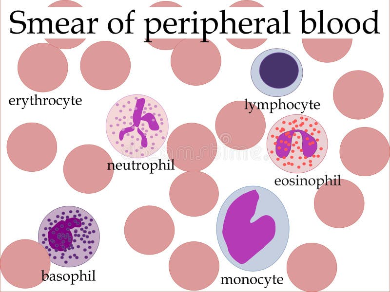
Free with trial Smear of peripheral blood cells under microscope - red blood cells and five types of white blood cells. With writings. Histology vectors Smear of peripheral blood
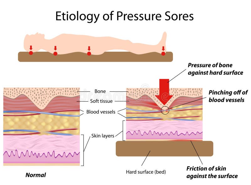
Free with trial Causes of pressure sores (bed sores), eps8. Histology vectors Etiology of pressure sores. Causes of pressure sores (bed sores), eps8
Free with trial Diagram of the alkaline mucous layer in the stomach. Diagram of the histological cross-section of the stomach Layers of the Stomach. Histology vectors Mucous layer in the stomach
Free with trial Microscopic view human tissue structure. Photo displays cells. Histology shows epithelial with connective tissue. Study of internal organs, for medical research. Healthcare for. Histology illustrations Microscopic view human tissue structure. Photo displays cells. Histology shows epithelial with connective tissue. Study of
Free with trial Small intestine wall anatomy, a fold of the intestinal lining, villi and epithelial cell with microvilli detailed illustrations. Histology vectors Small intestine wall anatomy
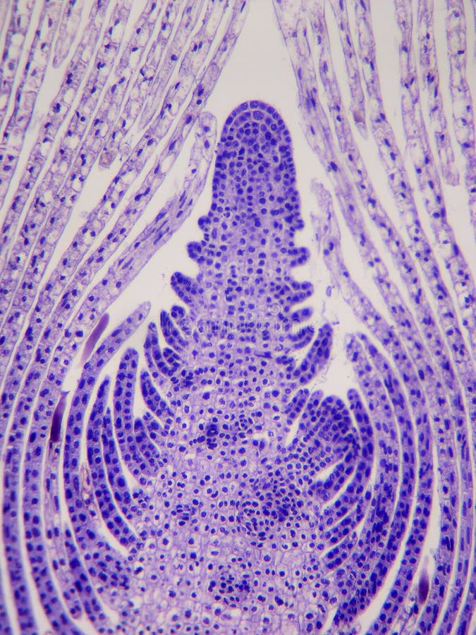
Free with trial Apical meristem of a terminal bud-microscopic longitudinal section. Histology illustrations Apical meristem, terminal bud. Apical meristem of a terminal bud-microscopic longitudinal section
Free with trial Multiple myeloma is a cancer of the bone marrow. healthy plasma cells in the bone marrow mutate and multiply uncontrollably. Myeloma cells suppress the growth of healthy cells that make blood. malignant plasma cells produce a paraprotein inactive antibody or M protein. Histology vectors Multiple myeloma. plasma cell myeloma. Multiple myeloma is a cancer of the bone marrow. healthy plasma cells in the bone marrow mutate and multiply uncontrollably. Myeloma cells suppress the growth of healthy cells that make blood. malignant plasma cells produce a paraprotein inactive antibody or M protein
Free with trial Histology slide displays human tissue sample under microscope. Cells appear purple, pink. Microscopic view of cells. Medical research illustration of cellular biology physiology at. Histology illustrations Histology slide displays human tissue sample under microscope. Cells appear purple, pink. Microscopic view of cells. Medical
Free with trial Intestinal villi and microvilli detailed anatomy on a white background. Histology vectors Intestinal villi and microvilli
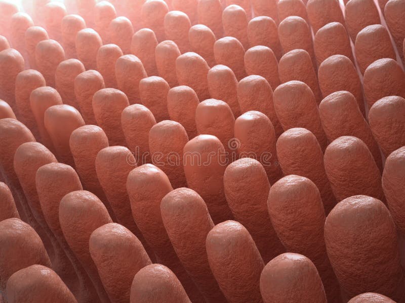
Free with trial Inside the small intestine villi - 3d medical illustration. Histology illustrations Inside the small intestine villi
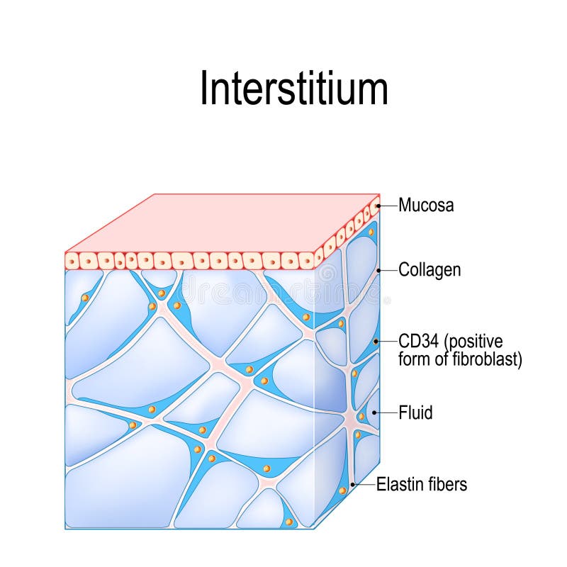
Free with trial Structure of Interstitium. new organ. interstitial is a reservoir and transportation system for nutrients and solutes distributing among organs, and cells. immune regulation. Human Tissues. Histology vectors Structure of Interstitium. Human Tissues. Structure of Interstitium. new organ. interstitial is a reservoir and transportation system for nutrients and solutes distributing among organs, and cells. immune regulation. Human Tissues
Free with trial Histology illustration of human tissue for science research. Microscopic view shows cells structure. Micro tissue with nucleus. Biology medical illustration for, health anatomy. Histology illustrations Histology illustration of human tissue for science research. Microscopic view shows cells structure. Micro tissue with nucleus.
Free with trial Papilloma. Human papilloma virus (HPV). This HPV is the causative agent of warts and birthmarks. Papilloma of skin (wart). Intraductal Papilloma. Process of papillomas Formation. Portrait of woman. Histology vectors Papilloma
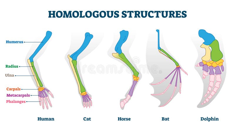
Free with trial Homologous structures vector illustration. Biological species example scheme. Labeled structural diagram with bone titles. Humerus, ulna and carpals in various creature skeletons from common ancestry. Histology vectors Homologous structure vector illustration. Biological species example scheme. Homologous structures vector illustration. Biological species example scheme. Labeled structural diagram with bone titles. Humerus, ulna and carpals in various creature skeletons from common ancestry.
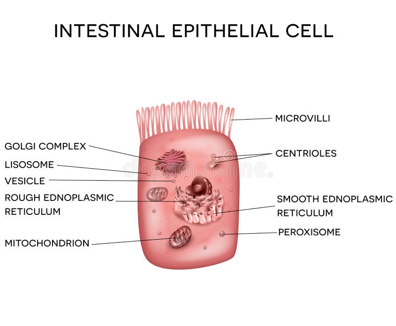
Free with trial Intestinal epithelial cell with microvilli, detailed bright illustation. Histology illustrations Intestinal epithelial cell
Free with trial Illustration of human histology tissue. Microscopic view reveals cells with pink and purple color scheme. Scientific art for medical or, biology study publications. Histology illustrations Illustration of human histology tissue. Microscopic view reveals cells with pink and purple color scheme. Scientific art for
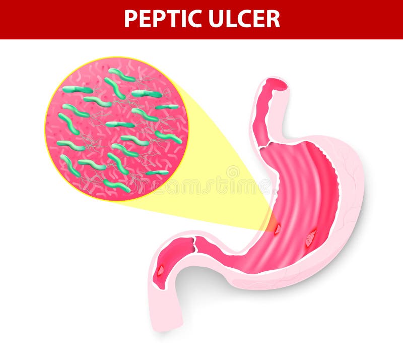
Free with trial A peptic ulcer is a sore on the lining of the stomach or duodenum. Cause of peptic ulcer is bacterial infection. Helicobacter pylori. Vector diagram. Histology vectors Peptic ulcer
Free with trial Small intestine anatomy. Digital illustration. Histology illustrations Bowel and villi cross-section. Small intestine anatomy. Digital illustration.
Free with trial Detailed Microscopic View of Mammalian Ovary Histology Featuring Oocytes and Follicular Cells in a Beautiful Blue-Violet Stain. Generative AI. Histology illustrations Detailed Microscopic View of Mammalian Ovary Histology Featuring Oocytes and Follicular Cells in a Beautiful BlueViolet Stain
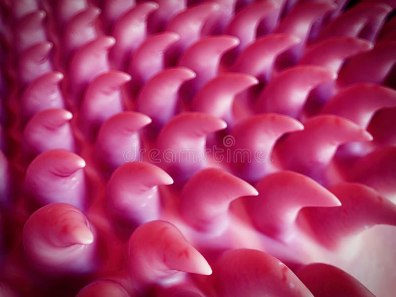
Free with trial Intestinal villi anatomy on a beautiful abstract technology background. Histology vectors Healthy intestine villi. Intestinal villi anatomy on a beautiful abstract technology background
Free with trial Gastrointestinal tract anatomy. Intestinal villi, small intestine lining, epithelial cells with microvilli detailed illustration. Histology vectors Gastrointestinal tract detailed anatomy. Gastrointestinal tract anatomy. Intestinal villi, small intestine lining, epithelial cells with microvilli detailed illustration.
Free with trial Epithelia are tissues consisting of closely apposed cells without intervening intercellular substances. Histology illustrations G Unicellular Epithelium. Epithelia are tissues consisting of closely apposed cells without intervening intercellular substances.
Free with trial Close up view shows histology tissue sample with microscope. Human cells examination in lab for scientific research. Used for health banner medical article. Lab testing equipment. Histology illustrations Close up view shows histology tissue sample with microscope. Human cells examination in lab for scientific research. Used for
Free with trial 3d illustration of a mestastising cancer cell from a malignant tumor crawling as it spreads through the body to infect secondary organs. Histology illustrations Metastasizing cancer cell. 3d illustration of a mestastising cancer cell from a malignant tumor crawling as it spreads through the body to infect secondary organs
Free with trial Celiac disease Small intestine lining damage. good and damaged villi. leaky gut progression on white. Histology vectors Celiac disease Small intestine lining damage. good and damaged villi . leaky gut progression
Free with trial Celiac disease Small intestine lining damage. good and damaged villi. leaky gut progression on white background. Histology vectors Celiac disease Small intestine lining damage. good and damaged villi . leaky gut progression
Free with trial Microscopic view of human tissue cells showing abnormal structures. Detailed histology image reveals cellular changes and potential disease indicators. Medical research and pathology,. Histology illustrations Microscopic view of human tissue cells showing abnormal structures. Detailed histology image reveals cellular changes and
Free with trial Amyloid plaques may damage and kill neurons by generating reactive oxygen species during its self-aggregation. Histology illustrations Neuron with amyloid plaques. Amyloid plaques may damage and kill neurons by generating reactive oxygen species during its self-aggregation.
Free with trial Celiac disease affected small intestine villi. Damaged cells by body's reaction to gluten. Intestinal villi do not absorb nutrients because of reduced surface area. Abstract grey background. Histology vectors Celiac disease
Free with trial Celiac disease affected small intestine villi. Healthy villi and unhealthy villi with damaged cells on a blue technology background. Histology vectors Celiac disease
Free with trial Microscopic view of human nerve tissue with layered structures. Shows detailed histology of axons, myelin sheath. Useful for medical research educational content. Offers insight into. Histology illustrations Microscopic view of human nerve tissue with layered structures. Shows detailed histology of axons, myelin sheath. Useful for
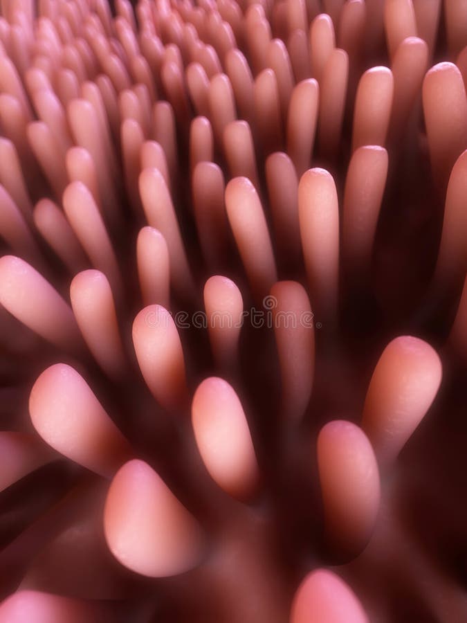
Free with trial SEM style illustration of the tongue surface. Histology illustrations The tongue surface
Free with trial 1. Healthy neuron. 2. Neuron with amyloid plaques yellow. 3. Dead neuron being digested by microglia cells red. Histology illustrations Neuron network with amyloid plaques. 1. Healthy neuron. 2. Neuron with amyloid plaques yellow. 3. Dead neuron being digested by microglia cells red.
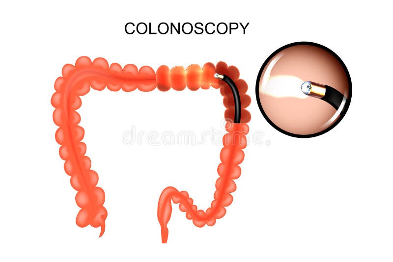
Free with trial Vector illustration of large intestine, colonoscop. medicine. Histology vectors The colon, colonoscopy. Vector illustration of large intestine, colonoscop. medicine
Free with trial Microscopic image shows purple-stained tissue structure with distinct cells, lobules. Histology slide cellular organization pattern for medical science education research purposes. Histology illustrations Microscopic image shows purple-stained tissue structure with distinct cells, lobules. Histology slide cellular organization
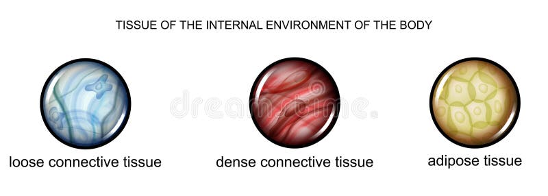
Free with trial Vector illustration of tissues of the internal environment of the body: loose, dense connective tissue and adipose tissue. Histology vectors Tissue of the internal environment of the body. Vector illustration of tissues of the internal environment of the body: loose, dense connective tissue and adipose tissue
Free with trial 3d rendered medically accurate illustration of too many white blood cells due to leukemia. Histology illustrations Leukemia