Free with trial Biotechnical organisms and objects in medicine and science, superimposed over technical paper. Human cell structures illustrations Biotechnical Abstract. Biotechnical organisms and objects in medicine and science, superimposed over technical paper.
Free with trial Typical structures of a bacterium (rod shape), eps8. Human cell structures vectors Structure of a bacterial cell. Typical structures of a bacterium (rod shape), eps8
Free with trial Skin male and female. Men tend to have more connective tissue that are arranged in such a way that it gives better support to the surrounding structures of the skin and fat. A womans fat cells are held in place below the skin by cube-like structures contained by collagen bands. Vector diagram. Human cell structures vectors Skin male and female
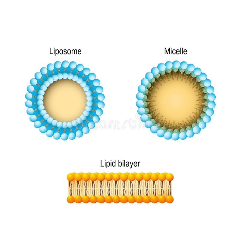
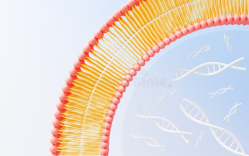
Free with trial Cell membrane Lipid bilayer, Micelle, Liposome. Phospholipids. Human cell structures vectors Cell membrane Lipid bilayer, Micelle, Liposome. Phospholipids
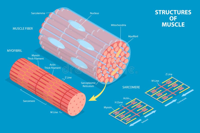
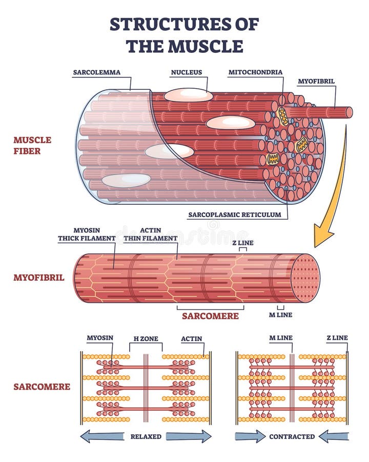
Free with trial Structures of muscle with fiber, myofibril and sarcomere contraction outline diagram. Labeled educational isolated parts closeup description from anatomical and physiology sides vector illustration. Human cell structures vectors Structures of muscle with fiber, myofibril and sarcomere outline diagram. Structures of muscle with fiber, myofibril and sarcomere contraction outline diagram. Labeled educational isolated parts closeup description from anatomical and physiology sides vector illustration.
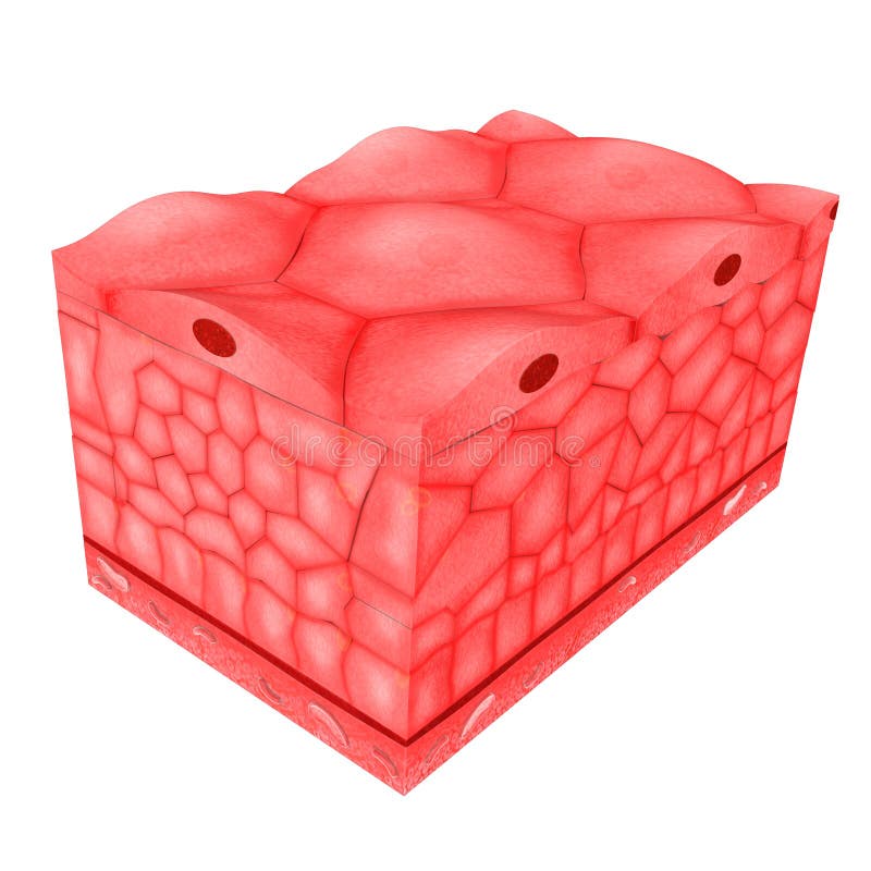
Free with trial Epithelial tissue covers the whole surface of the body. It is made up of cells closely packed and ranged in one or more layers. This tissue is specialised to form the covering or lining of all internal and external body surfaces. Epithelial tissue that occurs on surfaces on the interior of the body is known as endothelium. Epithelial cells are packed tightly together, with almost no intercellular spaces and only a small amount of intercellular substance. Epithelial tissue, regardless of the type, is usually separated from the underlying tissue by a thin sheet of connective tissue; basement membrane. The basement membrane provides structural support for the epithelium and also binds it to neighbouring structures. Human cell structures illustrations Epithelial Tissues. Epithelial tissue covers the whole surface of the body. It is made up of cells closely packed and ranged in one or more layers. This tissue is specialised to form the covering or lining of all internal and external body surfaces. Epithelial tissue that occurs on surfaces on the interior of the body is known as endothelium. Epithelial cells are packed tightly together, with almost no intercellular spaces and only a small amount of intercellular substance. Epithelial tissue, regardless of the type, is usually separated from the underlying tissue by a thin sheet of connective tissue; basement membrane. The basement membrane provides structural support for the epithelium and also binds it to neighbouring structures.
Free with trial Epithelial tissue covers the whole surface of the body. It is made up of cells closely packed and ranged in one or more layers. This tissue is specialised to form the covering or lining of all internal and external body surfaces. Epithelial tissue that occurs on surfaces on the interior of the body is known as endothelium. Epithelial cells are packed tightly together, with almost no intercellular spaces and only a small amount of intercellular substance. Epithelial tissue, regardless of the type, is usually separated from the underlying tissue by a thin sheet of connective tissue; basement membrane. The basement membrane provides structural support for the epithelium and also binds it to neighbouring structures. Human cell structures illustrations Epithelial Tissues. Epithelial tissue covers the whole surface of the body. It is made up of cells closely packed and ranged in one or more layers. This tissue is specialised to form the covering or lining of all internal and external body surfaces. Epithelial tissue that occurs on surfaces on the interior of the body is known as endothelium. Epithelial cells are packed tightly together, with almost no intercellular spaces and only a small amount of intercellular substance. Epithelial tissue, regardless of the type, is usually separated from the underlying tissue by a thin sheet of connective tissue; basement membrane. The basement membrane provides structural support for the epithelium and also binds it to neighbouring structures.
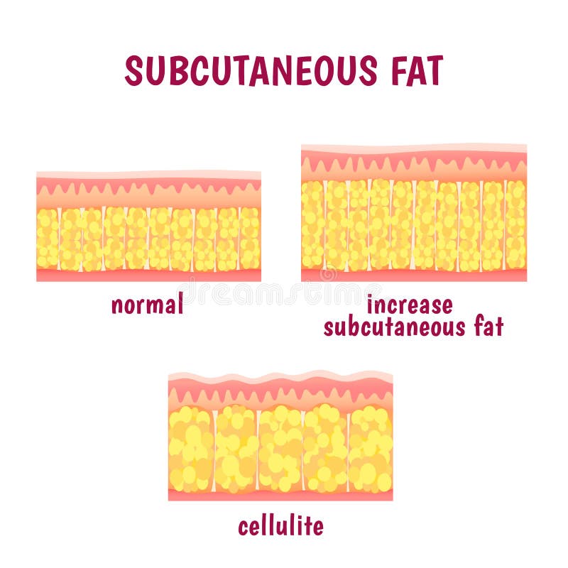
Free with trial Leather sectional layer of subcutaneous fat, cellulite scheme. Human cell structures vectors Leather sectional layer of subcutaneous fat
Free with trial DNA glass structures closeup against a blue backdrop - medical 3D illustration - rendering. Human cell structures illustrations Complex DNA - medical 3D illustration. DNA glass structures closeup against a blue backdrop - medical 3D illustration - rendering
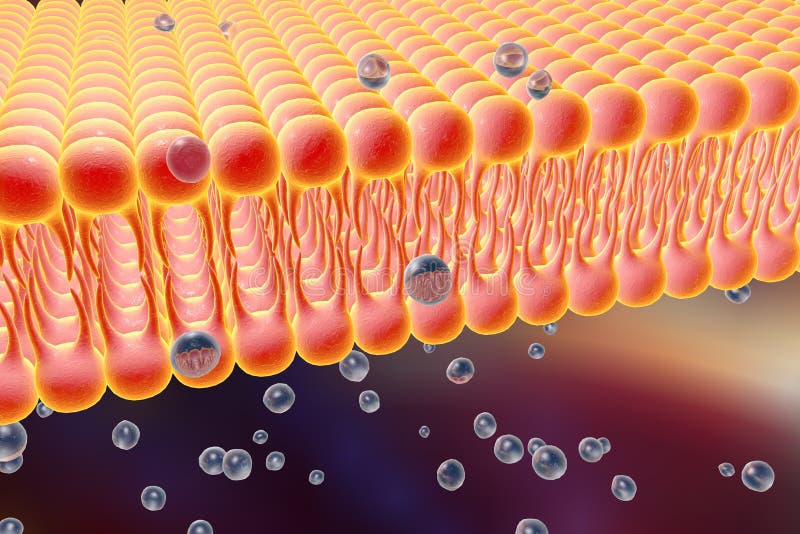
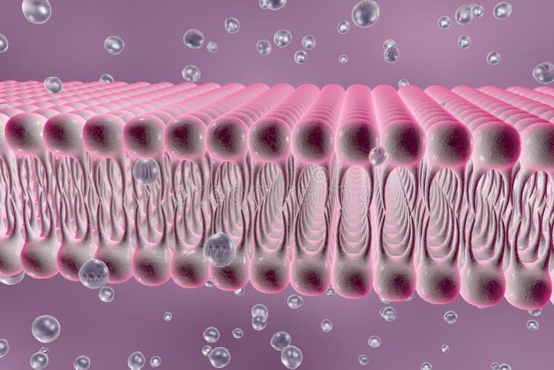
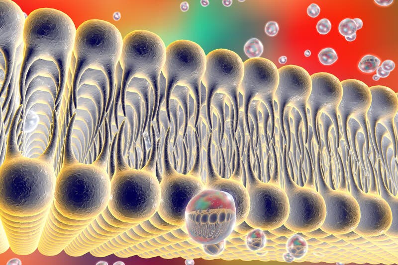
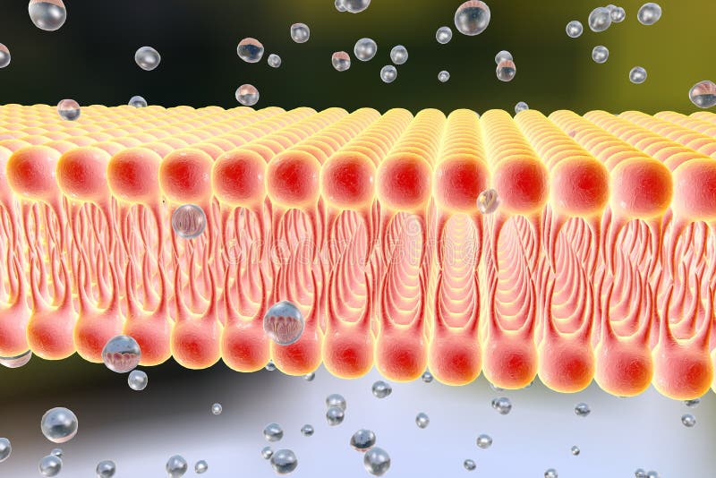
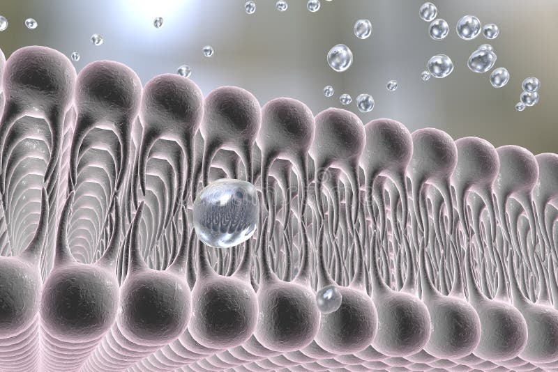
Free with trial Cell membrane, lipid bilayer, 3d illustration of a diffusion of liquid molecules through cell membrane. Human cell structures illustrations Cellular membrane with diffusion of molecules. Cell membrane, lipid bilayer, 3d illustration of a diffusion of liquid molecules through cell membrane
Free with trial Cell membrane, lipid bilayer, 3d illustration of a diffusion of liquid molecules through cell membrane. Human cell structures illustrations Cellular membrane with diffusion of molecules. Cell membrane, lipid bilayer, 3d illustration of a diffusion of liquid molecules through cell membrane
Free with trial Cell membrane, lipid bilayer, 3d illustration of a diffusion of liquid molecules through cell membrane. Human cell structures illustrations Cellular membrane with diffusion of molecules. Cell membrane, lipid bilayer, 3d illustration of a diffusion of liquid molecules through cell membrane
Free with trial Centrioles are cylindrical organelles. Found in most eukaryotic cells. The pairs of adjacent centrioles make up structures called centrosomes. Human cell structures vectors Anatomy of Centrioles, Centrioles are cylindrical organelles. Centrioles are cylindrical organelles.Found in most eukaryotic cells. The pairs of adjacent centrioles make up structures called centrosomes.
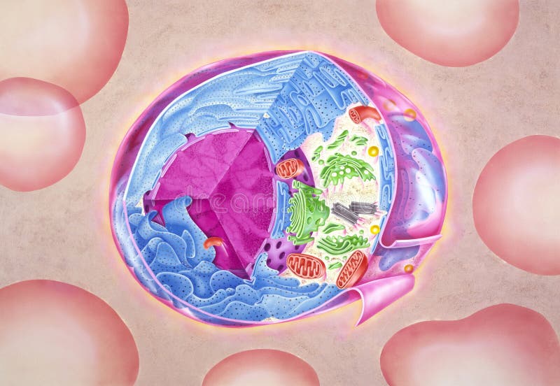
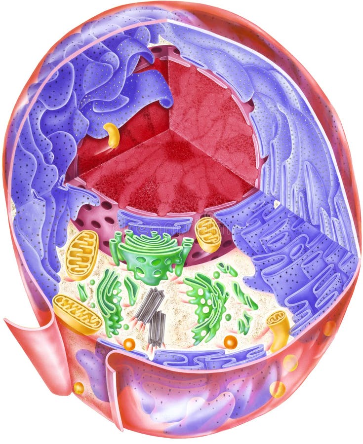
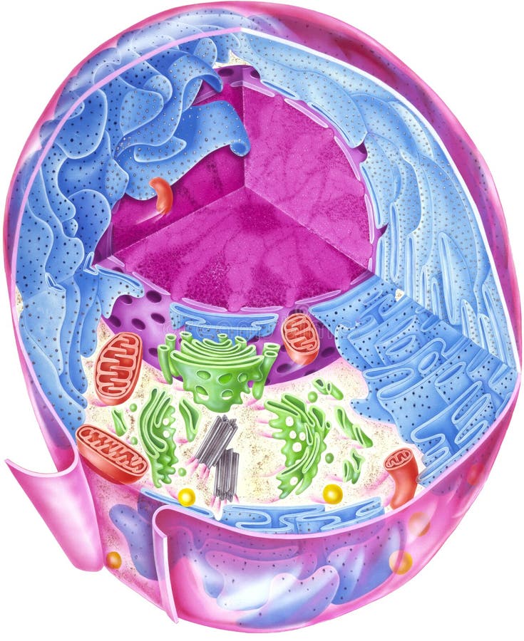
Free with trial Human cell showing internal structures. Human cell structures illustrations Cell - Showing Internal Structures. Human cell showing internal structures.
Free with trial Human cell showing internal structures. Human cell structures illustrations Cell - Showing Internal Structures. Human cell showing internal structures.
Free with trial A human cell showing an array of internal structures. Human cell structures illustrations Cell - Showing Internal Structures. A human cell showing an array of internal structures.
Free with trial This microscopic image showcases the intricate architecture of areolar connective tissue, a crucial component of the human body. Areolar tissue, with its delicate network of fibers and cells, plays a vital role in supporting various structures and facilitating essential bodily functions. Observe the interwoven collagen and elastic fibers, the interspersed ground substance, and the various cell. Human cell structures illustrations Unveiling the Structure and Location of Areolar Connective Tissue A Microscopic Exploration of Its Role in the Human. This microscopic image showcases the intricate architecture of areolar connective tissue, a crucial component of the human body. Areolar tissue, with its delicate network of fibers and cells, plays a vital role in supporting various structures and facilitating essential bodily functions. Observe the interwoven collagen and elastic fibers, the interspersed ground substance, and the various cell
Free with trial 3d render cell membrane Phospholipid Bilayer. Layers of molecules. Gene expression. nucleotide database. Fluid mosaic model for education. Human gene. Plasma membrane structures. Human cell structures illustrations 3d render cell membrane Phospholipid Bilayer. Layers of molecules. Gene expression. nucleotide database. Fluid mosaic model for
Free with trial 3d render cell membrane Phospholipid Bilayer. Layers of molecules. Gene expression. nucleotide database. Fluid mosaic model for education. Human gene. Plasma membrane structures. Human cell structures illustrations 3d render cell membrane Phospholipid Bilayer. Layers of molecules. Gene expression. nucleotide database. Fluid mosaic model for
Free with trial 3d render cell membrane Phospholipid Bilayer. Layers of molecules. Gene expression. nucleotide database. Fluid mosaic model for education. Human gene. Plasma membrane structures. Human cell structures illustrations 3d render cell membrane Phospholipid Bilayer. Layers of molecules. Gene expression. nucleotide database. Fluid mosaic model for
Free with trial 3d render cell membrane Phospholipid Bilayer. Layers of molecules. Gene expression. nucleotide database. Fluid mosaic model for education. Human gene. Plasma membrane structures. Human cell structures illustrations 3d render cell membrane Phospholipid Bilayer. Layers of molecules. Gene expression. nucleotide database. Fluid mosaic model for
Free with trial 3d render cell membrane Phospholipid Bilayer. Layers of molecules. Gene expression. nucleotide database. Fluid mosaic model for education. Human gene. Plasma membrane structures. Human cell structures illustrations 3d render cell membrane Phospholipid Bilayer. Layers of molecules. Gene expression. nucleotide database. Fluid mosaic model for
Free with trial Detailed medical illustration showing the anatomy of the human trachea and esophagus. The image includes a cross-sectional view with histological details of the tracheal wall and surrounding structures. Human cell structures vectors Detailed medical illustration showing the anatomy of the human trachea and esophagus. The image includes a cross-sectional view
Free with trial Close-up of 3D rendered human DNA spiral structures. Human cell structures illustrations Genetics concept depicting magnified double helix DNA strands. Close-up of 3D rendered human DNA spiral structures.
Free with trial Illustration of monocyte cell to macrophage cell differentiation. Human cell structures vectors Monocyte cell to macrophage cell differentiation
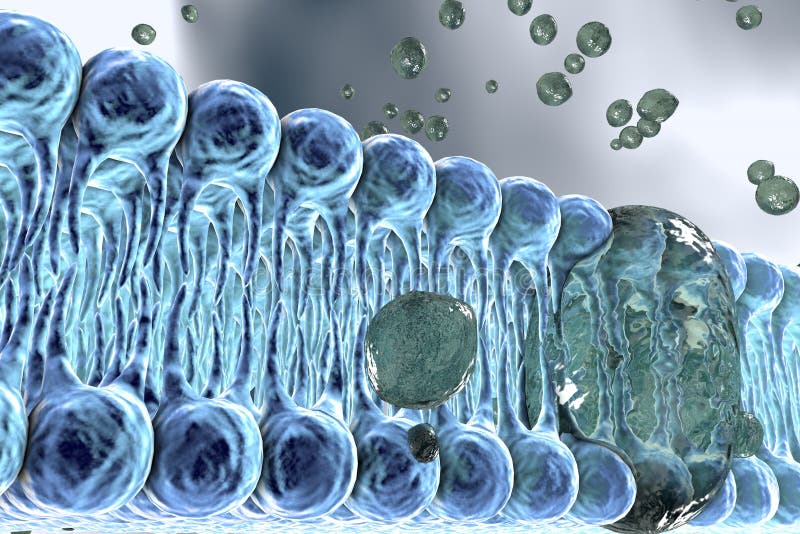
Free with trial Cell membrane, lipid bilayer, close-up view. 3D illustration. Human cell structures illustrations Cell membrane, lipid bilayer
Free with trial Cell membrane, lipid bilayer, close-up view. 3D illustration. Human cell structures illustrations Cell membrane, lipid bilayer
Free with trial Cell membrane, lipid bilayer, close-up view. 3D illustration of a diffusion of liquid molecules through cell membrane. Human cell structures illustrations Cell membrane, lipid bilayer
Free with trial Parts and organs of human body, isolated icons with inscriptions. Heart and brain, kidneys and lungs, stomach and pancreas, skin and reproductive systems of men and women. Vector in flat style. Human cell structures vectors Organs of human body, systems and structures part. Parts and organs of human body, isolated icons with inscriptions. Heart and brain, kidneys and lungs, stomach and pancreas, skin and reproductive systems of men and women. Vector in flat style
Free with trial This digital illustration presents a striking close-up view of a nerve cell, drawing viewers into the fascinating world of biology and neuroscience. Occupying the majority of the frame, the nerve cell stands out against the deep black background, offering an intimate glimpse into the intricate structures that make up the human nervous system. The nerve cell, also known as a neuron, exhibits a captivating blue hue that infuses the image with an otherworldly quality. The cell's intricate architecture is on full display, with its numerous dendrites extending outwards like radiant beams of light. The image provides a profound visual representation of the complexity and beauty of life at its most microscopic level. Despite its simplicity, the image subtly evokes the grandeur and mystery of the universe, with the neuron resembling a glowing sun against the vast black backdrop. The neuron's radiant blue glow brings an element of vibrancy and energy to the image, further emphasizing the cell's vital role in transmitting information throughout the body. The image is not just a close-up of a nerve cell, it's a journey into the microscopic universe within us, a homage to the intricate complexity of life. Reminiscent of celestial bodies in the endless expanse of space, this image serves as a potent reminder of the magical and complex world that resides within each one of us. Human cell structures illustrations A close up digital illustration of a nerve cell. This digital illustration presents a striking close-up view of a nerve cell, drawing viewers into the fascinating world of biology and neuroscience. Occupying the majority of the frame, the nerve cell stands out against the deep black background, offering an intimate glimpse into the intricate structures that make up the human nervous system. The nerve cell, also known as a neuron, exhibits a captivating blue hue that infuses the image with an otherworldly quality. The cell's intricate architecture is on full display, with its numerous dendrites extending outwards like radiant beams of light. The image provides a profound visual representation of the complexity and beauty of life at its most microscopic level. Despite its simplicity, the image subtly evokes the grandeur and mystery of the universe, with the neuron resembling a glowing sun against the vast black backdrop. The neuron's radiant blue glow brings an element of vibrancy and energy to the image, further emphasizing the cell's vital role in transmitting information throughout the body. The image is not just a close-up of a nerve cell, it's a journey into the microscopic universe within us, a homage to the intricate complexity of life. Reminiscent of celestial bodies in the endless expanse of space, this image serves as a potent reminder of the magical and complex world that resides within each one of us.
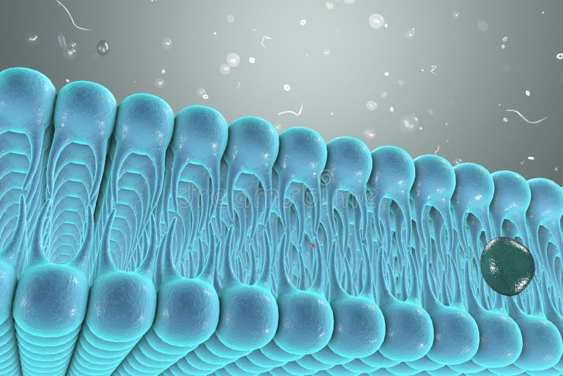
Free with trial In biology, the extracellular matrix (ECM) is a collection of extracellular molecules secreted by cells that provides structural and biochemical support to the surrounding cells. [1] Because multicellularity evolved independently in different multicellular lineages, the composition of ECM varies between multicellular structures; however, cell adhesion, cell-to-cell communication and differentiation are common functions of the ECM. Human cell structures illustrations G Multicellular Epithelium. In biology, the extracellular matrix (ECM) is a collection of extracellular molecules secreted by cells that provides structural and biochemical support to the surrounding cells.[1] Because multicellularity evolved independently in different multicellular lineages, the composition of ECM varies between multicellular structures; however, cell adhesion, cell-to-cell communication and differentiation are common functions of the ECM.
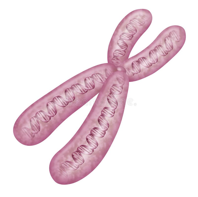
Free with trial In the nucleus of each cell, the DNA molecule is packaged into thread-like structures called chromosomes. Each chromosome is made up of DNA tightly coiled many times around proteins called histones that support its structure. Chromosomes are not visible in the cell’s nucleus—not even under a microscope—when the cell is not dividing. However, the DNA that makes up chromosomes becomes more tightly packed during cell division and is then visible under a microscope. Most of what researchers know about chromosomes was learned by observing chromosomes during cell division. Each chromosome has a constriction point called the centromere, which divides the chromosome into two sections, or “arms. ” The short arm of the chromosome is labeled the “p arm. ” The location of the centromere on each chromosome gives the chromosome its characteristic shape, and can be used to help describe the location of specific genes. Human cell structures illustrations Chromosome
Free with trial In biology, the extracellular matrix (ECM) is a collection of extracellular molecules secreted by cells that provides structural and biochemical support to the surrounding cells. [1] Because multicellularity evolved independently in different multicellular lineages, the composition of ECM varies between multicellular structures; however, cell adhesion, cell-to-cell communication and differentiation are common functions of the ECM. Human cell structures illustrations G Multicellular Epithelium. In biology, the extracellular matrix (ECM) is a collection of extracellular molecules secreted by cells that provides structural and biochemical support to the surrounding cells.[1] Because multicellularity evolved independently in different multicellular lineages, the composition of ECM varies between multicellular structures; however, cell adhesion, cell-to-cell communication and differentiation are common functions of the ECM.
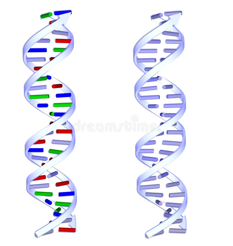
Free with trial Two 3D DNA structures on white background. Human cell structures illustrations Two DNA structures on white background
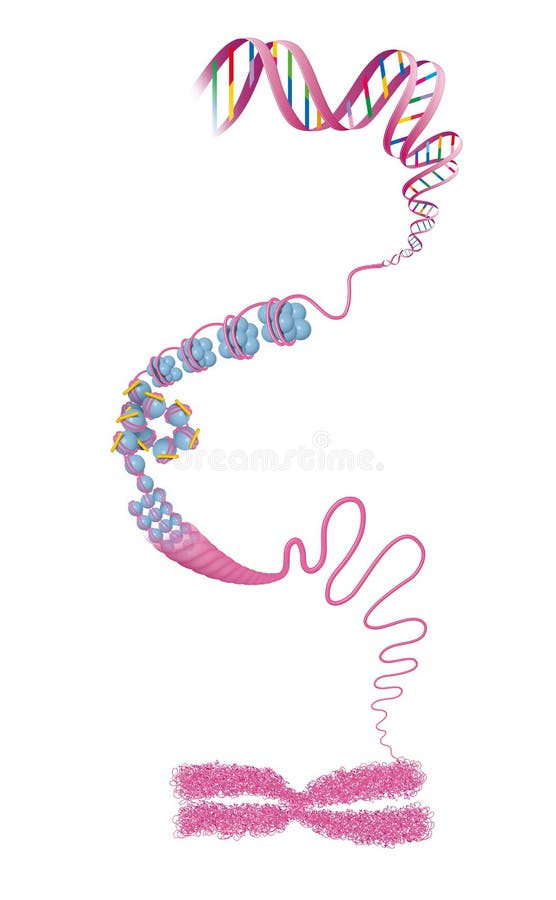
Free with trial In the nucleus of each cell, the DNA molecule is packaged into thread-like structures called chromosomes. Each chromosome is made up of DNA tightly coiled many times around proteins called histones that support its structure. DNA and histone proteins are packaged into structures called chromosomes. Human cell structures illustrations DNA and histone proteins are packaged into chromosomes. In the nucleus of each cell, the DNA molecule is packaged into thread-like structures called chromosomes. Each chromosome is made up of DNA tightly coiled many times around proteins called histones that support its structure. DNA and histone proteins are packaged into structures called chromosomes
Free with trial In the nucleus of each cell, the DNA molecule is packaged into thread-like structures called chromosomes. Each chromosome is made up of DNA tightly coiled many times around proteins called histones that support its structure. Human cell structures illustrations Chromosome - DNA molecule. Illustration. In the nucleus of each cell, the DNA molecule is packaged into thread-like structures called chromosomes. Each chromosome is made up of DNA tightly coiled many times around proteins called histones that support its structure
Free with trial Cell membrane, lipid bilayer, 3d illustration of a diffusion of liquid molecules through cell membrane. Human cell structures illustrations Cellular membrane with diffusion of molecules. Cell membrane, lipid bilayer, 3d illustration of a diffusion of liquid molecules through cell membrane
Free with trial Each tip of the “Y” of an antibody contains a paratope that is specific for one particular epitope analogous to a lock and key on an antigen, allowing these two structures to bind together with precision. Using this binding mechanism, an antibody can tag a microbe or an infected cell. Human cell structures illustrations Each antibody binds to a specific antigen; an interaction similar to a lock and key. Each tip of the “Y” of an antibody contains a paratope that is specific for one particular epitope analogous to a lock and key on an antigen, allowing these two structures to bind together with precision. Using this binding mechanism, an antibody can tag a microbe or an infected cell.
Free with trial DNA structures closeup against a blue backdrop - medical 3D illustration - rendering. Human cell structures illustrations Complex DNA - medical 3D illustration. DNA structures closeup against a blue backdrop - medical 3D illustration - rendering
Free with trial Taste buds contain the taste receptor cells, which are also known as gustatory cells. The taste receptors are located around the small structures known as papillae found on the upper surface of the tongue, soft palate, upper esophagus, the cheek, and epiglottis. Human cell structures illustrations Taste bud structure in the human tongue. Taste buds contain the taste receptor cells, which are also known as gustatory cells. The taste receptors are located around the small structures known as papillae found on the upper surface of the tongue, soft palate, upper esophagus, the cheek, and epiglottis.
Free with trial Cell membrane, lipid bilayer, 3d illustration of a diffusion of liquid molecules through cell membrane. Human cell structures illustrations Cellular membrane with diffusion of molecules. Cell membrane, lipid bilayer, 3d illustration of a diffusion of liquid molecules through cell membrane
Free with trial A striking close-up image revealing a microscopic environment. It shows spherical cells invaded by a viscous substance, a biofilm or a layer of mucus, from which fine filamentous structures emerge. Ideal for illustrating complex topics such as microbiology, medical research, infection control, cell therapies, or health issues related to bacterial biofilms and the body's defenses. Human cell structures illustrations The Threat of Biofilm: Microbiology and Cellular Defense. A striking close-up image revealing a microscopic environment. It shows spherical cells invaded by a viscous substance, a biofilm or a layer of mucus, from which fine filamentous structures emerge. Ideal for illustrating complex topics such as microbiology, medical research, infection control, cell therapies, or health issues related to bacterial biofilms and the body's defenses.
Free with trial Cell membrane, lipid bilayer, 3d illustration of a diffusion of liquid molecules through cell membrane. Human cell structures illustrations Cellular membrane with diffusion of molecules. Cell membrane, lipid bilayer, 3d illustration of a diffusion of liquid molecules through cell membrane
Free with trial Cell membrane, lipid bilayer, 3d illustration of a diffusion of liquid molecules through cell membrane. Human cell structures illustrations Cellular membrane with diffusion of molecules. Cell membrane, lipid bilayer, 3d illustration of a diffusion of liquid molecules through cell membrane
Free with trial Cell membrane, lipid bilayer, 3d illustration of a diffusion of liquid molecules through cell membrane. Human cell structures illustrations Cellular membrane with diffusion of molecules. Cell membrane, lipid bilayer, 3d illustration of a diffusion of liquid molecules through cell membrane
Free with trial Cell membrane, lipid bilayer, 3d illustration of a diffusion of liquid molecules through cell membrane. Human cell structures illustrations Cellular membrane with diffusion of molecules. Cell membrane, lipid bilayer, 3d illustration of a diffusion of liquid molecules through cell membrane
Free with trial The arrangement of flagella refers to how these whip-like structures are positioned on the surface of a cell. Human cell structures vectors Flagella arrangement. The arrangement of flagella refers to how these whip-like structures are positioned on the surface of a cell.
Free with trial Cell membrane, lipid bilayer, 3d illustration of a diffusion of liquid molecules through cell membrane. Human cell structures illustrations Cellular membrane with diffusion of molecules. Cell membrane, lipid bilayer, 3d illustration of a diffusion of liquid molecules through cell membrane
Free with trial Cell membrane, lipid bilayer, 3d illustration of a diffusion of liquid molecules through cell membrane. Human cell structures illustrations Cellular membrane with diffusion of molecules. Cell membrane, lipid bilayer, 3d illustration of a diffusion of liquid molecules through cell membrane
Free with trial Cell membrane, lipid bilayer, 3d illustration of a diffusion of liquid molecules through cell membrane. Human cell structures illustrations Cellular membrane with diffusion of molecules. Cell membrane, lipid bilayer, 3d illustration of a diffusion of liquid molecules through cell membrane
Free with trial Antibody icon. Types of antibodies and immunoglobulin structures. IgE concept. Object for use in medicine, education and science. Vector illustration flat design. Isolated on white background. Human cell structures vectors Antibody icon. Types of antibodies and immunoglobulin structures
Free with trial Ovarian cancer is a cancerous tumor of an ovary. [10] It may originate from the ovary itself or more commonly from communicating nearby structures such as fallopian tubes or the inner lining of the abdomen. Human cell structures illustrations CAR T cell therapy in Ovarian cancer - closeup view 3d illustration. Ovarian cancer is a cancerous tumor of an ovary.[10] It may originate from the ovary itself or more commonly from communicating nearby structures such as fallopian tubes or the inner lining of the abdomen.
Free with trial Sympathetic trunk ganglia. Paravertebral ganglia of the sympathetic nervous system. Nerve cell bodies of the spinal cord. Brain and spinal cord and body communication. Flat vector illustration. Human cell structures vectors Sympathetic trunk ganglia. Paravertebral ganglia of the sympathetic
Free with trial Chromatin consists of long strands of DNA wrapped around proteins called histones. The primary structure of chromatin is the nucleosome, which consists of DNA wound around a core of eight histone proteins. These nucleosomes are further compacted and organized into higher-order structures, ultimately forming chromosomes during cell division. Human cell structures vectors Chromatin
Free with trial 3D Isometric Flat Vector Conceptual Illustration of Structures Of Muscle , Medical Educational Diagram. Human cell structures vectors 3D Isometric Flat Vector Conceptual Illustration of Structures Of Muscle
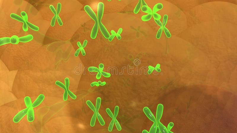
Free with trial Chromosomes are thread-like structures found within the nucleus of eukaryotic cells that contain the genetic material (DNA) of an organism. They are responsible for storing and transmitting genetic information from one generation to the next. Chromosomes are visible under a microscope during cell division when they condense into distinct, easily observable structures. Human cell structures vectors Couple of chromosomes. Chromosomes are thread-like structures found within the nucleus of eukaryotic cells that contain the genetic material (DNA) of an organism. They are responsible for storing and transmitting genetic information from one generation to the next. Chromosomes are visible under a microscope during cell division when they condense into distinct, easily observable structures.
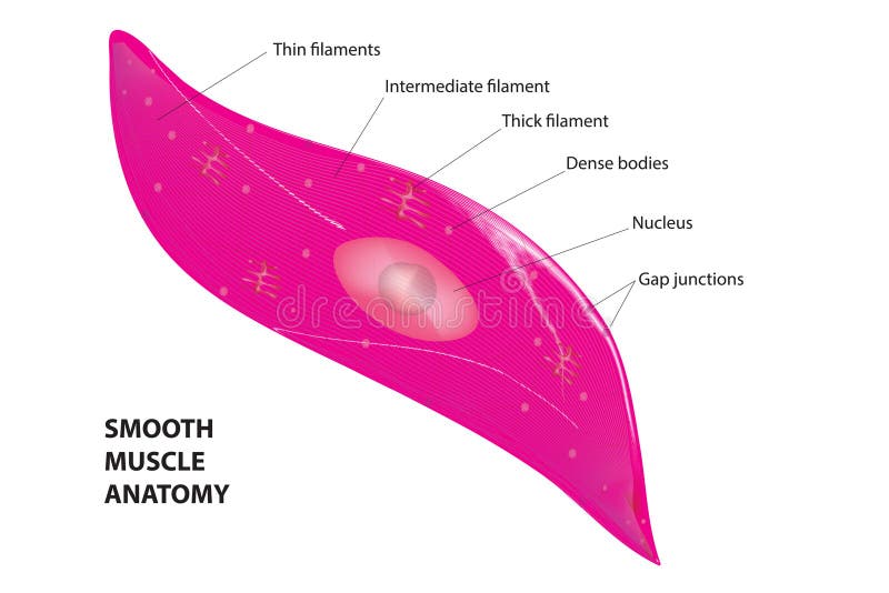
Free with trial Smooth muscle cells, also known as smooth muscle fibers or myocytes, are specialized cells found in the walls of various organs and structures throughout the body. These cells play crucial roles in involuntary movements, such as the contraction and relaxation of hollow organs, blood vessels, and other structures involved in the regulation of physiological processes. Human cell structures vectors Smooth muscle cell diagram. Smooth muscle cells, also known as smooth muscle fibers or myocytes, are specialized cells found in the walls of various organs and structures throughout the body. These cells play crucial roles in involuntary movements, such as the contraction and relaxation of hollow organs, blood vessels, and other structures involved in the regulation of physiological processes.
Free with trial A large set of multi-colored beautiful medical scientific twisted structures of spirals of abstract models of DNA genes on a white background. Vector illustration. Human cell structures vectors A large set of multi-colored beautiful medical scientific twisted structures of spirals of abstract models of DNA genes on a white
Free with trial High-magnification microscopic view of a hair follicle and surrounding cells. The image shows intricate details of the structures and components of this biological system, illuminated with a soft, warm light. The image is suitable for scientific and educational materials, or a general microstock presentation of cellular biology, scientific research, and biological topics. The image is suitable. Human cell structures illustrations Microscopic hair follicle and cells. High-magnification microscopic view of a hair follicle and surrounding cells. The image shows intricate details of the structures and components of this biological system, illuminated with a soft, warm light. The image is suitable for scientific and educational materials, or a general microstock presentation of cellular biology, scientific research, and biological topics. The image is suitable
Free with trial Neuron Network 3D Rendering of Complex Neuron Connections Within Human Brain, Blue Background. , Generated by AI. Human cell structures illustrations Neuron Network 3D Rendering of Complex Neuron Connections Within Human Brain, Blue Background.
Free with trial This image showcases a fascinating perspective on the lungs, revealing the intricate interplay between cellular structures and microscopic organisms such as bacteria, viruses, fungi, and pathogens. It creates awareness and understanding of how these tiny beings impact human health and respiratory systems. Human cell structures illustrations Unveiling Microscopic Worlds: Exploring the Intricacies of Bacteria and Viruses in Lungs and Beyond. This image showcases a fascinating perspective on the lungs, revealing the intricate interplay between cellular structures and microscopic organisms such as bacteria, viruses, fungi, and pathogens. It creates awareness and understanding of how these tiny beings impact human health and respiratory systems
Free with trial Abstract biology. Microscopy of colorful organic structures, microorganisms, cells. Microbiology concept. Generative AI. Human cell structures illustrations Abstract biology. Microscopy of colorful organic structures, microorganisms, cells. Microbiology concept. Generative AI
Free with trial Liposuction illustration phased introduction of drugs into the skin, removing fat cross-sectional image. Human cell structures vectors Scheme of cosmetic surgery. Liposuction illustration phased introduction of drugs into the skin, removing fat cross-sectional image
Free with trial Nerve structure on white background -- 3D Rendering. Human cell structures illustrations Nerve structure on wihite background- 3D Rendering
Free with trial Sample picture Illustrate explain DNA detection procedure used scrape the tissue on the buccal mucosa cell to send the lab. Illustration infographic. Human cell structures vectors Diagnostic methods for DNA. 3D Illustration infographic. Sample picture Illustrate explain DNA detection procedure used scrape the tissue on the buccal mucosa cell to send the lab. Illustration infographic
Free with trial DNA molecule spiral structures on abstract white background. Biology, science and medical technology concept. 3D illustration. Human cell structures illustrations DNA molecules on white background. DNA molecule spiral structures on abstract white background. Biology, science and medical technology concept. 3D illustration
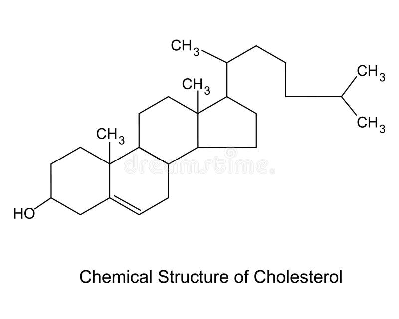
Free with trial Chemical Structure of Cholesterol. Model of the cholesterol structure. Human cell structures vectors Chemical Structure of Cholesterol.
Free with trial Neuronal communication. The dendrites contain receptors for neurotransmitters released by nearby neurons. Soma, dendrites, axons, terminal buttons, and synaptic vesicles. Human cell structures vectors Neuronal communication. The dendrites contain receptors for neurotransmitters released by nearby neurons.
Free with trial Difference between dominant and recessive genes. Homozygous and Heterozygous. Genotype. Vector illustration. Didactic illustration. Human cell structures vectors Difference between dominant and recessive genes
Free with trial Lungs are displayed in a striking presentation, showcasing intricate details and structures, with a distinct tumor located on one lobe. Human cell structures illustrations Lungs depicting anatomical details with a tumor in vibrant colors. Lungs are displayed in a striking presentation, showcasing intricate details and structures. Lungs are displayed in a striking presentation, showcasing intricate details and structures, with a distinct tumor located on one lobe.
Free with trial Taste buds contain the taste receptor cells, which are also known as gustatory cells. The taste receptors are located around the small structures known as papillae found on the upper surface of the tongue, soft palate, upper esophagus, the cheek, and epiglottis. Human cell structures illustrations A taste receptor is a type of cellular receptor which facilitates the sensation of taste. Taste buds contain the taste receptor cells, which are also known as gustatory cells. The taste receptors are located around the small structures known as papillae found on the upper surface of the tongue, soft palate, upper esophagus, the cheek, and epiglottis
Free with trial A 3D illustration representing flu virus attack. Human cell structures illustrations Virus Attack
Free with trial An image of a DNA strand. A 3D illustration of a genetic code chain with grid background. Abstract close up of a chromosome helix and science structures. Human cell structures illustrations Genetic DNA with science background. An image of a DNA strand. A 3D illustration of a genetic code chain with grid background. Abstract close up of a chromosome helix and science structures.
Free with trial The centrosome consists of two main components: a pair of cylindrical structures called centrioles and the pericentriolar material (PCM) that surrounds them. Each centriole is composed of nine sets of triplet microtubules arranged in a cylindrical structure. The PCM contains proteins that are involved in microtubule nucleation and organization. Human cell structures vectors Centrosome
Free with trial This captivating microscopic image delves into the fascinating world of microcirculation. Observe the intricate network of blood vessels, specifically capillaries, showcasing the remarkable structure and function of the circulatory system. Red blood cells, crucial for oxygen transport, are clearly visible traversing the narrow channels of these vessels, alongside plasma, the fluid component of. Human cell structures illustrations Unveiling the Intricate Microcirculation A Detailed Microscopic View of Blood Vessels Red Blood Cells and Plasma. This captivating microscopic image delves into the fascinating world of microcirculation. Observe the intricate network of blood vessels, specifically capillaries, showcasing the remarkable structure and function of the circulatory system. Red blood cells, crucial for oxygen transport, are clearly visible traversing the narrow channels of these vessels, alongside plasma, the fluid component of
Free with trial A detailed illustration combining a human eye with microscopic cell structures, symbolizing biological research, vision science, or medical studies. Ideal for ophthalmology, genetics, or scientific education, representing the intricate layers of human biology and the focus on cellular understanding, vector design Generative AI. Human cell structures vectors Biological Research: Human Eye and Cell Structures, vector design Generative AI. A detailed illustration combining a human eye with microscopic cell structures, symbolizing biological research, vision science, or medical studies. Ideal for ophthalmology, genetics, or scientific education, representing the intricate layers of human biology and the focus on cellular understanding, vector design Generative AI
Free with trial Microscopic view of human cell biology highlighting cellular structures in a vibrant, colorful, and detailed scientific illustration ,Generative AI. Human cell structures illustrations Microscopic view of human cell biology highlighting cellular structures in a vibrant, colorful, and detailed scientific
Free with trial Microscopic view of human or embryonic stem cell revealing intricate details and structures, Generated by AI. Human cell structures illustrations Microscopic view of human or embryonic stem cell revealing intricate details and structures
Free with trial Highly detailed 3d rendering of a human cell against a microscope background showcasing structures, Generated by AI. Human cell structures illustrations Highly detailed 3d rendering of a human cell against a microscope background showcasing structures
Free with trial Magnified view of human or embryonic stem cell showcasing intricate details and cellular structures, Generated by AI. Human cell structures illustrations Magnified view of human or embryonic stem cell showcasing intricate details and cellular structures


![In biology, the extracellular matrix (ECM) is a collection of extracellular molecules secreted by cells that provides structural and biochemical support to the surrounding cells. [1] Because multicellularity evolved independently in different multicellular lineages, the composition of ECM varies between multicellular structures; however, cell adhesion, cell-to-cell communication and differentiation are common functions of the ECM. Human cell structures illustrations](https://thumbs.dreamstime.com/b/g-multicellular-epithelium-biology-extracellular-matrix-ecm-collection-extracellular-molecules-secreted-cells-55455706.jpg)
![In biology, the extracellular matrix (ECM) is a collection of extracellular molecules secreted by cells that provides structural and biochemical support to the surrounding cells. [1] Because multicellularity evolved independently in different multicellular lineages, the composition of ECM varies between multicellular structures; however, cell adhesion, cell-to-cell communication and differentiation are common functions of the ECM. Human cell structures illustrations](https://thumbs.dreamstime.com/b/g-multicellular-epithelium-biology-extracellular-matrix-ecm-collection-extracellular-molecules-secreted-cells-55455697.jpg)


![Ovarian cancer is a cancerous tumor of an ovary. [10] It may originate from the ovary itself or more commonly from communicating nearby structures such as fallopian tubes or the inner lining of the abdomen. Human cell structures illustrations](https://thumbs.dreamstime.com/b/car-t-cell-therapy-ovarian-cancer-closeup-view-d-illustration-cancerous-tumor-ovary-may-originate-itself-323970673.jpg)