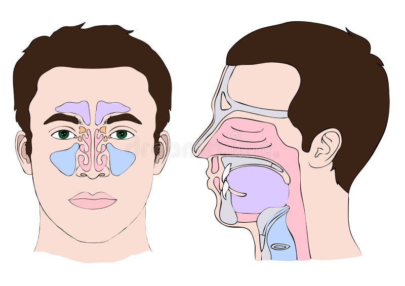
Free with trial Anatomy of the nose and throat. Median section of human head. Human nose cross section vectors Nose anatomy. Anatomy of the nose and throat. Median section of human head
Free with trial Cross section of nose and throat showing major structures including the tongue and epiglottis. Human nose cross section vectors Cross section of nose and throat
Free with trial Nose external anatomy for medical. Human nose cross section vectors Nose external anatomy
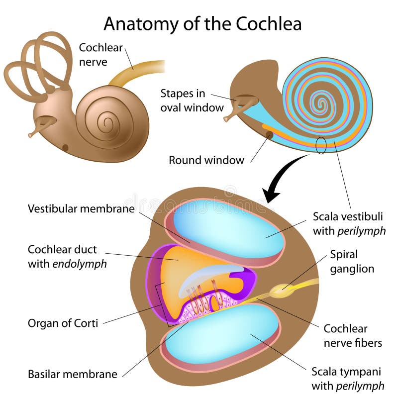
Free with trial Anatomy of the cochlea of human ear, eps10. Human nose cross section vectors Anatomy of the cochlea
Free with trial Nose, throat anatomy, human mouth, respiratory system. Human nose cross section vectors Nose, throat anatomy
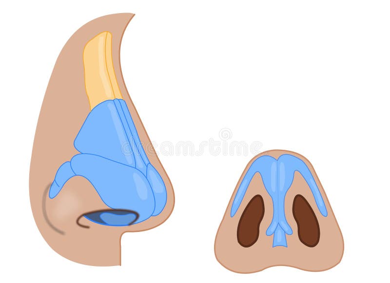
Free with trial Nose anatomy isolated on white photo-realistic vector illustration. Human nose cross section vectors Nose anatomy isolated on white vector. Nose anatomy isolated on white photo-realistic vector illustration
Free with trial Respiratory system: bronchiole and bronchi, diaphragm, trachea, alveoli and cross-section of the lungs. Vector illustration for your design and medical use. human anatomy. silhouette of a man on white background. Human nose cross section vectors Respiratory system. human anatomy. Respiratory system: bronchiole and bronchi, diaphragm, trachea, alveoli and cross-section of the lungs. Vector illustration for your design and medical use. human anatomy. silhouette of a man on white background.
Free with trial Snoring anatomy medical vector diagram with nose, mouth, tongue and air passage. Human nose cross section vectors Snoring anatomy medical vector diagram with air passage. Snoring anatomy medical vector diagram with nose, mouth, tongue and air passage.
Free with trial The oral cavity represents the first part of the digestive tube. Its primary function is to serve as the entrance of the alimentary tract and to initiate the digestive process by salivation and propulsion of the alimentary bolus into the pharynx. [1, 5] It also serves as a secondary respiratory conduit, a site of sound modification for the production of speech, and a chemosensory organ. Human nose cross section illustrations Mouth anatomy. The oral cavity represents the first part of the digestive tube. Its primary function is to serve as the entrance of the alimentary tract and to initiate the digestive process by salivation and propulsion of the alimentary bolus into the pharynx.[1, 5] It also serves as a secondary respiratory conduit, a site of sound modification for the production of speech, and a chemosensory organ.
Free with trial The oral cavity represents the first part of the digestive tube. Its primary function is to serve as the entrance of the alimentary tract and to initiate the digestive process by salivation and propulsion of the alimentary bolus into the pharynx. [1, 5] It also serves as a secondary respiratory conduit, a site of sound modification for the production of speech, and a chemosensory organ. Human nose cross section illustrations Mouth anatomy. The oral cavity represents the first part of the digestive tube. Its primary function is to serve as the entrance of the alimentary tract and to initiate the digestive process by salivation and propulsion of the alimentary bolus into the pharynx.[1, 5] It also serves as a secondary respiratory conduit, a site of sound modification for the production of speech, and a chemosensory organ.
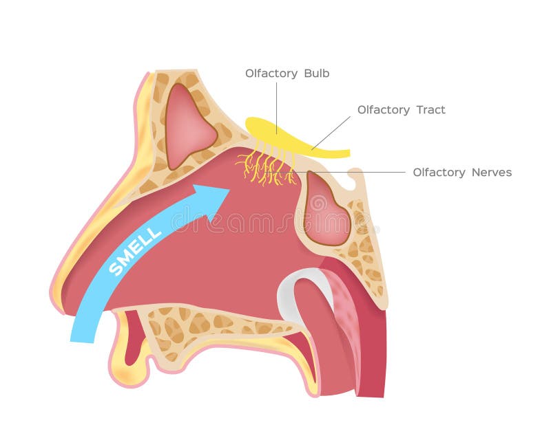
Free with trial Nasal receptors olfactory bulb on white background. Human nose cross section vectors Nasal receptors olfactory bulb
Free with trial Nose sinuses anatomical vector illustration cross section. Educational information. Human nose cross section vectors Nose sinuses anatomical vector illustration cross section.
Free with trial Vector cross section of human face with deviated and normal nasal septum. Human nose cross section vectors Vector cross section of face with deviated and normal nasal septum
Free with trial Nose, throat anatomy, human mouth, respiratory system. Human nose cross section vectors Nose, throat anatomy
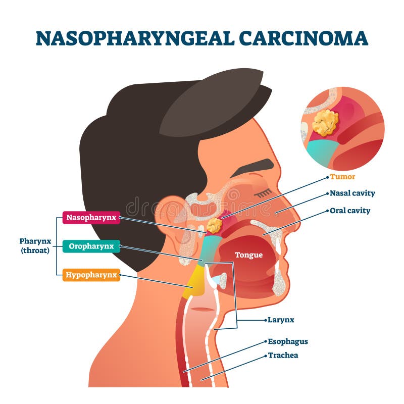
Free with trial Nasopharyngeal carcinoma tumor, vector illustration labeled diagram. Medical nose, mouth and throat cross section scheme with problem area. Health care educational information. Nasal and oral cavity. Human nose cross section vectors Nasopharyngeal carcinoma tumor, vector illustration
Free with trial Sinusitis disease, vector nose illustration, sinus anatomy, human respiratory system. Human nose cross section vectors Sinusitis disease
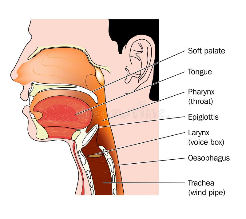
Free with trial Human head and neck, shown from the nose to the shoulder in cross section. Included are the nasal cavity, mouth, tongue, epiglottis, larynx, vocal cords, esophagus, and trachea (windpipe). Human nose cross section illustrations Head and Neck - Cutaway View. Human head and neck, shown from the nose to the shoulder in cross section. Included are the nasal cavity, mouth, tongue, epiglottis, larynx, vocal cords, esophagus, and trachea (windpipe).
Free with trial Cross section through the head showing the nasopharynx, oropharynx and larynx. Human nose cross section vectors Nose mouth and throat. Cross section through the head showing the nasopharynx, oropharynx and larynx
Free with trial Vector nose illustration, sinus anatomy, human respiratory system. Human nose cross section vectors Sinus anatomy, human respiratory system
Free with trial Oral cavity. Illustration isolated on white background. Human nose cross section vectors Oral cavity
Free with trial Throat Head Face Anatomy Vector illustration. Human nose cross section vectors Throat Head Face Anatomy Vector Diagram. Throat Head Face Anatomy Vector illustration
Free with trial Longitudinal section of the diagram of the anatomy of the human nose vector. Human nose cross section vectors Longitudinal section of the diagram of the anatomy of the human nose vector
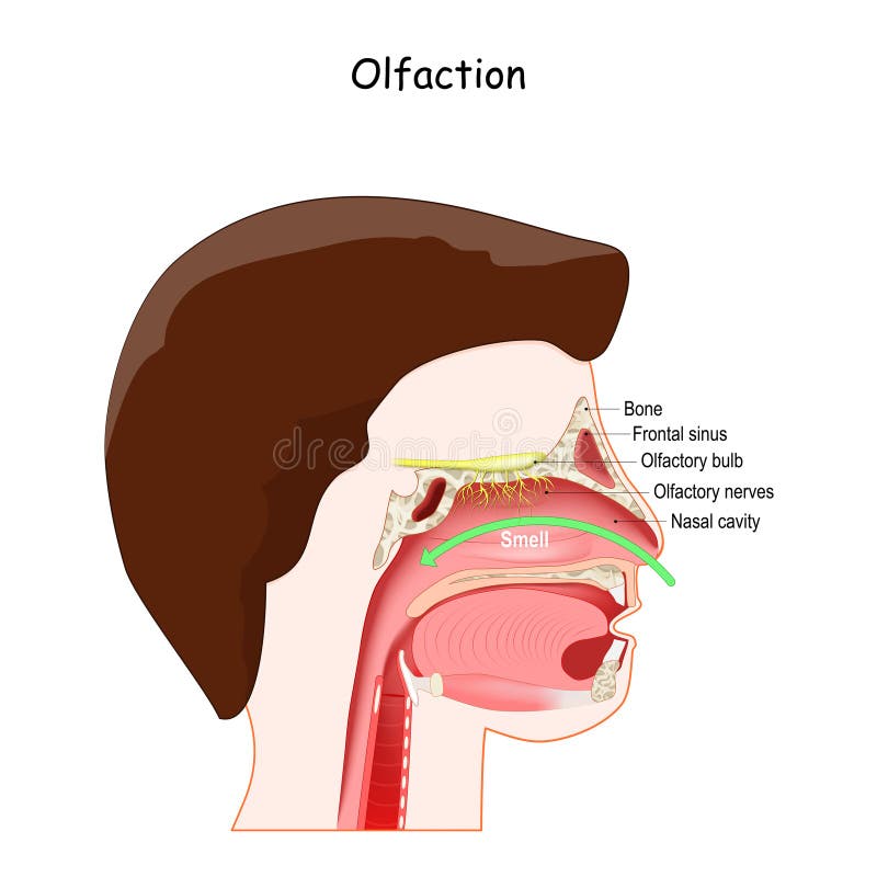
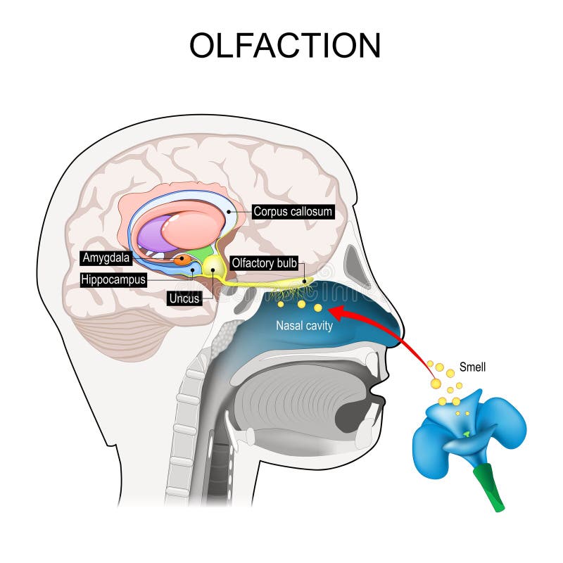
Free with trial Olfaction, sense of smell, and Olfactory system. Cross section of Human`s head with Nasal cavity, Frontal sinus, Olfactory bulb and nerve. vector illustration for education and medical use. Human nose cross section vectors Cross section of head with Nasal cavity, and Olfactory bulb. Olfaction, sense of smell, and Olfactory system. Cross section of Human`s head with Nasal cavity, Frontal sinus, Olfactory bulb and nerve. vector illustration for education and medical use
Free with trial Allergic rhinitis. Inflammation in the nose that occurs when the immune system reacts to allergens in the air. triggers: dust, pollen, and pet hair. Cross section of human nose. Human nose cross section vectors Allergic rhinitis triggers. Allergic rhinitis. Inflammation in the nose that occurs when the immune system reacts to allergens in the air. triggers: dust, pollen, and pet hair. Cross section of human nose
Free with trial Olfaction. Olfactory nerves. Cross section of a human head with part of the brain involved with smell. smell-brain. educational scheme. Vector illustration. Human nose cross section vectors Olfaction. Olfactory nerves. Cross section of the brain. olfaction. Olfactory nerves. Cross section of a human head with part of the brain involved with smell. smell-brain. educational scheme. Vector illustration
Free with trial A detailed illustration showcases the anatomy of the human upper respiratory system. The image depicts a cross-section of the head and neck, revealing the nasal cavity, oral cavity, pharynx, larynx, trachea, and other related structures. It's suitable for medical education, healthcare publications, and scientific presentations. Human nose cross section vectors Human Upper Respiratory System Anatomy. A detailed illustration showcases the anatomy of the human upper respiratory system. The image depicts a cross-section of the head and neck, revealing the nasal cavity, oral cavity, pharynx, larynx, trachea, and other related structures. It's suitable for medical education, healthcare publications, and scientific presentations.
Free with trial Kidney stone disease. Cross section of Ureter with Stone and Stent. Renal calculi. Anatomy of a human urinary system includes the kidneys, ureters, urinary bladder, and urethra. Vector illustration. Human nose cross section vectors Kidney stone disease. Cross section of Ureter with Stone and Stent
Free with trial Nose, throat anatomy, human mouth, respiratory system, Anatomy model of human head, Nasal cavity. vector illustration of Human Nose diagram. organ anatomy. Human nose cross section vectors Nose anatomy cross section diagram showing soft palate paranasal sinuses elements flat vector illustration. nose, throat anatomy, human mouth, respiratory system, Anatomy model of human head, Nasal cavity . vector illustration of Human Nose diagram . organ anatomy
Free with trial Nose, throat anatomy, human mouth, respiratory system, Anatomy model of human head, Nasal cavity. vector illustration of Human Nose diagram. organ anatomy. Human nose cross section vectors Nose anatomy cross section diagram showing soft palate paranasal sinuses elements flat vector illustration. nose, throat anatomy, human mouth, respiratory system, Anatomy model of human head, Nasal cavity . vector illustration of Human Nose diagram . organ anatomy
Free with trial Anatomy of the nose and throat. Human organ structure. tonsil anatomy, teeth, polyps, rhinitis, sore throat, 3d render, 2d graphic, illustration. Human nose cross section illustrations Anatomy of the nose and throat. Human organ structure. tonsil anatomy, teeth, polyps, rhinitis, sore throat, 3d render, 2d graphic
Free with trial Upper respiratory tract infographic diagram. for medical anatomy physiology science education. details part of human body respiratory system. respiration concept. cross section chart scheme. Human nose cross section vectors Upper respiratory tract infographic diagram
Free with trial Obstructive Sleep Apnea. normal breathing, and anatomy of Snoring. Cross section of human`s head. Blocked airway with Tongue. Human nose cross section vectors Obstructive Sleep Apnea. normal breathing, and anatomy of Snoring. Obstructive Sleep Apnea. normal breathing, and anatomy of Snoring. Cross section of human`s head. Blocked airway with Tongue
Free with trial Adenoid hypertrophy. Cross section of the human`s head with healthy and inflammation enlarged adenoids. vector illustration. Human nose cross section vectors Adenoid hypertrophy
Free with trial Olfactory nerve. location of the first cranial nerve. Cross section of a human head with brain, nasal cavity, olfactory bulb, and smell receptors relating to the sense of smell. Vector poster. Human nose cross section vectors Olfactory nerve. location of the first cranial nerve
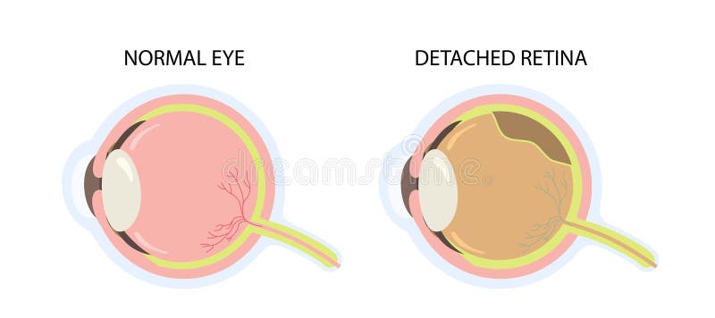
Free with trial Cross section of eye with retinal detachment and normal condition. Retina peels on schematic diagram. Human nose cross section vectors Cross Section Retinal Detachment Eye Vector Illustration. Cross section of eye with retinal detachment and normal condition. Retina peels on schematic diagram
Free with trial Stye. Cross section of a humans eyeball with Infection of the oil gland. Close-up of eye with Hordeolum of lower lid. Vector illustration. Human nose cross section vectors Stye. Hordeolum. Cross section of a humans eyeball with Infection. Stye. Cross section of a humans eyeball with Infection of the oil gland. Close-up of eye with Hordeolum of lower lid. Vector illustration
Free with trial Upper respiratory tract anatomy. Pharynx cross section diagram with descriptions. Structure of the bones, muscles and tissue. Medical education poster. Flat vector illustration. Human nose cross section illustrations Upper respiratory tract anatomy. Pharynx cross section diagram
Free with trial A detailed 3D-rendered medical illustration of a human stomach in a cut-section view. The internal muscular layers, including the longitudinal, circular, and oblique layers, should be distinctly visible and labeled. Human nose cross section illustrations A detailed 3D-rendered medical illustration of a human stomach in a cut-section view
Free with trial A detailed 3D-rendered medical illustration of a human stomach in a cut-section view. The internal muscular layers, including the longitudinal, circular, and oblique layers, should be distinctly visible and labeled. Human nose cross section illustrations A detailed 3D-rendered medical illustration of a human stomach in a cut-section view
Free with trial A detailed 3D-rendered medical illustration of a human stomach in a cut-section view. The internal muscular layers, including the longitudinal, circular, and oblique layers, should be distinctly visible and labeled. Human nose cross section illustrations A detailed 3D-rendered medical illustration of a human stomach in a cut-section view
Free with trial A detailed 3D-rendered medical illustration of a human stomach in a cut-section view. The internal muscular layers, including the longitudinal, circular, and oblique layers, should be distinctly visible and labeled. Human nose cross section illustrations A detailed 3D-rendered medical illustration of a human stomach in a cut-section view
Free with trial Dysphagia. Swallowing difficulty. Esophageal dysphagia. Aspiration risk. Cross section of a human head with Esophagus, Trachea, Nasal cavity, Tongue, Epiglottis, and Food bolus. Healthy and Unhealthy people that have difficulty in swallowing, and the inhalation of fluid while drinking. Medical diagnosis. Detailed vector. Isometric Flat illustration. Schematic diagram. Human nose cross section vectors Dysphagia. Swallowing difficulty, aspiration. Dysphagia. Swallowing difficulty. Esophageal dysphagia. Aspiration risk. Cross section of a human head with Esophagus, Trachea, Nasal cavity, Tongue, Epiglottis, and Food bolus. Healthy and Unhealthy people that have difficulty in swallowing, and the inhalation of fluid while drinking. Medical diagnosis. Detailed vector. Isometric Flat illustration. Schematic diagram
Free with trial Antique engraving illustration of human oral cavity black and white clipart isolate on white background,Human anatomy for medical education. Human nose cross section vectors Antique engraving illustration of human oral cavity black and white clipart isolate on white background,Human anatomy for medical
Free with trial Abstract vector Throat n Nose diagram. Human nose cross section vectors Abstract Artwork Throat n Nose diagram. Abstract vector Throat n Nose diagram
Free with trial Human mouth anatomy infographic diagram structure and parts including nasal cavity pharynx tonsil uvula tongue and lips vector system scheme illustration upper respiratory tract chart for medical physiology biology science education. Human nose cross section vectors Human mouth anatomy infographic diagram
Free with trial Human pharynx sagittal cross section side view showing connection between nasal cavity oral cavity and esophagus with realistic anatomical detail isolated on white background for medical study. Human nose cross section vectors Detailed Pharynx Anatomy Side View Nasal Oral Esophagus Passage. Human pharynx sagittal cross section side view showing connection between nasal cavity oral cavity and esophagus with realistic anatomical detail isolated on white background for medical study.
Free with trial Detailed illustration of the human ear anatomy, showcasing the outer, middle, and inner ear structures. The diagram clearly labels the pinna, ear canal, middle ear, ossicles, semicircular canals, cochlea, and other relevant parts. This image is suitable for educational materials, medical publications, and healthcare-related content. Human nose cross section illustrations Human Ear Anatomy Diagram. Detailed illustration of the human ear anatomy, showcasing the outer, middle, and inner ear structures. The diagram clearly labels the pinna, ear canal, middle ear, ossicles, semicircular canals, cochlea, and other relevant parts. This image is suitable for educational materials, medical publications, and healthcare-related content.
Free with trial Abstract polygonal light of nasal cavity structure. Business wireframe mesh spheres from flying debris. Human nose anatomy concept. Blue structure style vector illustration with geometry triangles. Human nose cross section vectors Nasal cavity structure
Free with trial Human biology, organs anatomy illustration. Set of human internal organs: liver, lungs, heart, kidney, brain, eyes, stomach, trachea etc. Engraved hand drawn in old sketch and vintage style. Human nose cross section vectors Human biology, organs anatomy illustration. Set of human internal organs: liver, lungs, heart, kidney, brain, eyes, stomach
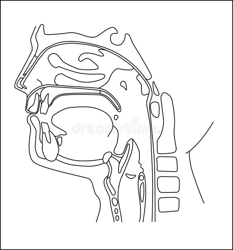
Free with trial Healthy people Nasopharyngeal part on white background. Linear black ink hand drawn picture logo sketch in doodle vintage engraving style pen on paper. View closeup with space for text. Human nose cross section vectors Nasopharyngeal section. Vector drawing. Healthy people Nasopharyngeal part on white background. Linear black ink hand drawn picture logo sketch in doodle vintage engraving style pen on paper. View closeup with space for text
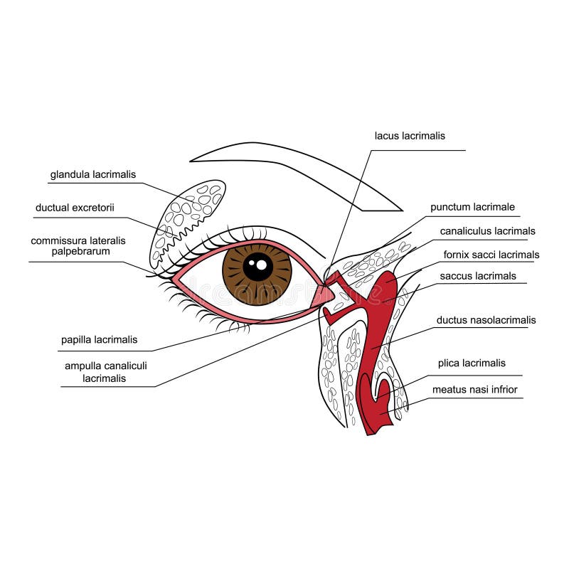
Free with trial Illustration of the lacrimal apparatus. Human nose cross section vectors Lacrimal apparatus
Free with trial Illustration of the lacrimal apparatus. Human nose cross section vectors Anatomy of the lacrimal apparatus. Illustration of the lacrimal apparatus
Free with trial Vector illutration, anatomy of the Lacrimal apparatus. Human nose cross section vectors Vector illutration, anatomy of the Lacrimal apparatus
Free with trial Parts and functions of the ear organ in humans with a white background. Human nose cross section vectors Parts and functions of the ear organ in humans
Free with trial An illustrative diagram shows a human hand highlighting the anatomical aspects of carpal tunnel syndrome. The median nerve and flexor tendons are labeled, with a highlighted area indicating numbness. The carpal tunnel, with detailed inset showing cross-section views of the median nerve, flexor tendons, flexor retinaculum, and carpal bones, is emphasized. Shaded regions and labeling annotations provide clarity on nerve pathways and affected zones. The illustration serves as an educational tool for understanding this medical condition. Human nose cross section vectors Carpal Tunnel Syndrome Illustration. An illustrative diagram shows a human hand highlighting the anatomical aspects of carpal tunnel syndrome. The median nerve and flexor tendons are labeled, with a highlighted area indicating numbness. The carpal tunnel, with detailed inset showing cross-section views of the median nerve, flexor tendons, flexor retinaculum, and carpal bones, is emphasized. Shaded regions and labeling annotations provide clarity on nerve pathways and affected zones. The illustration serves as an educational tool for understanding this medical condition.
Free with trial Rhinitis. Irritation and inflammation of the mucous membrane of nasal cavity. Nonallergic or allergic rhinitis symptoms. Flat vector illustration. Human nose cross section vectors Rhinitis. Irritation and inflammation of the mucous membrane of nasal
Free with trial Illustration shows two types of uveal melanoma. On the left, choroidal melanoma is depicted in an eye cross-section, highlighting the tumor beneath the retina within the choroid layer. Key parts labeled include the lens, cornea, retina, and optic nerve. On the right, iris melanoma is shown on the iris as a dark spot or growth, with labels for the pupil and iris. This educational diagram provides visual aid for understanding eye melanoma's anatomical impact. Human nose cross section vectors Uveal melanoma (Eye melanoma) eye flashcard illustration. Illustration shows two types of uveal melanoma. On the left, choroidal melanoma is depicted in an eye cross-section, highlighting the tumor beneath the retina within the choroid layer. Key parts labeled include the lens, cornea, retina, and optic nerve. On the right, iris melanoma is shown on the iris as a dark spot or growth, with labels for the pupil and iris. This educational diagram provides visual aid for understanding eye melanoma's anatomical impact.
Free with trial A detailed anatomical vector illustration showing the internal cross-section of the human nose and nasal cavity. The graphic depicts structures such as the nasal conchae, septum, and olfactory region in a clean, flat design. Human nose cross section vectors Human Nose Internal Anatomy Cross Section Vector Illustration. A detailed anatomical vector illustration showing the internal cross-section of the human nose and. A detailed anatomical vector illustration showing the internal cross-section of the human nose and nasal cavity. The graphic depicts structures such as the nasal conchae, septum, and olfactory region in a clean, flat design.
Free with trial 3D medical illustration of a human nose cross-section. The illustration highlights the internal structures and anatomy of the nasal cavity. It features soft pink tones with details of internal structures. It is presented against a dark blue gradient background, with a light beige band across the nose. The image likely aims to showcase human anatomy education or medical resources. Human nose cross section illustrations 3 d human nose cross section medical illustration. 3D medical illustration of a human nose cross-section. The illustration highlights the internal structures and anatomy of the nasal cavity. It features soft pink tones with details of internal structures. It is presented against a dark blue gradient background, with a light beige band across the nose. The image likely aims to showcase human anatomy education or medical resources
Free with trial Cross-section of a human nose showing the internal structure, including the nasal cavity, turbinates, and olfactory bulb ,Generative AI. Human nose cross section illustrations Cross-section of a human nose showing the internal structure, including the nasal cavity, turbinates, and olfactory bulb
Free with trial Detailed anatomical representation of a human nose, showing internal structures and vasculature. The image displays a cross-section view, revealing the intricate details of the nasal passages and blood vessels. It presents a vivid depiction for educational or research purposes in a studio setting. Human nose cross section illustrations Human nose anatomy cross section illustration. Detailed anatomical representation of a human nose, showing internal structures and vasculature. The image displays a cross-section view, revealing the intricate details of the nasal passages and blood vessels. It presents a vivid depiction for educational or research purposes in a studio setting
Free with trial Detailed anatomical cross-section of a human nose. The image displays the internal structures with different colors and shading. The style is highly detailed and realistic. It is suitable for educational or medical purposes. The image could be used in textbooks, medical journals, or scientific presentations. It has a dark background. Human nose cross section illustrations Human nose cross section anatomy diagram. Detailed anatomical cross-section of a human nose. The image displays the internal structures with different colors and shading. The style is highly detailed and realistic. It is suitable for educational or medical purposes. The image could be used in textbooks, medical journals, or scientific presentations. It has a dark background
Free with trial Cross section of human pancreas showing internal structure and enzymes. Human nose cross section illustrations Cross section of human pancreas showing internal structure and enzymes.
Free with trial Detailed cross section illustration of human nose anatomy revealing nasal cavity and sinus structures Useful for medical education and healthcare visuals. Human nose cross section illustrations Nose Anatomy Cross Section Depicting Nasal Cavity and Sinus. Detailed cross section illustration of human nose anatomy revealing nasal cavity and sinus structures Useful for medical education and healthcare visuals
Free with trial A detailed digital illustration showcasing the human senses and skin anatomy, featuring a realistic eye, ear, lips, nose, and cross-section of skin with hair follicles, all set against a clean white background. Human nose cross section illustrations Human Senses and Skin Anatomy
Free with trial Black and white illustration of the human brain's cross-section. The cerebrum features distinctive gyri and sulci, while the cerebellum is depicted with its unique, folded texture. The brainstem extends downward, showing the midbrain, pons, and medulla oblongata. The intricate details highlight the brain's complex structure, emphasizing functional areas. This anatomical depiction is often used for educational purposes to illustrate neuroanatomy. Human nose cross section illustrations Human Brain Cross Section Anatomy. Black and white illustration of the human brain's cross-section. The cerebrum features distinctive gyri and sulci, while the cerebellum is depicted with its unique, folded texture. The brainstem extends downward, showing the midbrain, pons, and medulla oblongata. The intricate details highlight the brain's complex structure, emphasizing functional areas. This anatomical depiction is often used for educational purposes to illustrate neuroanatomy.
Free with trial A detailed cross-section illustration of the human eye. Great for ophthalmology clinics, biology education, vision science, and medical diagrams, vector design Generative AI. Human nose cross section vectors Human Eye Anatomy Cross Section, vector design Generative AI. A detailed cross-section illustration of the human eye. Great for ophthalmology clinics, biology education, vision science, and medical diagrams, vector design Generative AI
Free with trial An educational cross-section illustration of the human ear showing the inner ear structure. Suitable for hearing clinics, medical textbooks, and audiology presentations, vector design Generative AI. Human nose cross section vectors Human Ear Anatomy Medical Cross Section Diagram, vector design Generative AI. An educational cross-section illustration of the human ear showing the inner ear structure. Suitable for hearing clinics, medical textbooks, and audiology presentations, vector design Generative AI


![The oral cavity represents the first part of the digestive tube. Its primary function is to serve as the entrance of the alimentary tract and to initiate the digestive process by salivation and propulsion of the alimentary bolus into the pharynx. [1, 5] It also serves as a secondary respiratory conduit, a site of sound modification for the production of speech, and a chemosensory organ. Human nose cross section illustrations](https://thumbs.dreamstime.com/b/mouth-anatomy-oral-cavity-represents-first-part-digestive-tube-its-primary-function-to-serve-as-entrance-43014702.jpg)
![The oral cavity represents the first part of the digestive tube. Its primary function is to serve as the entrance of the alimentary tract and to initiate the digestive process by salivation and propulsion of the alimentary bolus into the pharynx. [1, 5] It also serves as a secondary respiratory conduit, a site of sound modification for the production of speech, and a chemosensory organ. Human nose cross section illustrations](https://thumbs.dreamstime.com/b/mouth-anatomy-oral-cavity-represents-first-part-digestive-tube-its-primary-function-to-serve-as-entrance-43014656.jpg)