Free with trial Banner Upper Recession gums illustration vector on blue background. Dental concept. Human periodontitis vectors Banner Upper Recession gums illustration vector on blue background. Dental concept.
Free with trial Teeth infographic. Gum disease stages. Editable vector illustration in modern style. Medical concept in natural colors on a light green background. Keep your teeth healthy. Human periodontitis vectors Teeth infographic Gum disease stages. Teeth infographic. Gum disease stages. Editable vector illustration in modern style. Medical concept in natural colors on a light green background. Keep your teeth healthy
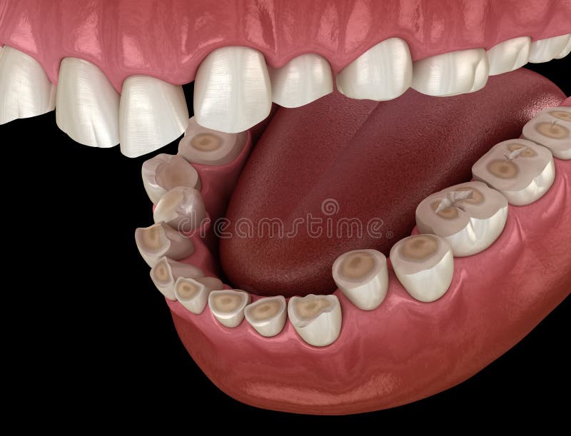
Free with trial Dental attrition Bruxism resulting in loss of tooth tissue. Medically accurate tooth 3D illustration. Human periodontitis illustrations Dental attrition Bruxism resulting in loss of tooth tissue. Medically accurate tooth. Dental attrition Bruxism resulting in loss of tooth tissue. Medically accurate tooth 3D illustration
Free with trial Gum disease, periodontitis. Healthy white tooth. Vector stock illustration. Human periodontitis vectors Gum disease, periodontitis. Healthy white tooth. Vector stock illustration
Free with trial Tooth and periodontium anatomical structure. Sectional human central incisor showing the structures that form the dental tissue and the periodontal tissues. Infographic, 3D illustration with subtitles. Human periodontitis illustrations Tooth and periodontium anatomical structure. Sectional human central incisor showing the structures of the tooth ans periodontium. tooth and periodontium anatomical structure. Sectional human central incisor showing the structures that form the dental tissue and the periodontal tissues. Infographic, 3D illustration with subtitles
Free with trial Tooth and periodontium anatomical structure. Sectional human central incisor showing the structures that form the dental tissue and the periodontal tissues. Infographic, 3D illustration. Human periodontitis illustrations Tooth and periodontium anatomical structure. Sectional human central incisor showing the structures of the tooth ans periodontium. Tooth and periodontium anatomical structure. Sectional human central incisor showing the structures that form the dental tissue and the periodontal tissues. Infographic, 3D illustration
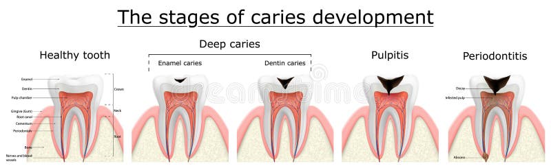
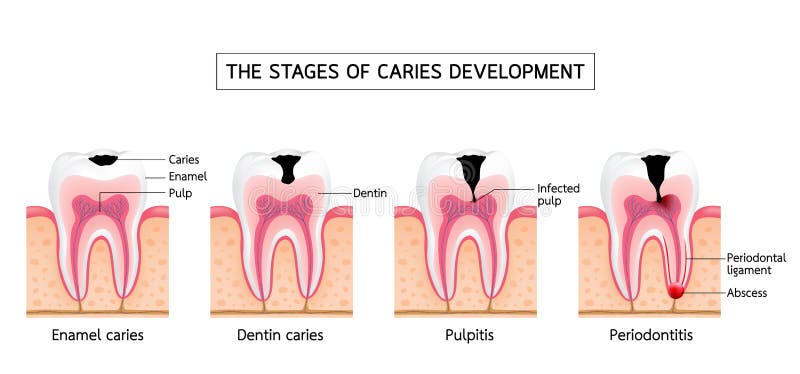
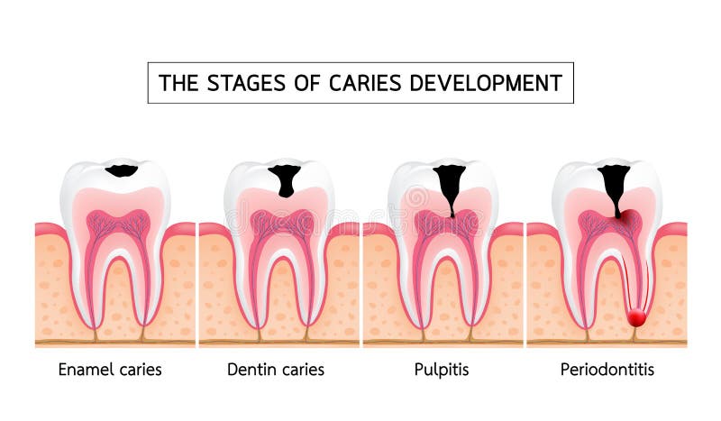
Free with trial Stages of caries development. Dental disease: caries, pulpitis and periodontitis, realistic vector illustration. Human periodontitis vectors Stages of caries development. Dental disease: caries, pulpitis and periodontitis, realistic vector illustration
Free with trial Dental disease set. Dentin caries, gingivitis and periodontitis. Oral cavity problem. Dental education poster. Flat vector illustration. Human periodontitis vectors Dental disease set. Dentin caries, gingivitis and periodontitis. Oral cavity
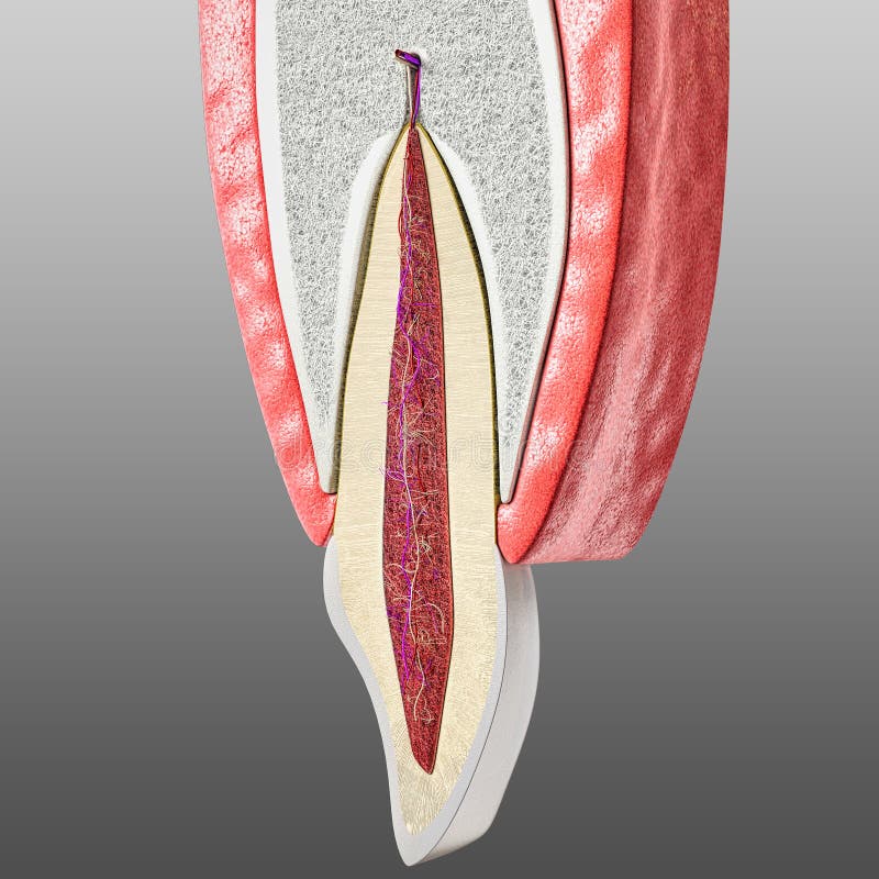
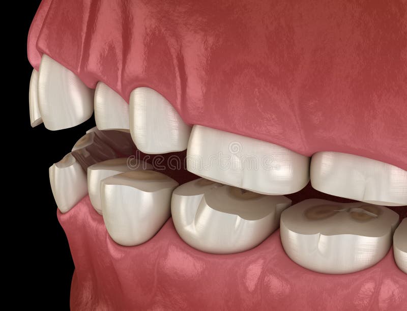
Free with trial A highly detailed 3D rendering of a human tooth, presented in a cross-section view to reveal its intricate internal anatomy. The illustration clearly shows the white enamel, yellow dentin, and the red pulp chamber containing nerves and blood vessels. Surrounding the tooth are the pink gums (gingiva) and the supporting alveolar bone, also depicted with their vascular and nervous supply. This anatomically accurate representation is ideal for educational materials, dental presentations, medical textbooks, and any content related to oral health, dentistry, or biological sciences, set against a clean black background for maximum clarity. Human periodontitis illustrations Detailed Human Tooth Anatomy Cross-Section. A highly detailed 3D rendering of a human tooth, presented in a cross-section view to reveal its intricate internal. A highly detailed 3D rendering of a human tooth, presented in a cross-section view to reveal its intricate internal anatomy. The illustration clearly shows the white enamel, yellow dentin, and the red pulp chamber containing nerves and blood vessels. Surrounding the tooth are the pink gums (gingiva) and the supporting alveolar bone, also depicted with their vascular and nervous supply. This anatomically accurate representation is ideal for educational materials, dental presentations, medical textbooks, and any content related to oral health, dentistry, or biological sciences, set against a clean black background for maximum clarity.
Free with trial 3d rendering of human teeth showing receding gums and bone loss. This could illustrate the need for a dentist appointment. Human periodontitis illustrations Human teeth showing receding gums and bone loss
Free with trial Tooth anatomy and decay chart. Vector biomedical illustration. Side view. Stages of teeth periodontitis illness isolated on white background. Design for healthcare, dentistry. Human periodontitis vectors Tooth anatomy and decay chart. Vector biomedical illustration. Side view. Stages of teeth periodontitis illness isolated on white
Free with trial Gum disease inflammation bacteria can enter in to the blood stream an affect heart. Periodontitis disease disease anatomy on an abstract blue background. Human periodontitis vectors Dental disease. Gum disease inflammation bacteria can enter in to the blood stream an affect heart. Periodontitis disease disease anatomy on an abstract blue background
Free with trial Dental treatment abstract concept design, gum disease Periodontitis healing to prevent bacteria from inflamed gums enter in to the blood stream and affect other organs. Human periodontitis vectors Dental treatment
Free with trial Teeth problems complications, Gum disease inflammation bacteria can enter in to the blood stream an affect brain. Periodontitis disease info poster on a white background. Human periodontitis vectors Teeth problems complications
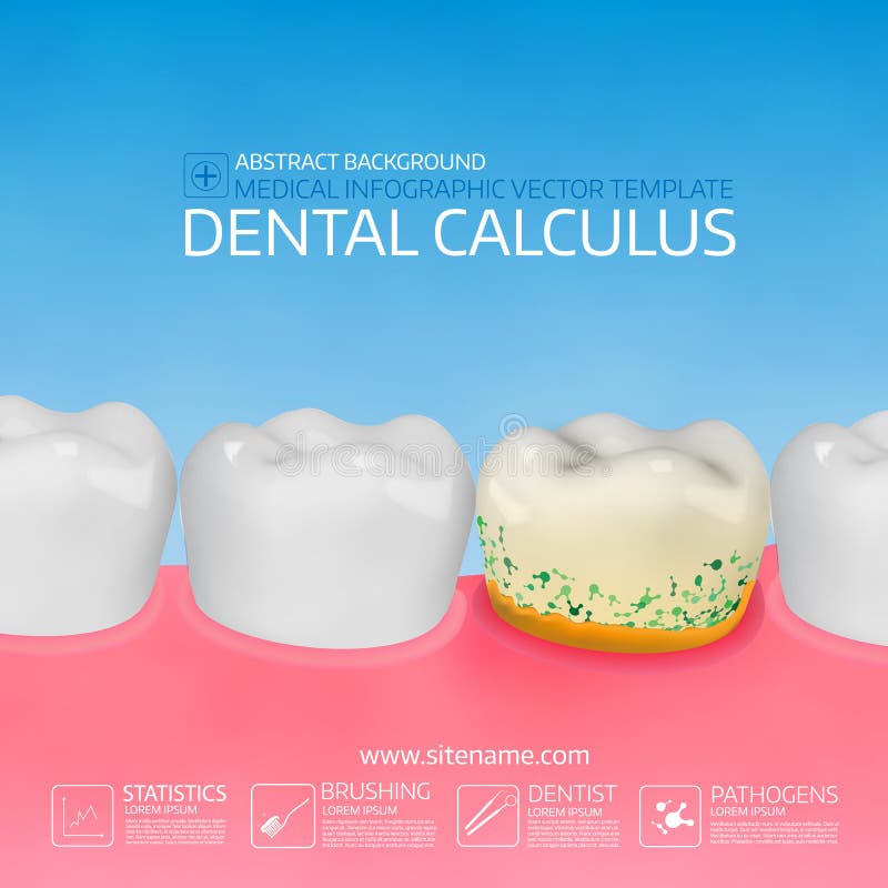
Free with trial The development of dental disease, plaque and calculus. Human periodontitis vectors Structure of human teeth. The development of dental disease, plaque and calculus
Free with trial Gingivitis vector illustration. Medical oral mouth illness symptoms example. Compared healthy and tooth with inflammation. Isolated anatomy disease diagnostics. State before periodontitis with biofilm. Human periodontitis vectors Gingivitis vector illustration. Medical oral mouth illness symptoms example
Free with trial Periodontitis, dental disease. Inflammation of the gums. Human periodontitis vectors Periodontitis, dental disease.
Free with trial The development of dental disease, plaque and calculus. Human periodontitis vectors Structure of human teeth. The development of dental disease, plaque and calculus
Free with trial Human tooth dental infographic. Editable vector illustration. Medical image in white, pink and dark blue colors on a light blue background useful for poster, leaflet or brochure graphic design. Human periodontitis vectors Teeth vector anatomy. Human tooth dental infographic. Editable vector illustration. Medical image in white, pink and dark blue colors on a light blue background useful for poster, leaflet or brochure graphic design.
Free with trial Stages of caries development. Tooth structure in flat style. Tooth decay with enamel. Dental disease caries, pulpitis and periodontitis, realistic vector illustration. Human periodontitis illustrations Stages of caries development. Tooth structure in flat style. Tooth decay with enamel. Dental disease caries, pulpitis
Free with trial Graphic illustration of the stages of periodontal disease, showing the progression from healthy teeth to teeth with advanced bone destruction due to inflammation and plaque buildup. Human periodontitis illustrations Stages of periodontitis illustrated by different tooth conditions. Graphic illustration of the stages of periodontal disease, showing the progression from healthy teeth to teeth with advanced bone destruction due to inflammation and plaque buildup
Free with trial Enamel caries, Dentin caries, Pulpitis and Periodontitis. Dental care info-graphic, illustration on white background. Human periodontitis vectors Stages of caries development. Enamel caries, Dentin caries, Pulpitis and Periodontitis. Dental care info-graphic, illustration on white background.
Free with trial Enamel caries, Dentin caries, Pulpitis and Periodontitis. Dental care info-graphic, illustration on white background. Human periodontitis vectors Stages of caries development. Enamel caries, Dentin caries, Pulpitis and Periodontitis. Dental care info-graphic, illustration on white background.
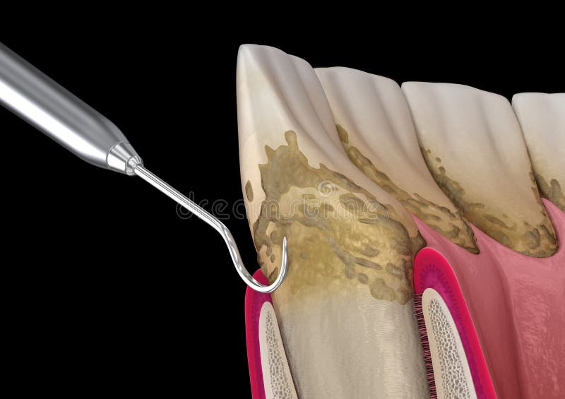
Free with trial Oral hygiene: Scaling and root planing conventional periodontal therapy. Medically accurate 3D illustration of human teeth treatment. Human periodontitis illustrations Oral hygiene: Scaling and root planing conventional periodontal therapy. Medically accurate 3D illustration of human teeth
Free with trial Vector flat isolated illustration of periodontitis. Tooth anatomy infographics. Medical banner or poster illustration. Dental problem. Medical Oral Health. Template of Periodontal Disease. Human periodontitis vectors Vector isolated illustration of tooth. Vector flat isolated illustration of periodontitis. Tooth anatomy infographics. Medical banner or poster illustration. Dental problem. Medical Oral Health. Template of Periodontal Disease
Free with trial Vector flat isolated illustration of periodontitis. Tooth anatomy infographics. Medical banner or poster illustration. Dental problem. Medical Oral Health. Template of Periodontal Disease. Human periodontitis vectors Vector isolated illustration of tooth. Vector flat isolated illustration of periodontitis. Tooth anatomy infographics. Medical banner or poster illustration. Dental problem. Medical Oral Health. Template of Periodontal Disease
Free with trial Caries types stages and prevention poster with text vector. Enamel and dental problems, pulpitis and periodontitis, decay and involvement of pulp. Brush teeth, avoid hot and hot meal, visit doctor. Human periodontitis vectors Caries types stages and prevention poster with text vector
Free with trial Black toothbrush and three digital drawn blood drops above it. Dental care and propblems of teeth and gums. Periodontitis, gingivitis, bleeding gums, hygiene illustration of periodontal disease. Human periodontitis illustrations Black toothbrush and three digital drawn blood drops above it. Dental care and propblems of teeth and gums. Periodontitis, gingivi
Free with trial Vector flat isolated illustration of periodontitis. Tooth anatomy infographics. Medical banner or poster illustration. Dental problem. Medical Oral Health. Template of Periodontal Disease. Human periodontitis vectors Vector isolated illustration of tooth. Vector flat isolated illustration of periodontitis. Tooth anatomy infographics. Medical banner or poster illustration. Dental problem. Medical Oral Health. Template of Periodontal Disease
Free with trial Vector flat isolated illustration of periodontitis. Tooth anatomy infographics. Medical banner or poster illustration. Dental problem. Medical Oral Health. Template of Periodontal Disease. Human periodontitis vectors Vector isolated illustration of tooth. Vector flat isolated illustration of periodontitis. Tooth anatomy infographics. Medical banner or poster illustration. Dental problem. Medical Oral Health. Template of Periodontal Disease
Free with trial Vector flat isolated illustration of periodontitis. Tooth anatomy infographics. Medical banner or poster illustration. Dental problem. Medical Oral Health. Template of Periodontal Disease. Human periodontitis vectors Vector isolated illustration of tooth. Vector flat isolated illustration of periodontitis. Tooth anatomy infographics. Medical banner or poster illustration. Dental problem. Medical Oral Health. Template of Periodontal Disease
Free with trial Vector flat isolated illustration of periodontitis. Tooth anatomy infographics. Medical banner or poster illustration. Dental problem. Medical Oral Health. Template of Periodontal Disease. Human periodontitis vectors Vector isolated illustration of tooth. Vector flat isolated illustration of periodontitis. Tooth anatomy infographics. Medical banner or poster illustration. Dental problem. Medical Oral Health. Template of Periodontal Disease
Free with trial Teeth infographic Gum disease stages gingivitis and periodontitis. Editable vector illustration in flat style. Medical concept in natural colors on background. Keep your teeth healthy. Vector illustration. Human periodontitis illustrations Teeth infographic Gum disease stages gingivitis
Free with trial Tooth cross section scientific modern design icon on an abstract blue background. Human periodontitis vectors Tooth cross section icon. Tooth cross section scientific modern design icon on an abstract blue background
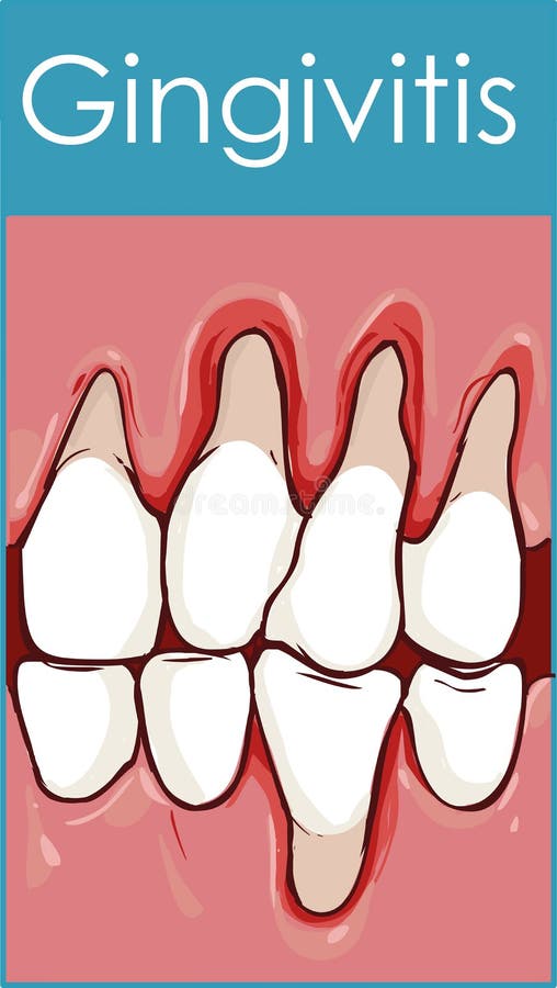
Free with trial Gingivitis. Inflammation of the gums. Vector illustration on background. Human periodontitis vectors Gingivitis.
Free with trial Tooth decay disease, dental root tinfection. Human periodontitis vectors Tooth disease. Tooth decay disease, dental root tinfection
Free with trial Vector detailed illustration of the stages of gum disease. Image of teeth and gums in a section of bone tissue with affected areas and description. Human periodontitis vectors Stage of Gum Disease. Vector detailed illustration of the stages of gum disease. Image of teeth and gums in a section of bone tissue with affected areas and description.
Free with trial Common dental problems: caries, plaque and gum disease, with healthy teeth for comparison. Modern medical infographic chart. Vector illustration. Human periodontitis illustrations Dental health illustration. Common dental problems: caries, plaque and gum disease, with healthy teeth for comparison. Modern medical infographic chart. Vector illustration.
Free with trial Complications of gum disease. Bacteria from inflamed gums can enter in to the blood stream and affect other organs such as brain, heart and cause diabetes. Abstract blue scientific background. Human periodontitis vectors Dental complications. Complications of gum disease. Bacteria from inflamed gums can enter in to the blood stream and affect other organs such as brain, heart and cause diabetes. Abstract blue scientific background
Free with trial Stages of tooth decay illustration. Development of dental caries illustration. Human periodontitis vectors Development of dental caries illustration.
Free with trial Dental attrition Bruxism resulting in loss of tooth tissue. Medically accurate tooth 3D illustration. Human periodontitis illustrations Dental attrition Bruxism resulting in loss of tooth tissue. Medically accurate tooth illustration
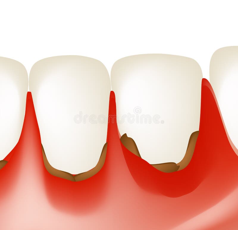
Free with trial Vector illustration of healthy and diseased tooth. bleeding gums. tooth loss. Human periodontitis vectors Healthy and diseased tooth. bleeding gums. tooth loss
Free with trial Vector illustration of abrasion of enamel. the affected tooth. Human periodontitis vectors Tooth, the abrasion of enamel. Vector illustration of abrasion of enamel. the affected tooth.
Free with trial Tooth structure, training medical anatomical poster. Vector illustration. Human periodontitis vectors Tooth structure, training medical anatomical poster, vector illustration.
Free with trial Vector illustration of oral mucosal papillomatosis. cytomegalovirus. Human periodontitis vectors Papillomatosis of the oral mucosa. Vector illustration of oral mucosal papillomatosis. cytomegalovirus
Free with trial Vector illustration of dental plaque. microbes under magnification. Human periodontitis vectors Plaque. microbes under magnification
Free with trial Teeth make bad breath and bacteria. Stomatology dentistry vector. Concept. Dirty teeth, stuck in teeth, decay teeth and gingivitis. Human periodontitis vectors Bad breath and bacteria. Stomatology dentistry vector concept. Teeth make bad breath and bacteria. Stomatology dentistry vector. Concept. Dirty teeth, stuck in teeth, decay teeth and gingivitis.
Free with trial Caries removing process. Medically accurate tooth 3D illustration. Human periodontitis illustrations Caries removing process. Medically accurate tooth illustration
Free with trial Teeth with caries, treatment. Medically accurate tooth 3D illustration. Human periodontitis illustrations Teeth with caries, treatment. Medically accurate tooth illustration
Free with trial Tooth cavity chronic disease painful concept. Medical help therapy treatment. Innovation laser dentistry pain. Oral dental medical care poster template. 3D vector illustration. Human periodontitis vectors Tooth cavity chronic disease painful concept. Medical help therapy treatment. Innovation laser dentistry pain. Oral
Free with trial Molar teeth damaged by caries. Medically accurate tooth 3D illustration. Human periodontitis illustrations Molar teeth damaged by caries. Medically accurate tooth 3D illustration
Free with trial Teeth infographic. Gum disease chart. Editable vector illustration in modern style. Medical concept in natural colors on a dark violet background. Keep your teeth healthy. Human periodontitis vectors Gum disease vector. Teeth infographic. Gum disease chart. Editable vector illustration in modern style. Medical concept in natural colors on a dark violet background. Keep your teeth healthy
Free with trial Dental attrition Bruxism resulting in loss of tooth tissue. Medically accurate tooth 3D illustration. Human periodontitis illustrations Dental attrition Bruxism resulting in loss of tooth tissue. Medically accurate tooth 3D illustration
Free with trial Teeth infographic. Gum disease stages. Editable vector illustration in modern style. Medical concept in natural colors on a light green background. Keep your teeth healthy. Human periodontitis vectors Teeth infographic vector. Teeth infographic. Gum disease stages. Editable vector illustration in modern style. Medical concept in natural colors on a light green background. Keep your teeth healthy
Free with trial Dental attrition Bruxism resulting in loss of tooth tissue. Medically accurate tooth 3D illustration. Human periodontitis illustrations Dental attrition Bruxism resulting in loss of tooth tissue. Medically accurate tooth illustration
Free with trial Teeth disease infographic. Gum disease stages. Editable vector illustration in modern style. Medical concept in natural colors on a light green background. Keep your teeth healthy. Human periodontitis vectors Teeth vector Anatomy. Teeth disease infographic. Gum disease stages. Editable vector illustration in modern style. Medical concept in natural colors on a light green background. Keep your teeth healthy
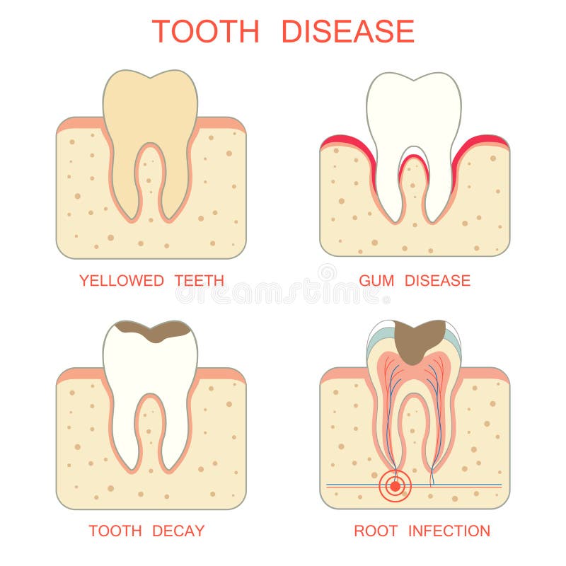
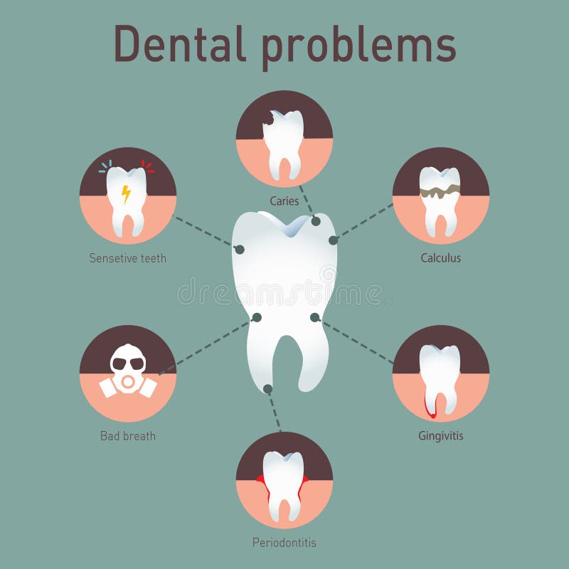
Free with trial Tooth disease. Medical vector infografics. Dental problems. Human periodontitis vectors Medical vector infografics Dental problems. Tooth disease. Medical vector infografics. Dental problems
Free with trial Stage of caries development. Tooth structure in flat style. Tooth decay with enamel. Dental disease realistic vector illustration. Human periodontitis vectors Stage of caries development. Tooth structure in flat style. Tooth decay with enamel. Dental disease realistic vector
Free with trial Caries removing process. Medically accurate tooth 3D illustration. Human periodontitis illustrations Caries removing process. Medically accurate tooth 3D illustration
Free with trial Tooth diseases: dental calculus. Vector cartoon illustration of molar affected by tartar. Human periodontitis illustrations Dental calculus
Free with trial Periostitis tooth - Lump on Gum Above Tooth. Medically accurate dental 3D illustration. Human periodontitis illustrations Periostitis tooth - Lump on Gum Above Tooth. Medically accurate dental 3D illustration
Free with trial Banner Upper White spots on teeth illustration vector on blue background. Dental concept. Human periodontitis vectors Banner Upper White spots on teeth illustration vector on blue ba
Free with trial Cute cartoon sad tooth and bacterial plaque. Human periodontitis vectors Cute cartoon sad tooth and bacterial plaque
Free with trial Tooth Cystectomy Surgery - recovery after Periostitis. Medically accurate 3D illustration. Human periodontitis illustrations Tooth Cystectomy Surgery - recovery after Periostitis . Medically accurate 3D illustration
Free with trial Tooth Cystectomy Surgery - recovery after Periostitis. Medically accurate 3D illustration. Human periodontitis illustrations Tooth Cystectomy Surgery - recovery after Periostitis . Medically accurate 3D illustration
Free with trial Caries stage, tooth decay scheme with caries, stomatological illustration with dental diseases,infographicsn. Human periodontitis vectors Caries stage, tooth decay scheme with caries, stomatological illustration with dental diseases, point by point schematic. Caries stage, tooth decay scheme with caries, stomatological illustration with dental diseases,infographicsn
Free with trial Tooth diseases: periodontosis. Vector cartoon illustration of healthy and affected molar. Human periodontitis illustrations Tooth with periodontosis. Tooth diseases: periodontosis. Vector cartoon illustration of healthy and affected molar
Free with trial Toothache, tooth disease symptoms concept. Caries, sickness, treatment need, medical assistance necessity, young woman suffering from pain, negative emotions facial expression. Simple flat vector. Human periodontitis vectors Toothache, tooth disease symptoms concept
Free with trial Gingivitis. Inflammation of the gums. Dental calculus. Infographics. Vector illustration on isolated background. Human periodontitis vectors Gingivitis. Inflammation of the gums. Dental calculus. Infographics. Vector illustration on isolated background
Free with trial Dental calculus with bacteria. Colorful vector illustration. Infographic template. Human periodontitis vectors Dental calculus with bacteria. Colorful vector illustration.
Free with trial Tooth and teeth oral care vector icon set. Dental hygiene, dentist therapy icons. Human periodontitis vectors Tooth and teeth oral care vector icon set
Free with trial Dental care and therapy line vector icon set. Healthy tooth, caries, implants and oral hygiene set. Teeth icons. Human periodontitis vectors Dental care and therapy line vector icon set
Free with trial Periodontal disease icon. Trendy flat vector Periodontal disease icon on white background from Diseases collection, vector illustration can be use for web and mobile, eps10. Human periodontitis vectors Periodontal disease icon. Trendy flat vector Periodontal disease
Free with trial Friends healthy teeth and bad tooth drills a medical drill. concept stomatology of pediatric. dental diseases such as periodontitis, caries, tartar, fissure. illustration. Human periodontitis illustrations Friends healthy teeth and bad tooth drills a medical drill. concept stomatology of pediatric
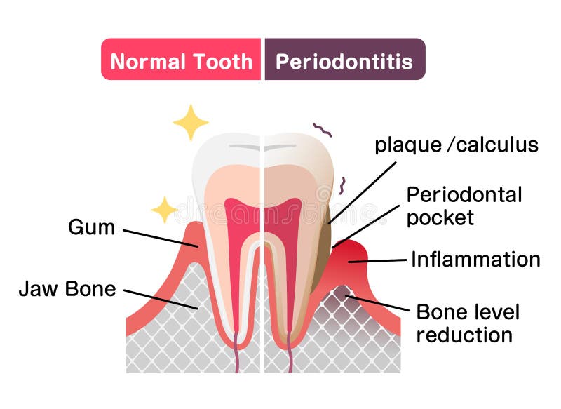
Free with trial Comparison of normal teeth and periodontal disease. flat vector illustration. Human periodontitis vectors Comparison of normal teeth and periodontal disease. flat vector illustration
Free with trial Medical poster about the stages of development of periodontal disease. Human periodontitis vectors Periodontal disease Vector illustration. Medical poster about the stages of development of periodontal disease
Free with trial Types of bruxism teeth grinding vector illustration. Human periodontitis vectors Types of bruxism teeth grinding vector illustration