Free with trial Plantar fasciitis, a common foot problem, eps8. Human talus vectors Plantar fasciitis
Free with trial Different grades of lateral ankle sprain, eps8. Human talus vectors Ankle sprain grading. Different grades of lateral ankle sprain, eps8
Free with trial Types of ankle sprains with indicated ligaments, eps8. Human talus illustrations Ankle sprains
Free with trial The ankle joint, tendons of the ankle joint foot anatomy vector illustration eps 10 infographic. Human talus vectors The ankle joint, tendons of the ankle joint foot anatomy vector illustration
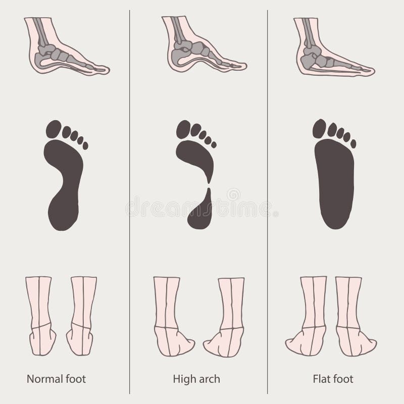
Free with trial Normal arch and fallen arch. Human talus vectors Flat foot. Normal arch and fallen arch
Free with trial Heel Spur. Bones of the Foot and Ankle. Calcaneal spur. Calcaneal spur a bone excrescence on the lower surface of the calcaneus which frequently causes pain on walking. Human talus vectors Heel Spur.
Free with trial Lateral and front views of the ankle joint, eps8. Human talus vectors The ankle joint
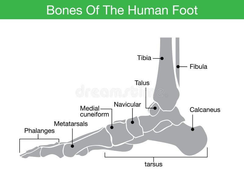
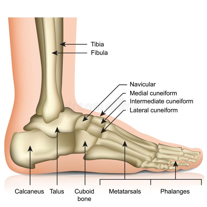
Free with trial Illustration about bones of the human foot which This simple style and have describe to name of all bone. Human talus vectors Bones of the human foot
Free with trial Plantar fasciitis vector illustration. Labeled human feet sport disorder diagram. Educational medical scheme with orthopedic leg disease. Painful plantar fascia inflammation and irritation infographic. Human talus vectors Plantar fasciitis vector illustration. Labeled human feet disorder diagram. Plantar fasciitis vector illustration. Labeled human feet sport disorder diagram. Educational medical scheme with orthopedic leg disease. Painful plantar fascia inflammation and irritation infographic
Free with trial Different views of a human foot front, back, side, lateral, medial, dorsal and plantar isolated on white background. Vector illustration for medical health care use. EPS 10. Human talus vectors Different views of a human foot on white background_Anatomy. Different views of a human foot front, back, side, lateral, medial, dorsal and plantar isolated on white background. Vector illustration for medical health care use. EPS 10.
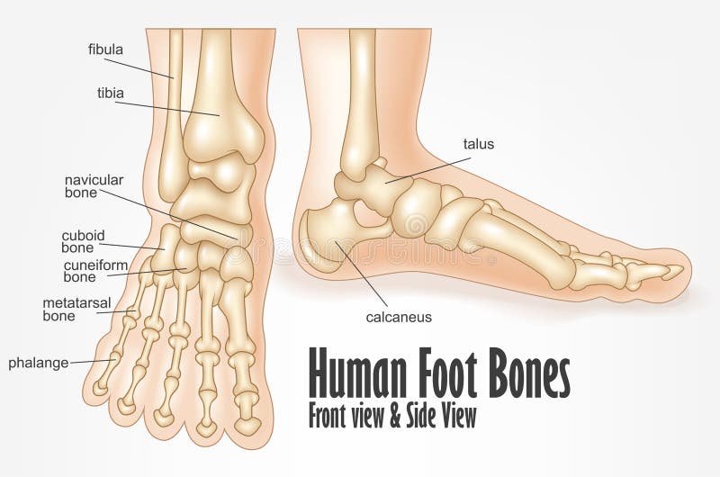
Free with trial Illustration Of Human foot bones front and side view anatomy. Human talus vectors Human foot bones front and side view anatomy
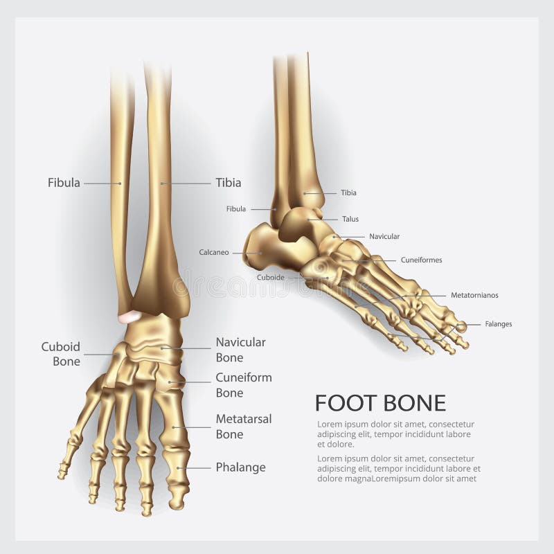
Free with trial Bones of the foot, lateral aspect and dorsal aspect. Created in Adobe Illustrator. Contains transparencies. EPS 10. Human talus vectors Foot bones. Bones of the foot, lateral aspect and dorsal aspect. Created in Adobe Illustrator. Contains transparencies. EPS 10.
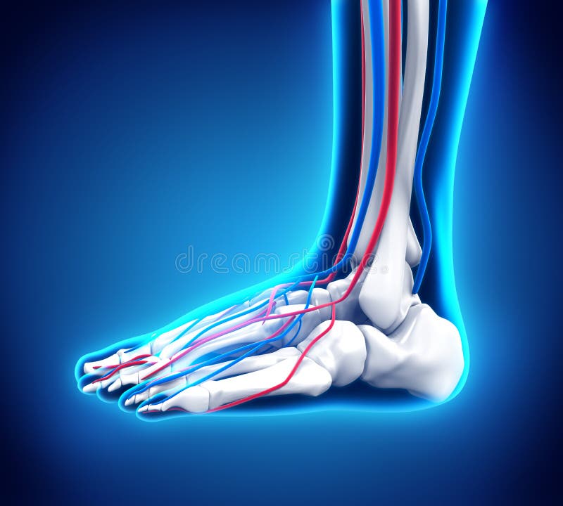
Free with trial Calcaneus Bone Anatomy with Ciculatory System with highlighted zone - pain concept. Human talus illustrations Calcaneus Bone Anatomy with Ciculatory System
Free with trial A common sport injury of the ankle joint, eps8. Human talus vectors Pott fracture. A common sport injury of the ankle joint, eps8
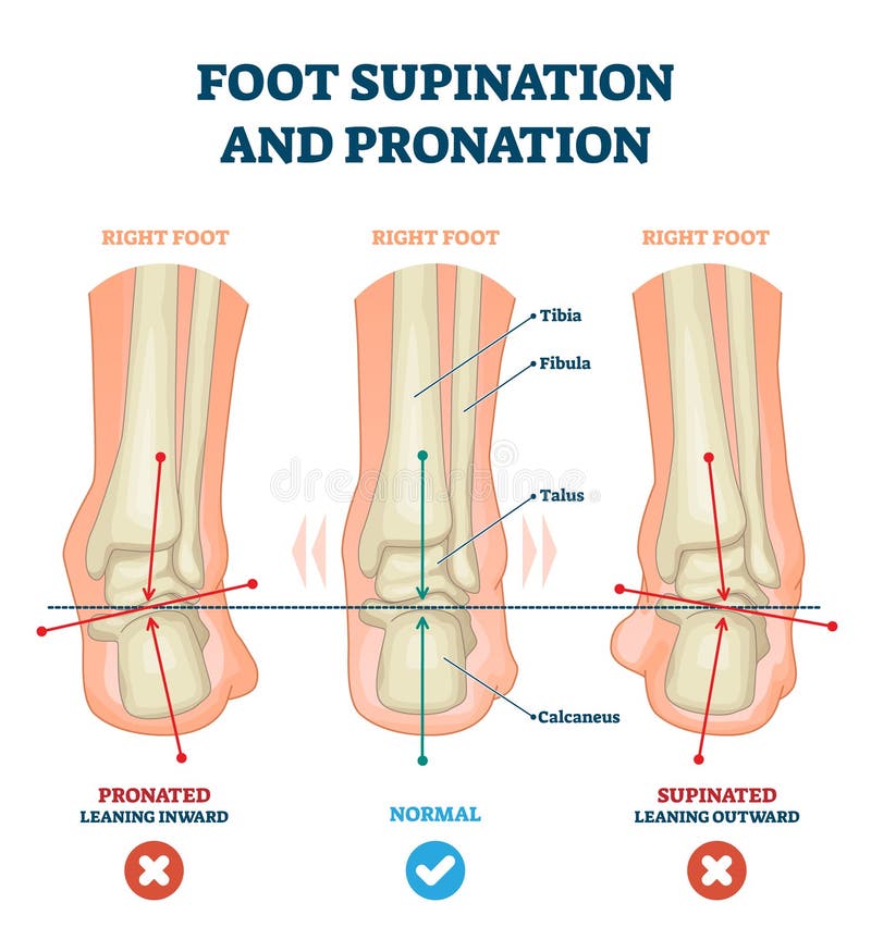
Free with trial Foot supination and pronation vector illustration. Labeled medical scheme with incorrect leg joint movement. Educational diagram with pronated, normal and supinated compared examples with bone titles. Human talus vectors Foot supination and pronation vector illustration. Labeled medical scheme. Foot supination and pronation vector illustration. Labeled medical scheme with incorrect leg joint movement. Educational diagram with pronated, normal and supinated compared examples with bone titles.
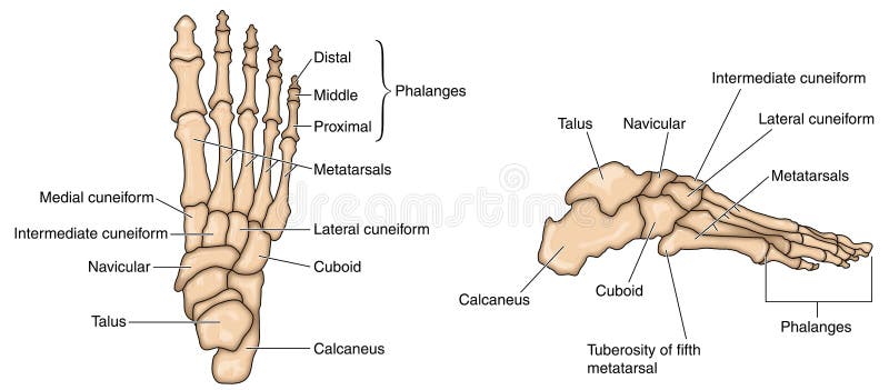
Free with trial Vector illustration of anatomy of the skeleton of the foot. Human talus vectors Anatomy of a skeleton foot. Vector illustration of anatomy of the skeleton of the foot
Free with trial Plantar fasciitis 3d medical illustration on white background eps 10. Human talus illustrations Plantar fasciitis 3d medical illustration on white background
Free with trial Metatarsals Bones Anatomy with Circulatory System with highlighted zone - pain concept. Human talus illustrations Metatarsals Bones Anatomy with Circulatory System
Free with trial Set of xray of human,human joints,knee joint,elbow joint, ankle joint, wrist, skeletal spinal bone structure of Human Spine, emblem or sign of medical diagnostic center ,flat vector illustration. Human talus vectors Set of xray of human. Set of xray of human,human joints,knee joint,elbow joint, ankle joint, wrist, skeletal spinal bone structure of Human Spine, emblem or sign of medical diagnostic center ,flat vector illustration.
Free with trial Human Foot Anatomy Illustration. 3D render. Human talus illustrations Human Foot Anatomy
Free with trial Human Foot Anatomy Illustration. 3D render. Human talus illustrations Human Foot Anatomy
Free with trial Different views of a human foot front, back, side, lateral, medial, dorsal and plantar isolated on white background. Vector illustration for medical health care use. EPS 10. Human talus vectors Different views of a human foot on white background_Skeleton. Different views of a human foot front, back, side, lateral, medial, dorsal and plantar isolated on white background. Vector illustration for medical health care use. EPS 10.
Free with trial Human Foot Anatomy illustration. 3D render. Human talus illustrations Human Foot Anatomy
Free with trial Human Foot Anatomy Illustration. 3D render. Human talus illustrations Human Foot Anatomy
Free with trial Human Anatomy Foot Bone Vector Illustration. Human talus vectors Human Anatomy Foot Bone
Free with trial Human bones icon set. Cartoon set of human bones vector icons for web design isolated on white background. Human talus vectors Human bones icon set, cartoon style. Human bones icon set. Cartoon set of human bones vector icons for web design isolated on white background
Free with trial Human bones icon set. Cartoon set of human bones vector icons for web design isolated on white background. Human talus vectors Human bones icon set, cartoon style. Human bones icon set. Cartoon set of human bones vector icons for web design isolated on white background
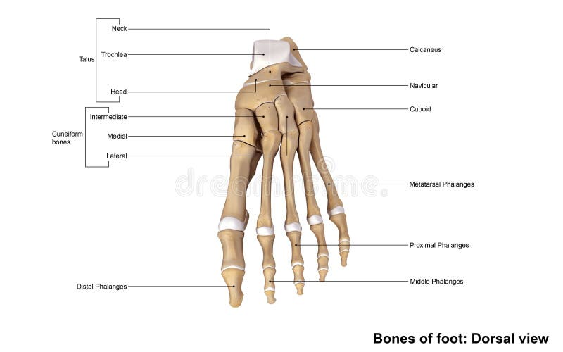
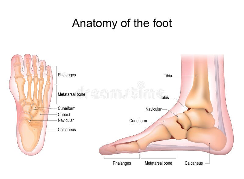
Free with trial Bones of foot. Human Anatomy. The diagram shows the placement and names of all bones of foot. Human talus vectors Bones of foot
Free with trial Vector illustration of human ankle anatomy. bones and tendons with description of every part of human foot. Human talus vectors Ankle anatomy
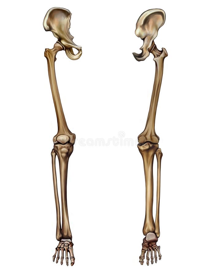
Free with trial The hip joint is one of the most important joints in the human body. It allows us to walk, run, and jump. It bears our body’s weight and the force of the strong muscles of the hip and leg. Yet the hip joint is also one of our most flexible joints and allows a greater range of motion than all other joints in the body except for the shoulder. The hip joint is a ball-and-socket synovial joint formed between the os coxa (hip bone) and the femur. A round, cup-shaped structure on the os coax, known as the acetabulum, forms the socket for the hip joint. The rounded head of the femur forms the ball of the joint. The tibia, sometimes known as the shin bone, is the larger and stronger of the two lower leg bones. It forms the knee joint with the femur and the ankle joint with the fibula and tarsus. The fibula is the long, thin and lateral bone of the lower leg. It runs parallel to the tibia, or shin bone, and plays a significant role in stabilizing the ankle and supporting the muscles of the lower leg. The bones of the ankle and foot form the most distal region of the lower limb in the appendicular skeleton. These bones are responsible for the propulsion, balance, and support of the body’s weight through many diverse activities, such as standing, walking, running, and jumping. Human talus illustrations Skeleton: Hip, Femur, Tibia, Fibula, Ankle and Foot bones. The hip joint is one of the most important joints in the human body. It allows us to walk, run, and jump. It bears our body’s weight and the force of the strong muscles of the hip and leg. Yet the hip joint is also one of our most flexible joints and allows a greater range of motion than all other joints in the body except for the shoulder. The hip joint is a ball-and-socket synovial joint formed between the os coxa (hip bone) and the femur. A round, cup-shaped structure on the os coax, known as the acetabulum, forms the socket for the hip joint. The rounded head of the femur forms the ball of the joint. The tibia, sometimes known as the shin bone, is the larger and stronger of the two lower leg bones. It forms the knee joint with the femur and the ankle joint with the fibula and tarsus. The fibula is the long, thin and lateral bone of the lower leg. It runs parallel to the tibia, or shin bone, and plays a significant role in stabilizing the ankle and supporting the muscles of the lower leg. The bones of the ankle and foot form the most distal region of the lower limb in the appendicular skeleton. These bones are responsible for the propulsion, balance, and support of the body’s weight through many diverse activities, such as standing, walking, running, and jumping.
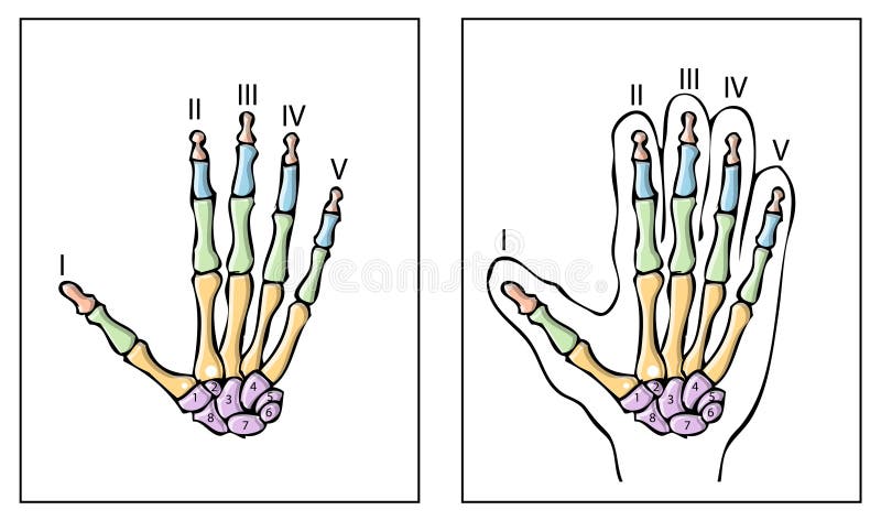
Free with trial The phalanges are the bones that make up the fingers of the hand and the toes of the foot. There are 56 phalanges in the human body, with fourteen on each hand and foot. Three phalanges are present on each finger and toe, with the exception of the thumb and large toe, which possess only two. The middle and far phalanges of the fourth and fifth toes are often fused together (symphalangism). The phalanges of the hand are commonly known as the finger bones. The phalanges of the foot differ from the hand in that they are often shorter and more compressed, especially in the proximal phalanges, those closest to the body. The phalanges are named according to whether they are proximal, intermediate or distal, and according to the finger or toe they are in. The proximal phalanges are those that are closest to the hand or foot. In the hand, the prominent, knobby ends of the proximal phalanx is often called the knuckle. The proximal phalanges join with the metacarpals of the hand or metatarsals of the foot at the metacarpophalangeal joint or metatarsophalangeal joint. The intermediate phalanx is not only intermediate in location, but usually also in size. The thumb and large toe do not possess a middle phalanx. The distal phalanges are the bones at the tips of the fingers or toes. The proximal, intermediate, and distal phalanges articulate with one another through interphalangeal articulations. Human talus illustrations Foot Dorsal view. The phalanges are the bones that make up the fingers of the hand and the toes of the foot. There are 56 phalanges in the human body, with fourteen on each hand and foot. Three phalanges are present on each finger and toe, with the exception of the thumb and large toe, which possess only two. The middle and far phalanges of the fourth and fifth toes are often fused together (symphalangism). The phalanges of the hand are commonly known as the finger bones. The phalanges of the foot differ from the hand in that they are often shorter and more compressed, especially in the proximal phalanges, those closest to the body. The phalanges are named according to whether they are proximal, intermediate or distal, and according to the finger or toe they are in. The proximal phalanges are those that are closest to the hand or foot. In the hand, the prominent, knobby ends of the proximal phalanx is often called the knuckle. The proximal phalanges join with the metacarpals of the hand or metatarsals of the foot at the metacarpophalangeal joint or metatarsophalangeal joint. The intermediate phalanx is not only intermediate in location, but usually also in size. The thumb and large toe do not possess a middle phalanx. The distal phalanges are the bones at the tips of the fingers or toes. The proximal, intermediate, and distal phalanges articulate with one another through interphalangeal articulations.
Free with trial Each foot is made up of 28 bones, 30 joints and more than 100 muscles, tendons and ligaments, all of which work together to provide support, balance and mobility. Human talus illustrations Foot Joints. Each foot is made up of 28 bones, 30 joints and more than 100 muscles, tendons and ligaments, all of which work together to provide support, balance and mobility.
Free with trial Bones of the foot and ankle joint medical vector illustration isolated on white background eps 10 infographic. Human talus vectors Bones of the foot and ankle joint medical vector illustration isolated on white background
Free with trial Vector illustration of Peroneal Tendon Injuries. Peroneal tendonitis. Inflammation of peroneal tendons. Lateral ankle injury. Human talus vectors Peroneal Tendon Injuries_Peroneal tendonitis. Vector illustration of Peroneal Tendon Injuries. Peroneal tendonitis. Inflammation of peroneal tendons. Lateral ankle injury.
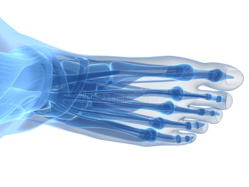
Free with trial Foot Bones Anatomy isolated on white background. 3D render. Human talus illustrations Foot Bones Anatomy Isolated
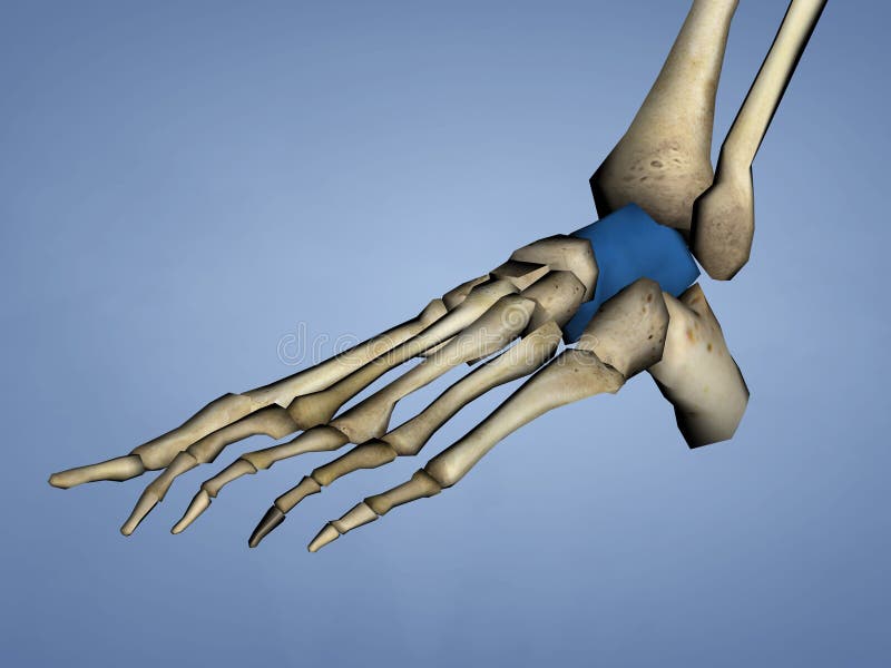
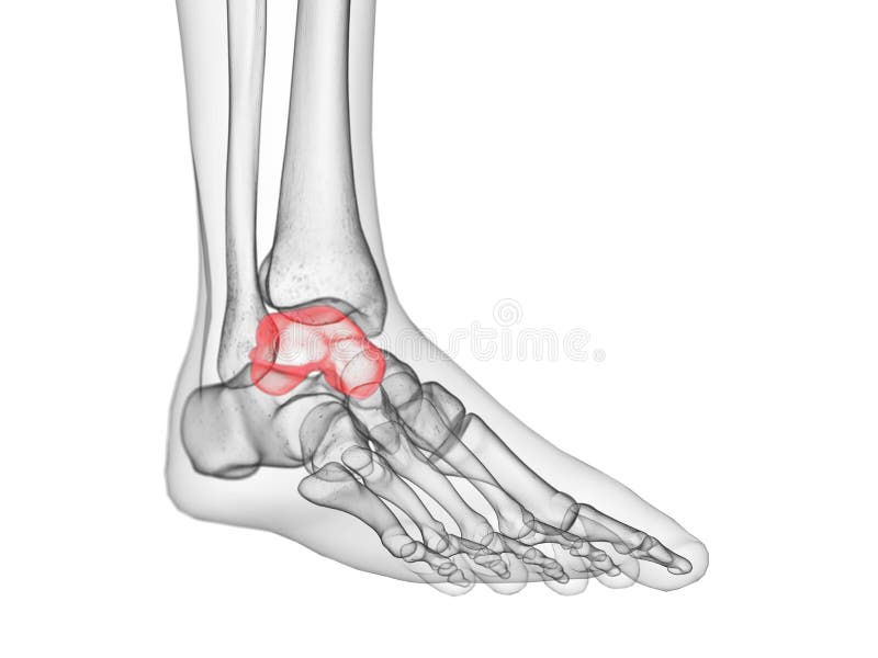
Free with trial Human Skeleton, Blue Background, 3D Model, Talus, Ankle Bone; Astragalus. Human talus illustrations Talus Bone, 3D Model. Human Skeleton, Blue Background, 3D Model, Talus, Ankle Bone; Astragalus
Free with trial Human Skeleton, Blue Background, 3D Model, Talus, Ankle Bone; Astragalus. Human talus illustrations Talus Bone, 3D Model. Human Skeleton, Blue Background, 3D Model, Talus, Ankle Bone; Astragalus
Free with trial Set of human joints, knee joint, elbow joint, ankle joint, wrist, skeletal spinal bone structure of Human Spine, emblem or sign of medical diagnostic center or clinic, flat vector illustration. Human talus vectors Set of human joints. Set of human joints, knee joint, elbow joint, ankle joint, wrist, skeletal spinal bone structure of Human Spine, emblem or sign of medical diagnostic center or clinic, flat vector illustration.
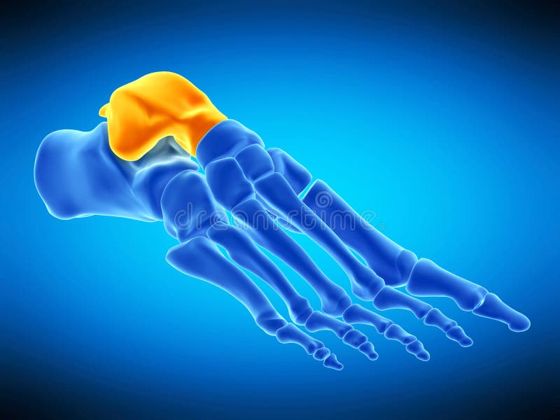
Free with trial Medically accurate illustration of the talus bone. Human talus illustrations The talus bone
Free with trial Medically accurate illustration of the talus bone. Human talus illustrations The talus bone
Free with trial The ankle joint anatomy. Talocrural region or the jumping bone. Part of human body where the foot and the leg meet. Plantar fascia and Achilles tendon. Lateral collateral ligaments of ankle joint Anterior talofibular, Posterior talofibular, and Calcaneofibular ligament. Bones of foot Talus, Fibula, Tibia, and Calcaneus. Vector illustration. Human talus vectors The ankle joint anatomy. Talocrural region or the jumping bone. Part of human body where the foot and the leg meet. Plantar fascia and Achilles tendon. Lateral
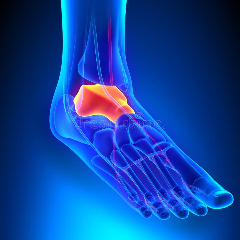
Free with trial Talus Bone Anatomy with Circulatory System with highlighted zone - pain concept. Human talus illustrations Talus Bone Anatomy with Circulatory System
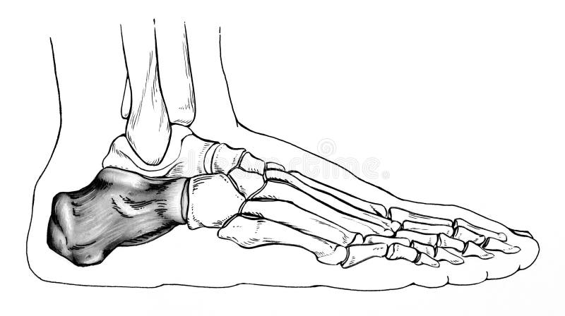
Free with trial Line drawing of a normal human foot, with an emphasis on the talus heel bone, ; side view. Human talus illustrations Foot - Bones Side View. Line drawing of a normal human foot, with an emphasis on the talus heel bone, ; side view.
Free with trial 3d rendered illustration of the human foot. Human talus illustrations Human foot anatomy. 3d rendered illustration of the human foot
Free with trial 3d rendered illustration of the human foot. Human talus illustrations Human foot anatomy. 3d rendered illustration of the human foot
Free with trial Human Foot Anatomy Illustration. 3D render. Human talus illustrations Human Foot Anatomy
Free with trial Human Foot Anatomy Illustration. 3D render. Human talus illustrations Human Foot Anatomy Illustration
Free with trial Foot anatomy. Human foot with the name and description of all bones and sites. top view and side view. Arches of the feet. skeleton anatomy. Vector illustration. Human talus vectors Foot anatomy. Human foot with the name and description of all bones and sites
Free with trial 3d rendered medically accurate illustration of the talus bone. Human talus illustrations The talus bone
Free with trial 3d rendered medically accurate illustration of the talus bone. Human talus illustrations The talus bone
Free with trial Graphic detailed black and white human skeleton bone hands. Isolated on white background. Vector icon set. Human talus vectors Graphic black and white human bone hands vector. Graphic detailed black and white human skeleton bone hands. Isolated on white background. Vector icon set
Free with trial Plantar Fasciitis. Illustration human foot anatomy explain on symptom plantar fasciitis. Human talus vectors Plantar Fasciitis. Illustration human foot. Plantar Fasciitis. Illustration human foot anatomy explain on symptom plantar fasciitis
Free with trial Bones of the human foot with the name and description of all sites. Lateral view. Human anatomy. Vector illustration isolated on a white background. Human talus illustrations Bones of the human foot with the name and description of all sites. Lateral view. Human anatomy.
Free with trial Medical accurate illustration of the intermediate talus bone. Human talus illustrations The intermediate talus bone
Free with trial Leg bones membrum inferius liberum, realistic drawing of the human skeleton, front and back view, femur, tibia, knee, patella, metatarsus, foot structure, anatomy, body parts, isolated image on a white background. Human talus illustrations Human leg bones membrum inferius liberum. Leg bones membrum inferius liberum, realistic drawing of the human skeleton, front and back view, femur, tibia, knee, patella, metatarsus, foot structure, anatomy, body parts, isolated image on a white background
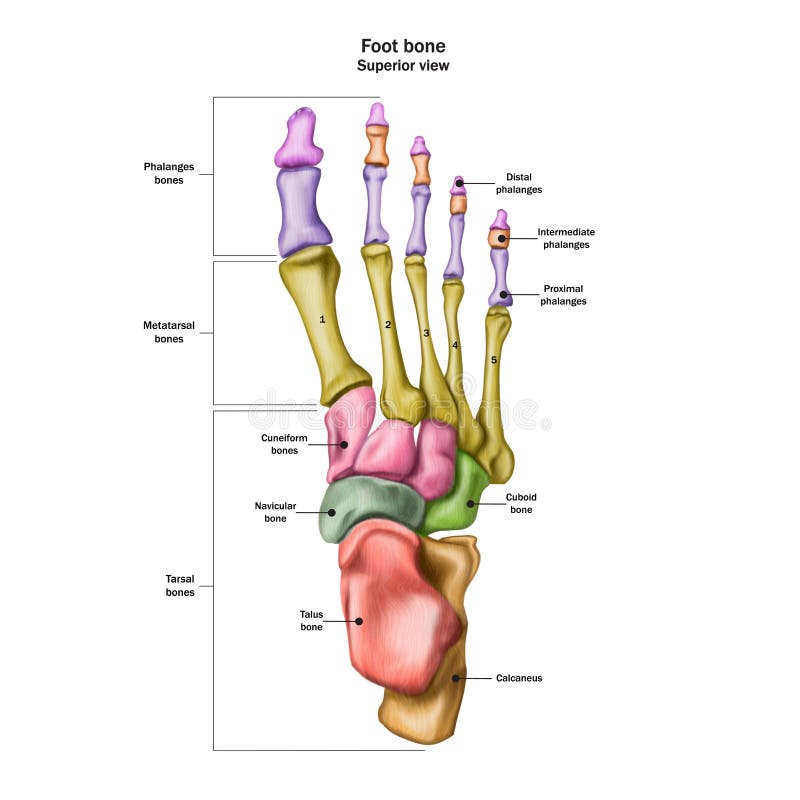
Free with trial Bones of the human foot with the name and description of all sites. Superior view. Human anatomy. Vector illustration isolated on a white background. Human talus illustrations Bones of the human foot with the name and description of all sites. Superior view. Human anatomy
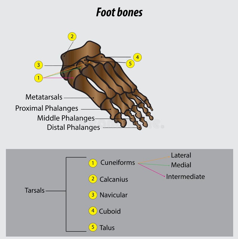
Free with trial Foot bones vector illustration with labels. Medical diagram with tibia, fibula, malleous, talus and navicular. Educational scheme with labeled cuneiforms, cuboid, lateral, calcanius and phalanges. Human talus vectors Vector illustration with foot bones. Medical diagram and educational scheme with tibia, fibula, malleous, talus and navicular. Foot bones vector illustration with labels. Medical diagram with tibia, fibula, malleous, talus and navicular. Educational scheme with labeled cuneiforms, cuboid, lateral, calcanius and phalanges.
Free with trial Human Foot Anatomy Illustration. 3D render. Human talus illustrations Human Foot Anatomy Illustration
Free with trial Talus fracture as broken leg with swelling ankle symptom outline diagram. Labeled educational scheme with medical bone trauma vector illustration. Human leg foot anatomical structure with painful part. Human talus vectors Talus fracture and broken leg with swelling ankle symptom outline diagram. Talus fracture as broken leg with swelling ankle symptom outline diagram. Labeled educational scheme with medical bone trauma vector illustration. Human leg foot anatomical structure with painful part
Free with trial Human Anatomy Foot Bone Vector Illustration. Human talus vectors Human Anatomy Foot Bone
Free with trial Cartoon Human Joints Set Health Care Medical Diagnostic X-ray. Flat Design Style Vector illustration. Human talus vectors Cartoon Human Joints Set. Vector. Cartoon Human Joints Set Health Care Medical Diagnostic X-ray. Flat Design Style Vector illustration
Free with trial Skeletal foot - injuryd talus bone. Xray view. Medically accurate 3D illustration. Human talus illustrations Skeletal foot - injuryd talus bone. Xray view. Medically accurate illustration
Free with trial Human Skeleton Hand Bones. Vector Illustration. Human talus illustrations Human Skeleton Hand Bones Vector. Human Skeleton Hand Bones. Vector Illustration.
Free with trial Human Foot Bone Anatomy Vector Illustration. Human talus vectors Human Foot Bone Anatomy
Free with trial Human foot bones and joints skeleton vector sketch body anatomy icon. Isolated symbol of feet and toes limbs structure of leg organ for anatomical orthopedic or medical surgery design element. Human talus vectors Vector sketch icon of human foot bones or joints. Human foot bones and joints skeleton vector sketch body anatomy icon. Isolated symbol of feet and toes limbs structure of leg organ for anatomical orthopedic or medical surgery design element
Free with trial 3d render illustration of the Human Foot Joint Pains. Human talus illustrations Human Foot Joint Pains
Free with trial Illustration on Human Anatomy, Foot Background. Human talus illustrations Illustration on Human Anatomy, Foot Background
Free with trial Set of human knee, elbow and ankle joints and wrist, emblem or sign of medical diagnostic center or clinic, flat design, vector. Human talus vectors Set of human knee, elbow and ankle joints and wrist, emblem or sign of medical diagnostic center or clinic, flat design
Free with trial Vector drawing of human foot human foot anatomy drawing labeled drawing with layers reedit able. Human talus vectors Vector drawing of human foot human foot anatomy drawing labeled
Free with trial 3d illustration of human body skeletal metatarsal. Anatomy parts of human body bones. Human talus illustrations 3d illustration of human body skeletal metatarsal
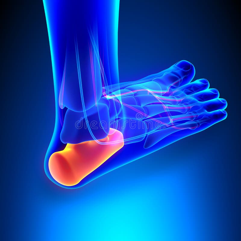
Free with trial 3D Illustration of Human Lrg Joint Pains (Calcaneus Bone). Human talus illustrations Human Leg Joint Pains (Calcaneus Bone). 3D Illustration of Human Lrg Joint Pains (Calcaneus Bone)
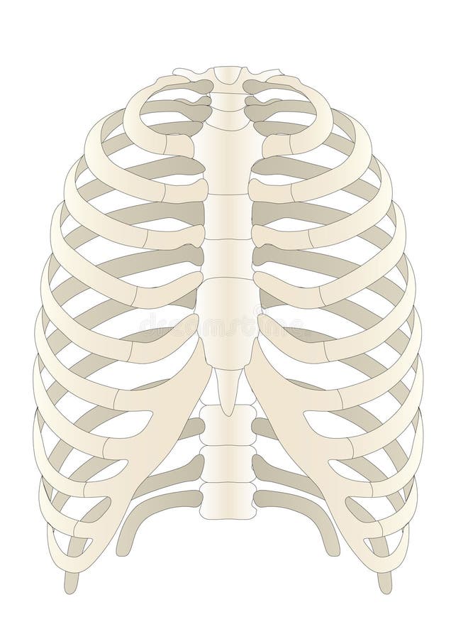
Free with trial 3d illustration of human body skeletal ribs. Anatomy parts of bone. Human talus illustrations 3d illustration of human body skeletal ribs
Free with trial 3d illustration of human body skeletal pelivs. metatarsal. Anatomy parts of human body bones. Human talus illustrations 3d illustration of human body skeletal pelivs
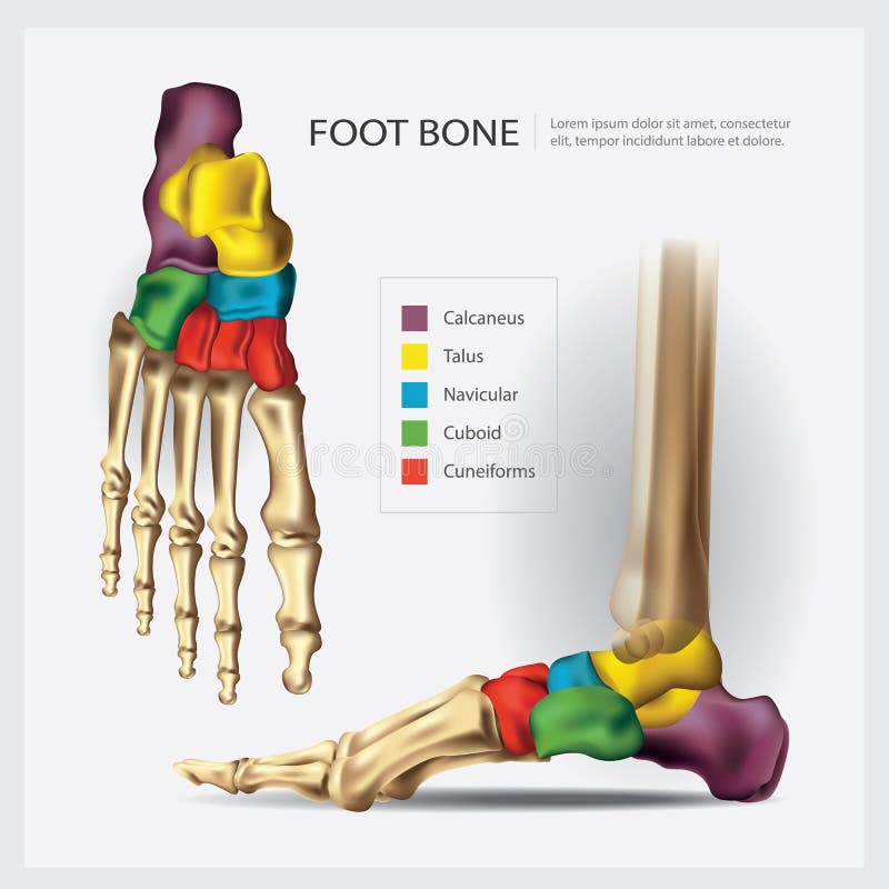
Free with trial Foot bone anatomy concept. Descriptions of the human feet bones and joints. Talus, ligaments, calcaneus, navicular anatomy. Skeletal medical anatomical poster. Ankle X ray isolated vector illustration. Human talus vectors Foot bones anatomy. Foot bone anatomy concept. Descriptions of the human feet bones and joints. Talus, ligaments, calcaneus, navicular anatomy. Skeletal medical anatomical poster. Ankle X ray isolated vector illustration
Free with trial 3D illustration of human body skeletal mandibula. Anatomy parts of human body bones. Human talus illustrations 3D illustration of human body skeletal mandibula