Free with trial Events from ovulation to implantation, eps8. Inner cell mass vectors Early human development. Events from ovulation to implantation, eps8
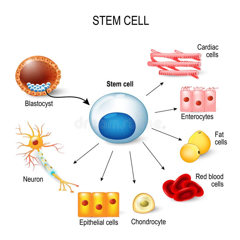
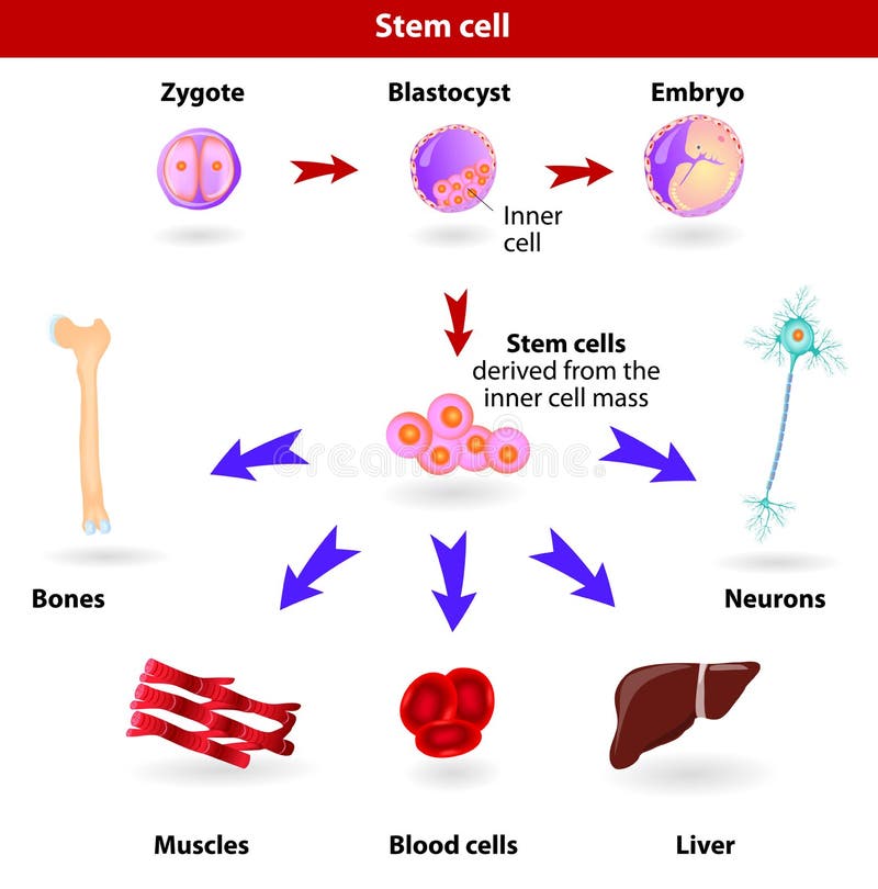
Free with trial Stem cells. These inner cell mass from a blastocyst. These stem cells can become any tissue in the body. for example: neuron, chondrocyte, enterocytes, red blood cells, muscle, fat or epithelial cells. Inner cell mass vectors Stem cells.
Free with trial Pluripotent, embryonic stem cells originate as inner cell mass cells within a blastocyst. These stem cells can become any tissue in the body, excluding a placenta. Inner cell mass vectors Stem cells
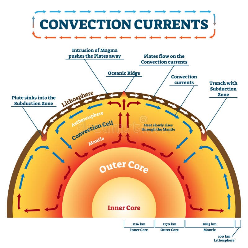
Free with trial Convection Currents vector illustration. Labeled educational process scheme. Geology land movement and heat transfer by mass motion as molten rock. Lithosphere, ocean ridge and subduction zone example. Inner cell mass vectors Convection Currents vector illustration. Labeled educational process scheme
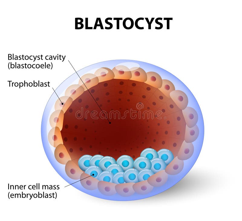
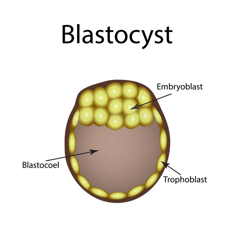
Free with trial Blastocyst. Human blastocyst, with inner cell mass. the mammalian conceptus in the post-morula stage, consisting of the trophoblast and an inner cell mass. Inner cell mass vectors Human blastocyst, with inner cell mass
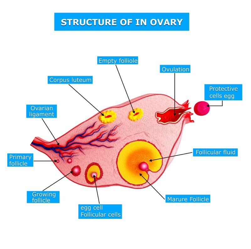
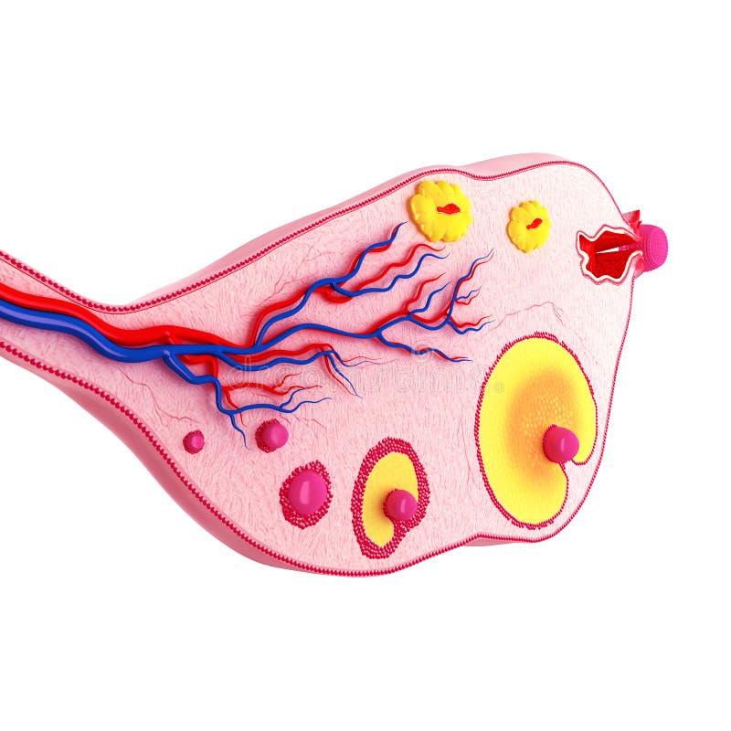
Free with trial 3d art illustration of side view of Ovarian cycle. Inner cell mass illustrations Side view of Ovarian cycle
Free with trial 3d art illustration of Anatomy of Ovarian cycle in blue. Inner cell mass illustrations Anatomy of Ovarian cycle in blue
Free with trial 3d rendered illustration of anatomy of ovarian cycle. Inner cell mass illustrations Anatomy of ovarian cycle
Free with trial 3d art illustration of Ovarian cycle with white background. Inner cell mass illustrations Ovarian cycle with white background
Free with trial Blastocyst vector illustration infographic diagram. Biological embryo early stage labeled scheme with ovulation, fertilization, zygote formation, cells and implantation. Inner cell mass vectors Blastocyst vector illustration infographic. Biological embryo early stage. Blastocyst vector illustration infographic diagram. Biological embryo early stage labeled scheme with ovulation, fertilization, zygote formation, cells and implantation.
Free with trial 3d art illustration of side view of Ovarian cycle. Inner cell mass illustrations Side view of Ovarian cycle
Free with trial Blastocyst. Human blastocyst, with inner cell mass. the mammalian conceptus in the post-morula stage, consisting of the trophoblast and an inner cell mass. Inner cell mass vectors Blastocyst with inner cell mass. Blastocyst. Human blastocyst, with inner cell mass. the mammalian conceptus in the post-morula stage, consisting of the trophoblast and an inner cell mass.
Free with trial Cross-section of human blastocyst showing inner mass and trophoblast layers, 3D illustration. Inner cell mass illustrations Cross-section of human blastocyst showing inner mass and trophoblast layers
Free with trial Cross-section of human blastocyst showing inner mass and trophoblast layers, 3D illustration. Inner cell mass illustrations Cross-section of human blastocyst showing inner mass and trophoblast layers
Free with trial Cross-section of human blastocyst showing inner mass and trophoblast layers, 3D illustration. Inner cell mass illustrations Cross-section of human blastocyst showing inner mass and trophoblast layers
Free with trial Cross-section of human blastocyst showing inner mass and trophoblast layers, 3D illustration. Inner cell mass illustrations Cross-section of human blastocyst showing inner mass and trophoblast layers
Free with trial Flat illustration of human blastocyst cell. Health, medical, and science concept. Inner cell mass vectors Flat illustration of human blastocyst cell
Free with trial Blastocyst with stem cell icon. Embryo development stage. Pregnancy, insemination and fertalization concept. Human sexual reproductive system. Flat vector illustration. Poster for clinic or education. Inner cell mass vectors Reproductive system concept. Blastocyst with stem cell icon. Embryo development stage. Pregnancy, insemination and fertalization concept. Human sexual reproductive system. Flat vector illustration. Poster for clinic or education.
Free with trial 3d art illustration of side view of Ovarian cycle. Inner cell mass illustrations Side view of Ovarian cycle
Free with trial 3d art illustration of side view of Ovarian cycle. Inner cell mass illustrations Side view of Ovarian cycle
Free with trial 3d art illustration of side view of Ovarian cycle. Inner cell mass illustrations Side view of Ovarian cycle
Free with trial 3d art illustration of side view of Ovarian cycle. Inner cell mass illustrations Side view of Ovarian cycle
Free with trial Human blastocyst. The structure of the blastocyst. Vector illustration on background. Inner cell mass vectors Human blastocyst. The structure of the blastocyst. Vector illustration on background
Free with trial Blastocyst cell structure anatomy diagram infographic showing early embryonic stage with trophoblast, blastocyst cavity, and inner cell mass, multicellular eukaryotic organism pre implantation phase. Inner cell mass vectors Blastocyst cell structure anatomy diagram infographic showing early embryonic stage with trophoblast, blastocyst cavity, and inner. Cell mass, multicellular. Blastocyst cell structure anatomy diagram infographic showing early embryonic stage with trophoblast, blastocyst cavity, and inner cell mass, multicellular eukaryotic organism pre implantation phase
Free with trial Blastocyst formation shown as stepwise early embryo development, moving from zygote to morula to blastocyst with labeled inner cell mass and trophoblast. Outline diagram. Inner cell mass vectors Blastocyst formation shown as stepwise early embryo development, moving from ... Blastocyst formation shown as stepwise early embryo development, moving from zygote to morula to blastocyst with labeled inner cell mass and trophoblast. Outline diagram
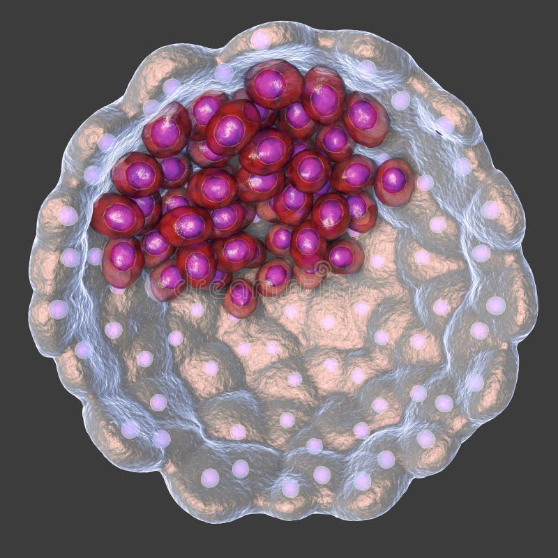
Free with trial This image depicts a detailed cellular structure with a labeled inner cell mass (ICM). The image showcases the intricate layers and patterns of the cell, highlighting the ICM at the center. The surrounding layers appear to be composed of various cellular components, possibly indicating a developing embryo or stem cell research context. Inner cell mass illustrations Cellular structure with icm label isolated on white background. This image depicts a detailed cellular structure with a labeled inner cell mass (ICM). The image showcases the intricate layers and patterns of the cell, highlighting the ICM at the center. The surrounding layers appear to be composed of various cellular components, possibly indicating a developing embryo or stem cell research context
Free with trial A detailed close-up of a blood vessel filled with red blood cells and a large white cell, possibly a mass or a blockage, set against a blurred background. This high-angle view emphasizes the intricate texture and colors of the cells, ideal for medical visualization, educational content, or symbolizing health concerns and the body's inner workings. Inner cell mass illustrations Detailed Blood Vessel Close Up with Red and White Blood Cells for Medical Visualization. A detailed close-up of a blood vessel filled with red blood cells and a large white cell, possibly a mass or a blockage, set against a blurred background. This high-angle view emphasizes the intricate texture and colors of the cells, ideal for medical visualization, educational content, or symbolizing health concerns and the body's inner workings
Free with trial A detailed 3D rendering of a human blastocyst, an early stage of embryonic development. The image showcases the inner cell mass, which will develop into the fetus, surrounded by the trophectoderm, which will form the placenta. The transparent outer layer represents the zona pellucida. This scientific illustration is ideal for educational materials, medical research, and concepts related to reproduction and fertility. Inner cell mass illustrations Early Human Embryo: Blastocyst Stage. A detailed 3D rendering of a human blastocyst, an early stage of embryonic development. The image showcases the inner cell mass, which will develop into the fetus, surrounded by the trophectoderm, which will form the placenta. The transparent outer layer represents the zona pellucida. This scientific illustration is ideal for educational materials, medical research, and concepts related to reproduction and fertility.
Free with trial Detailed illustration of a human blastocyst, a crucial stage in early embryonic development, showing the inner cell mass and trophectoderm. Inner cell mass illustrations Microscopic view of a human blastocyst embryo in early development. Detailed illustration of a human blastocyst, a crucial stage in early embryonic development, showing the inner cell mass and trophectoderm
Free with trial This detailed microscopic image reveals a blastocyst embryo suspended in amniotic fluid, showcasing intricate cellular activity. The image captures the dynamic electrical pulse, a crucial aspect of early embryonic development. The blastocyst, a crucial stage in the fertilization process, is characterized by its distinctive inner cell mass, which will eventually form the fetus. The surrounding. Inner cell mass illustrations Microscopic Blastocyst Embryo in Amniotic Fluid Observing Electrical Activity During Early Development. This detailed microscopic image reveals a blastocyst embryo suspended in amniotic fluid, showcasing intricate cellular activity. The image captures the dynamic electrical pulse, a crucial aspect of early embryonic development. The blastocyst, a crucial stage in the fertilization process, is characterized by its distinctive inner cell mass, which will eventually form the fetus. The surrounding
Free with trial A blastocyst is a human embryo about five days after fertilization. It is implanted in the uterine wall, which is shown in a microscopic image. The image is a scientific illustration depicting the complex biological processes of early human development. The inner cell mass, which will eventually form the fetus, is visible as a cluster of cells at the center of the blastocyst. The outer layer, called the trophoblast, will form the placenta. Inner cell mass illustrations This is a microscopic image of a blastocyst, a human embryo about five days after fertilization, implanted in the. A blastocyst is a human embryo about five days after fertilization. It is implanted in the uterine wall, which is shown in a microscopic image. The image is a scientific illustration depicting the complex biological processes of early human development. The inner cell mass, which will eventually form the fetus, is visible as a cluster of cells at the center of the blastocyst. The outer layer, called the trophoblast, will form the placenta.
Free with trial Embryonic Stage Cellular Growth on White A detailed close up of an embryo in its early stages of cellular growth, isolated on a stark white background. Emphasize intricate cellular detail and texture. Inner cell mass illustrations Embryonic Stage Cellular Growth on White A detailed close up of an embryo in its early stages of cellular growth, isolated on a
Free with trial Stem cell transplantation to the blood cell. Idea of medical treatment technology. Microbiology and medicine. Isolated vector illustration in cartoon style. Inner cell mass vectors Stem cell transplntation to the blood cell. Stem cell transplantation to the blood cell. Idea of medical treatment technology. Microbiology and medicine. Isolated vector illustration in cartoon style
Free with trial Blastocyst with stem cell icon. Embryo development stage. Doctor in clinic or laboratory. Pregnancy, insemination and fertilization concept. Human sexual reproductive system, flat vector illustration. Inner cell mass vectors Embryo cell anatomy. Blastocyst with stem cell icon. Embryo development stage. Doctor in clinic or laboratory. Pregnancy, insemination and fertilization concept. Human sexual reproductive system, flat vector illustration.
Free with trial Blastocyst with stem cell icon. Doctor in clinic or laboratory. Embryo development stage. Pregnancy, insemination and fertilization concept. Human sexual reproductive system. Flat vector illustration. Inner cell mass vectors Embryo cell anatomy. Blastocyst with stem cell icon. Doctor in clinic or laboratory. Embryo development stage. Pregnancy, insemination and fertilization concept. Human sexual reproductive system. Flat vector illustration.
Free with trial Principle of embryonic stem cell (ESC) isolation from a blastocyst. Inner cell mass illustrations ESC isolation. Principle of embryonic stem cell (ESC) isolation from a blastocyst
Free with trial Blastocyst with stem cell icon. Embryo development stage. Pregnancy, insemination and fertalization concept. Human sexual reproductive system. Flat vector illustration. Poster for clinic or education. Inner cell mass vectors Human fertility concept. Blastocyst with stem cell icon. Embryo development stage. Pregnancy, insemination and fertalization concept. Human sexual reproductive system. Flat vector illustration. Poster for clinic or education.
Free with trial Blastocyst with stem cell icon. Embryo development stage. Pregnancy, insemination and fertalization concept. Human sexual reproductive system. Flat vector illustration. Poster for clinic or education. Inner cell mass vectors Reproductive system concept. Blastocyst with stem cell icon. Embryo development stage. Pregnancy, insemination and fertalization concept. Human sexual reproductive system. Flat vector illustration. Poster for clinic or education.
Free with trial Blastocyst with stem cell icon. Embryo development stage. Pregnancy, insemination and fertalization concept. Human sexual reproductive system. Flat vector illustration. Poster for clinic or education. Inner cell mass vectors Reproductive system concept. Blastocyst with stem cell icon. Embryo development stage. Pregnancy, insemination and fertalization concept. Human sexual reproductive system. Flat vector illustration. Poster for clinic or education.
Free with trial Blastocyst with stem cell icon. Embryo development stage. Pregnancy, insemination and fertalization concept. Human sexual reproductive system. Flat vector illustration. Poster for clinic or education. Inner cell mass vectors Reproductive system concept. Blastocyst with stem cell icon. Embryo development stage. Pregnancy, insemination and fertalization concept. Human sexual reproductive system. Flat vector illustration. Poster for clinic or education.
Free with trial Insemination and fertilization concept. Female and male egg cell icon. Human sexual reproductive system and pregnancy flat vector illustration. Diagram or poster for clinic, laboratory and education. Inner cell mass vectors Insemination and fertilization
Free with trial Blastocyst with stem cell icon set. Embryo development stage. Pregnancy, insemination and fertalization concept. Human sexual reproductive system. Flat vector illustration. Medical poster for clinic. Inner cell mass vectors Reproductive system concept. Blastocyst with stem cell icon set. Embryo development stage. Pregnancy, insemination and fertalization concept. Human sexual reproductive system. Flat vector illustration. Medical poster for clinic.
Free with trial Blastocyst with stem cell icon. Embryo development stage. Pregnancy, insemination and fertalization concept. Human sexual reproductive system. Flat vector illustration. Poster for clinic or education. Inner cell mass vectors Reproductive system concept. Blastocyst with stem cell icon. Embryo development stage. Pregnancy, insemination and fertalization concept. Human sexual reproductive system. Flat vector illustration. Poster for clinic or education.
Free with trial Blastocyst. Embryo development stage. Stem cells. Fertilization, insemination and pregnancy. Human reproductive system. Poster for science, medical, clinic, and education use. vector illustration. Inner cell mass vectors Blastocyst. Embryo development stage. Stem cells
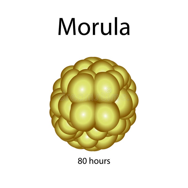
Free with trial Human morula. Vector illustration on background. Inner cell mass vectors Human morula. Vector illustration on background
Free with trial Embryonic stem cells. Embryo development stages. Pregnancy, insemination and fertalization concept. Ovum and spermatozoon. Human sexual reproductive system. Flat vector illustration. Medical poster. Inner cell mass vectors Reproductive system concept. Embryonic stem cells. Embryo development stages. Pregnancy, insemination and fertalization concept. Ovum and spermatozoon. Human sexual reproductive system. Flat vector illustration. Medical poster.