Free with trial A stunning microscopic visualization of red blood cells packed inside transparent cell membranes. This high-resolution image showcases the intricate details and vibrant colors of erythrocytes, making it ideal for medical, educational, and scientific purposes. The dynamic composition highlights the complexity of human biology, evoking wonder and curiosity about the inner workings of our bodies. Inner membranes illustrations Microscopic View of Red Blood Cells in Human Body. A stunning microscopic visualization of red blood cells packed inside transparent cell membranes. This high-resolution image showcases the intricate details and vibrant colors of erythrocytes, making it ideal for medical, educational, and scientific purposes. The dynamic composition highlights the complexity of human biology, evoking wonder and curiosity about the inner workings of our bodies.
Free with trial 3D visualization of numerous elongated mitochondria with transparent purple membranes and luminous pink folded cristae, representing bioenergetics and vitality in cell biology. Inner membranes illustrations Multiple purple mitochondria with pink cristae on dark background, conceptual ATP production, cellular respiration and metabolic. 3D visualization of numerous elongated mitochondria with transparent purple membranes and luminous pink folded cristae, representing bioenergetics and vitality in cell biology
Free with trial Artistic 3D visualization of numerous mitochondria with translucent green membranes and luminous yellow cristae, representing bioenergetics and metabolic health research. Inner membranes illustrations Multiple glowing green mitochondria with detailed yellow cristae on dark background, conceptual ATP energy production and cellular. Artistic 3D visualization of numerous mitochondria with translucent green membranes and luminous yellow cristae, representing bioenergetics and metabolic health research
Free with trial A collection of freshly cut green bell peppers arranged on a wooden surface. The peppers are vibrant green with their seeds and inner membranes exposed, ready for cooking or eating. The wooden surface provides a rustic background, highlighting the natural beauty of the vegetables. Inner membranes illustrations Freshly cut green bell peppers on a wooden surface. A collection of freshly cut green bell peppers arranged on a wooden surface. The peppers are vibrant green with their seeds and inner membranes exposed, ready for cooking or eating. The wooden surface provides a rustic background, highlighting the natural beauty of the vegetables
Free with trial This image showcases a collection of vibrant red chili peppers arranged in various forms. The central focus is a cluster of sliced chili peppers, revealing their inner seeds and membranes. Surrounding this are whole chili peppers, some of which are intact while others are partially cut. The background is a stark white, which accentuates the vivid red color of the peppers. The image captures the. Inner membranes illustrations Vibrant red chili peppers displayed in various forms. This image showcases a collection of vibrant red chili peppers arranged in various forms. The central focus is a cluster of sliced chili peppers, revealing their inner seeds and membranes. Surrounding this are whole chili peppers, some of which are intact while others are partially cut. The background is a stark white, which accentuates the vivid red color of the peppers. The image captures the
Free with trial A collection of sliced red bell pepper pieces are artfully arranged and appear to be floating against a stark black background. The vibrant red of the peppers contrasts sharply with the dark backdrop, highlighting their fresh texture and the details of their cut surfaces, including the seeds and inner membranes. Inner membranes illustrations Sliced Red Bell Peppers Floating on Black Background. A collection of sliced red bell pepper pieces are artfully arranged and appear to be floating against a stark black background. The vibrant red of the peppers contrasts sharply with the dark backdrop, highlighting their fresh texture and the details of their cut surfaces, including the seeds and inner membranes
Free with trial The image shows a cross-section of a lemon that has been sliced open to reveal its inner segments. The bright yellow flesh is divided into distinct wedges by white membranes, and the central core is visible in the middle. The texture appears juicy and moist, indicating freshness. Inner membranes illustrations A freshly cut cross-section of a bright yellow lemon. The image shows a cross-section of a lemon that has been sliced open to reveal its inner segments. The bright yellow flesh is divided into distinct wedges by white membranes, and the central core is visible in the middle. The texture appears juicy and moist, indicating freshness
Free with trial The image shows a collection of fresh red chili peppers. Some of the peppers are whole with stems attached, while others are sliced open, revealing their inner seeds and membranes. The vibrant red color of the peppers stands out vividly against the white background, highlighting their freshness and spicy nature. Inner membranes illustrations Fresh red chili peppers with seeds exposed and sliced. The image shows a collection of fresh red chili peppers. Some of the peppers are whole with stems attached, while others are sliced open, revealing their inner seeds and membranes. The vibrant red color of the peppers stands out vividly against the white background, highlighting their freshness and spicy nature
Free with trial Detailed illustration of a human cell showcasing various organelles. The nucleus is centrally located, depicted in purple with chromatin and a nucleolus. Surrounding it, the rough and smooth endoplasmic reticulum appear coiled and layered. Numerous mitochondria are seen, characterized by their double membranes and inner folds. The Golgi apparatus is nearby, illustrated in a stack-like form. Cytoskeletal elements are shown as fibrous structures. Red and purple circles represent vesicles and lysosomes, while small green dots likely depict ribosomes. The plasma membrane forms a clear boundary, maintaining the cell's shape. Inner membranes illustrations Detailed 3D illustration of a human cell with organelles. Detailed illustration of a human cell showcasing various organelles. The nucleus is centrally located. Detailed illustration of a human cell showcasing various organelles. The nucleus is centrally located, depicted in purple with chromatin and a nucleolus. Surrounding it, the rough and smooth endoplasmic reticulum appear coiled and layered. Numerous mitochondria are seen, characterized by their double membranes and inner folds. The Golgi apparatus is nearby, illustrated in a stack-like form. Cytoskeletal elements are shown as fibrous structures. Red and purple circles represent vesicles and lysosomes, while small green dots likely depict ribosomes. The plasma membrane forms a clear boundary, maintaining the cell's shape.
Free with trial The image shows a cut in half tomato on a white background. The tomato is cut in half longitudinally, revealing its inner structure. The cut surface shows the tomato's seeds and membranes. The tomato's outer skin is red and smooth. Inner membranes illustrations A cut in half tomato on a white background
Free with trial A group of vibrant red bell peppers with green stems, some whole and others sliced to reveal their inner seeds and membranes, all set against a clean white backdrop. Inner membranes illustrations Fresh red bell peppers and sliced peppers on a white background. A group of vibrant red bell peppers with green stems, some whole and others sliced to reveal their inner seeds and membranes, all set against a clean white backdrop
Free with trial This image features a vibrant yellow bell pepper that has been sliced into several rounds. The pepper is displayed against a dark background, highlighting its bright color and crisp texture. The slices reveal the inner white seeds and membranes, contrasting with the smooth, glossy exterior. Inner membranes illustrations Sliced yellow bell pepper on dark background isolated on white background. This image features a vibrant yellow bell pepper that has been sliced into several rounds. The pepper is displayed against a dark background, highlighting its bright color and crisp texture. The slices reveal the inner white seeds and membranes, contrasting with the smooth, glossy exterior
Free with trial This image features three green bell pepper halves, each displaying the inner structure with seeds and membranes. The peppers are arranged in a triangular formation against a transparent background, making the vibrant green color stand out prominently. The image is ideal for use in culinary or botanical contexts. Inner membranes illustrations Three green bell pepper halves isolated on white background. This image features three green bell pepper halves, each displaying the inner structure with seeds and membranes. The peppers are arranged in a triangular formation against a transparent background, making the vibrant green color stand out prominently. The image is ideal for use in culinary or botanical contexts
Free with trial The image shows a cross-sectional view of a lemon slice, highlighting its internal structure. The bright green outer flesh is clearly visible, radiating from the central core. The central core contains a white pith area surrounded by vascular bundles, with a light-colored, star-shaped arrangement. The texture of the flesh appears juicy and segmented by thin membranes, indicative of a ripe lemon's. Inner membranes illustrations Freshly sliced lemon cross-section revealing inner structure. The image shows a cross-sectional view of a lemon slice, highlighting its internal structure. The bright green outer flesh is clearly visible, radiating from the central core. The central core contains a white pith area surrounded by vascular bundles, with a light-colored, star-shaped arrangement. The texture of the flesh appears juicy and segmented by thin membranes, indicative of a ripe lemon's
Free with trial The image showcases a detailed 3D rendering of a pomegranate that has been split open to reveal its juicy, vibrant seeds inside. The rich red and pink hues highlight the texture and natural beauty of the fruit, emphasizing the intricate structure and glossy appearance of the seeds and inner membranes. Inner membranes illustrations A vivid 3d rendering of a split pomegranate revealing its seeds. The image showcases a detailed 3D rendering of a pomegranate that has been split open to reveal its juicy, vibrant seeds inside. The rich red and pink hues highlight the texture and natural beauty of the fruit, emphasizing the intricate structure and glossy appearance of the seeds and inner membranes
Free with trial This image shows a bunch of sliced red bell peppers neatly arranged in a transparent plastic bag. The bell peppers are vibrant red and appear fresh, with their inner white membranes and seeds visible. Inner membranes illustrations A bunch of sliced red bell peppers in a transparent plastic bag. This image shows a bunch of sliced red bell peppers neatly arranged in a transparent plastic bag. The bell peppers are vibrant red and appear fresh, with their inner white membranes and seeds visible
Free with trial The image displays a cross-section of a pomegranate. The outer rind is greenish and smooth, encasing the inner part of the fruit. The interior is divided into several compartments filled with juicy, red seeds. The seeds are arranged in a radial pattern around the central core, which is a lighter color. The vibrant red seeds contrast with the white membranes that separate each compartment. Inner membranes illustrations Cross section of a pomegranate showing seeds and juicy interior. The image displays a cross-section of a pomegranate. The outer rind is greenish and smooth, encasing the inner part of the fruit. The interior is divided into several compartments filled with juicy, red seeds. The seeds are arranged in a radial pattern around the central core, which is a lighter color. The vibrant red seeds contrast with the white membranes that separate each compartment
Free with trial A detailed illustration of a cell membrane showing its lipid bilayer composition with embedded proteins and a visible cross-section of the membrane's inner and outer layers. Inner membranes illustrations Cell membrane structure with lipid bilayer and embedded proteins. a detailed illustration of a cell membrane showing its lipid bilayer composition with embedded proteins and a visible cross-section of the membrane's inner and outer layers
Free with trial The image displays a neatly arranged pattern of sliced red bell peppers with seeds and membranes removed, showcasing their vibrant inner texture and fresh appearance against a clean white backdrop. Inner membranes illustrations Freshly sliced bell peppers arranged neatly on a white background. The image displays a neatly arranged pattern of sliced red bell peppers with seeds and membranes removed, showcasing their vibrant inner texture and fresh appearance against a clean white backdrop
Free with trial The image shows two split red chili peppers with visible seeds and inner membranes. Surrounding the peppers are scattered seeds and small spices, likely including coriander and peppercorns, against a plain background. Inner membranes illustrations Split red chili peppers and seeds displayed on a plain background. The image shows two split red chili peppers with visible seeds and inner membranes. Surrounding the peppers are scattered seeds and small spices, likely including coriander and peppercorns, against a plain background
Free with trial 3D render of biological cells featuring semi-transparent membranes and a glowing nucleus in warm hues. Cells are depicted in a fluid environment, suggesting movement and life. The glowing nucleus stands out, with textures indicating complexity and inner activity. The surroundings are darker, enhancing the nucleus's luminescent quality, with a sense of depth and dimension. The colors and lighting suggest a dynamic, almost ethereal quality, indicative of cellular processes. Inner membranes illustrations Dynamic 3D Render of a Vibrant Biological Cell with Glowing Nucleus. 3D render of biological cells featuring semi-transparent membranes and a glowing nucleus in warm hues. Cells are depicted in a fluid environment, suggesting movement and life. The glowing nucleus stands out, with textures indicating complexity and inner activity. The surroundings are darker, enhancing the nucleus's luminescent quality, with a sense of depth and dimension. The colors and lighting suggest a dynamic, almost ethereal quality, indicative of cellular processes.
Free with trial The image shows three cross-sectional slices of red bell peppers with their inner cores and white membranes clearly visible. The peppers are vibrant red with a slightly glossy surface, indicating freshness. The slices are evenly cut and placed on a plain white backdrop, highlighting their natural color and texture. Inner membranes illustrations Freshly sliced red bell peppers arranged neatly on a white background. The image shows three cross-sectional slices of red bell peppers with their inner cores and white membranes clearly visible. The peppers are vibrant red with a slightly glossy surface, indicating freshness. The slices are evenly cut and placed on a plain white backdrop, highlighting their natural color and texture
Free with trial The image showcases four distinct segments of a fresh orange, each peeled to reveal the juicy, vibrant orange flesh inside. The segments are arranged in a scattered formation on a plain white background, emphasizing their bright color and natural texture. The inner membranes are clearly visible, adding to the freshness and appeal of the fruit segments. The overall presentation highlights the. Inner membranes illustrations Freshly sliced orange segments displayed on white background isolated on white background. The image showcases four distinct segments of a fresh orange, each peeled to reveal the juicy, vibrant orange flesh inside. The segments are arranged in a scattered formation on a plain white background, emphasizing their bright color and natural texture. The inner membranes are clearly visible, adding to the freshness and appeal of the fruit segments. The overall presentation highlights the
Free with trial The image shows a cross-sectional view of an orange, highlighting its bright orange flesh and the central core with seeds. The outer peel is removed, exposing the juicy interior and showcasing the typical segmented structure of citrus fruits. Inner membranes illustrations A cross-section of a juicy orange revealing its inner flesh and central core. The image shows a cross-sectional view of an orange, highlighting its bright orange flesh and the central core with seeds. The outer peel is removed, exposing the juicy interior and showcasing the typical segmented structure of citrus fruits
Free with trial This image showcases a cross-section of a green bell pepper, highlighting its unique star-like shape and the intricate network of seeds and membranes within. Inner membranes illustrations A vibrant green bell pepper sliced to reveal its intricate inner structure. This image showcases a cross-section of a green bell pepper, highlighting its unique star-like shape and the intricate network of seeds and membranes within
Free with trial Three slices of red bell pepper (Capsicum annuum) are arranged on a white background. Each slice displays a vibrant red outer ring with inner white membranes and seeds. The glossy texture indicates freshness, ideal for culinary use. The cross-sectional shape reveals the natural symmetry and structure of the pepper, highlighting its versatile use in salads, stir-fries, or garnishes. Inner membranes illustrations Fresh red bell pepper slices isolated on white background for culinary use. Three slices of red bell pepper (Capsicum annuum) are arranged on a white background. Each slice displays a vibrant red outer ring with inner white membranes and seeds. The glossy texture indicates freshness, ideal for culinary use. The cross-sectional shape reveals the natural symmetry and structure of the pepper, highlighting its versatile use in salads, stir-fries, or garnishes.
Free with trial A luminous, organic sphere composed of delicate, layered membranes that twist and curl inwards, emitting a warm, gentle light from its core. Inner membranes illustrations Abstract swirling translucent sphere with soft internal glow and flowing texture. A luminous, organic sphere composed of delicate, layered membranes that twist and curl inwards, emitting a warm, gentle light from its core
Free with trial The image displays a whole green bell pepper on the left side and a halved green bell pepper on the right side. The halved pepper reveals its hollow interior with white membranes and seeds, showcasing the internal structure and freshness of the vegetable. Inner membranes illustrations Fresh green bell pepper shown whole and halved to reveal its inner structure. The image displays a whole green bell pepper on the left side and a halved green bell pepper on the right side. The halved pepper reveals its hollow interior with white membranes and seeds, showcasing the internal structure and freshness of the vegetable
Free with trial The image showcases three vibrant chili peppers sliced open to reveal their inner seeds and membranes. The peppers exhibit a gradient of colors from deep red to bright orange, indicating varying levels of ripeness and spiciness. Some seeds and small pieces of the peppers are scattered around, emphasizing freshness and natural presentation. The transparent background allows for versatile use in. Inner membranes vectors Freshly sliced chili peppers with seeds and stems on a transparent background. The image showcases three vibrant chili peppers sliced open to reveal their inner seeds and membranes. The peppers exhibit a gradient of colors from deep red to bright orange, indicating varying levels of ripeness and spiciness. Some seeds and small pieces of the peppers are scattered around, emphasizing freshness and natural presentation. The transparent background allows for versatile use in
Free with trial A detailed studio shot of a vibrant red bell pepper, sliced in half. The cut reveals the internal structure with visible seeds and membranes, highlighting its freshness and natural form. Isolated on a clean white background. Generative AI. Inner membranes illustrations Vibrant Red Bell Pepper Halved Revealing Seeds and Inner Texture Studio Shot. A detailed studio shot of a vibrant red bell pepper, sliced in half. The cut reveals the internal structure with visible seeds and membranes, highlighting its freshness and natural form. Isolated on a clean white background. Generative AI
Free with trial The image displays a freshly cut slice of lemon with a bright yellow interior, revealing the juicy segments and thin membranes. The outer skin is slightly textured, and the inner flesh appears moist and inviting, highlighting the natural freshness and tangy appeal of the citrus fruit. Inner membranes illustrations Freshly sliced lemon wedge showcasing vibrant citrus interior and juicy texture. The image displays a freshly cut slice of lemon with a bright yellow interior, revealing the juicy segments and thin membranes. The outer skin is slightly textured, and the inner flesh appears moist and inviting, highlighting the natural freshness and tangy appeal of the citrus fruit
Free with trial The image displays a freshly cut slice of lemon with a bright yellow interior, revealing the juicy segments and thin membranes. The outer skin is slightly textured, and the inner flesh appears moist and inviting, highlighting the natural freshness and tangy appeal of the citrus fruit. Inner membranes illustrations Freshly sliced lemon wedge showcasing vibrant citrus interior and juicy texture. The image displays a freshly cut slice of lemon with a bright yellow interior, revealing the juicy segments and thin membranes. The outer skin is slightly textured, and the inner flesh appears moist and inviting, highlighting the natural freshness and tangy appeal of the citrus fruit
Free with trial The image displays a freshly cut slice of lemon with a bright yellow interior, revealing the juicy segments and thin membranes. The outer skin is slightly textured, and the inner flesh appears moist and inviting, highlighting the natural freshness and tangy appeal of the citrus fruit. Inner membranes illustrations Freshly sliced lemon wedge showcasing vibrant citrus interior and juicy texture. The image displays a freshly cut slice of lemon with a bright yellow interior, revealing the juicy segments and thin membranes. The outer skin is slightly textured, and the inner flesh appears moist and inviting, highlighting the natural freshness and tangy appeal of the citrus fruit
Free with trial Close up watercolor illustration of two ripe red bell peppers against a white background. One pepper is whole, showcasing its glossy skin and green stem. The other is cut in half, revealing its internal structure with seeds and membranes, highlighting the vibrant red flesh and yellow interior. Generative AI. Inner membranes illustrations Vibrant Red Bell Peppers One Whole and One Halved Showcasing Seeds and Inner Texture. Close up watercolor illustration of two ripe red bell peppers against a white background. One pepper is whole, showcasing its glossy skin and green stem. The other is cut in half, revealing its internal structure with seeds and membranes, highlighting the vibrant red flesh and yellow interior. Generative AI
Free with trial The image shows three fresh yellow bell peppers, with one whole and two cut lengthwise open to reveal their inner cavities, seeds, and white membranes. The vibrant yellow color of the peppers contrasts with the white interior, highlighting their freshness and natural structure. The peppers appear to be ripe and ready for culinary use. Inner membranes illustrations Fresh yellow bell peppers cut open displaying their internal structure and seeds. The image shows three fresh yellow bell peppers, with one whole and two cut lengthwise open to reveal their inner cavities, seeds, and white membranes. The vibrant yellow color of the peppers contrasts with the white interior, highlighting their freshness and natural structure. The peppers appear to be ripe and ready for culinary use
Free with trial This image shows a detailed cross-sectional view of a green bell pepper. The outer skin is vibrant green, while the inner flesh and membranes are lighter in color. The central core contains numerous small, white seeds arranged in a circular pattern. The pepper’s segmented internal structure is clearly visible, showcasing its unique anatomy and the way it is divided into chambers. The image. Inner membranes illustrations A cross-section of a green bell pepper revealing its internal structure and seeds. This image shows a detailed cross-sectional view of a green bell pepper. The outer skin is vibrant green, while the inner flesh and membranes are lighter in color. The central core contains numerous small, white seeds arranged in a circular pattern. The pepper’s segmented internal structure is clearly visible, showcasing its unique anatomy and the way it is divided into chambers. The image
Free with trial The image showcases a variety of fresh red chili peppers in different shapes and sizes. Some whole peppers are displayed, while others are sliced open to reveal their inner seeds and membranes. The chilis range in color from deep red to a lighter, almost orange hue, indicating varying levels of ripeness and spiciness. The presentation is clean and simple, with the peppers arranged on a white. Inner membranes illustrations Assorted fresh red chili peppers and sliced varieties displayed on a plain background. The image showcases a variety of fresh red chili peppers in different shapes and sizes. Some whole peppers are displayed, while others are sliced open to reveal their inner seeds and membranes. The chilis range in color from deep red to a lighter, almost orange hue, indicating varying levels of ripeness and spiciness. The presentation is clean and simple, with the peppers arranged on a white
Free with trial The image shows three fresh red bell peppers. The first two peppers are displayed whole, showcasing their smooth, glossy skin and vibrant red color. The third pepper is cut in half, revealing its inner structure with white membranes and numerous seeds inside. The peppers are set against a clean, white background which highlights their color and texture prominently. Inner membranes illustrations Fresh red bell peppers displayed in whole and cross-section views against white background. The image shows three fresh red bell peppers. The first two peppers are displayed whole, showcasing their smooth, glossy skin and vibrant red color. The third pepper is cut in half, revealing its inner structure with white membranes and numerous seeds inside. The peppers are set against a clean, white background which highlights their color and texture prominently
Free with trial The image shows several slices of red bell pepper neatly arranged in a circular pattern on a plain white background. Each slice reveals the crisp texture and vibrant red color of the pepper, with visible seeds and inner white membranes. Inner membranes illustrations Freshly sliced red bell peppers arranged in a circular pattern isolated on white background. The image shows several slices of red bell pepper neatly arranged in a circular pattern on a plain white background. Each slice reveals the crisp texture and vibrant red color of the pepper, with visible seeds and inner white membranes
Free with trial This image features a collection of thinly sliced red and yellow bell peppers arranged neatly in a circular pattern on a white background. The slices reveal the crisp texture and vibrant colors of the peppers, with some seeds and inner membranes visible. Inner membranes illustrations Freshly sliced red and yellow bell peppers arranged in a circle isolated on white background. This image features a collection of thinly sliced red and yellow bell peppers arranged neatly in a circular pattern on a white background. The slices reveal the crisp texture and vibrant colors of the peppers, with some seeds and inner membranes visible
Free with trial The image shows three elongated red chili peppers that have been cut open, revealing their seeds and inner membranes. The peppers are placed on a white background with some scattered spices and dried herbs around them, creating a visually appealing contrast. Inner membranes illustrations Long red chili peppers with seeds exposed and scattered spices around isolated on white background. The image shows three elongated red chili peppers that have been cut open, revealing their seeds and inner membranes. The peppers are placed on a white background with some scattered spices and dried herbs around them, creating a visually appealing contrast
Free with trial A captivating close-up reveals the mesmerizing interior of slowly opening shells. Intricate spiraling irises, breathtaking in their delicate beauty, unfurl in a captivating array of colors. Shades of warm amber meet cool cream and deep indigo, creating a stunning color palette that shimmers with inner light. The irises are intricately patterned, showcasing a spiraling design that echoes nature'. Inner membranes illustrations Unveiling the Intricate Beauty of Shell Irises: Delicate Spirals in Amber, Cream, and Indigo. A captivating close-up reveals the mesmerizing interior of slowly opening shells. Intricate spiraling irises, breathtaking in their delicate beauty, unfurl in a captivating array of colors. Shades of warm amber meet cool cream and deep indigo, creating a stunning color palette that shimmers with inner light. The irises are intricately patterned, showcasing a spiraling design that echoes nature'
Free with trial This captivating microscopic image offers a detailed view of chloroplasts, the crucial organelles within plant cells responsible for photosynthesis. The intricate structure of these tiny powerhouses is clearly visible, showcasing the thylakoid membranes, grana stacks, and stroma. Chloroplasts are the fundamental components of plant metabolism, transforming light energy into chemical energy in. Inner membranes illustrations Unveiling the Inner Workings of Photosynthesis A Detailed Microscopic Look at Chloroplasts within a Plant Cell. This captivating microscopic image offers a detailed view of chloroplasts, the crucial organelles within plant cells responsible for photosynthesis. The intricate structure of these tiny powerhouses is clearly visible, showcasing the thylakoid membranes, grana stacks, and stroma. Chloroplasts are the fundamental components of plant metabolism, transforming light energy into chemical energy in
Free with trial Abstract illustration depicts glowing cells with prominent nuclei. The cells are shown in various sizes, layered against a gradient background transitioning through shades of blue, yellow, and brown. The cell membranes have a translucent halo effect, with nuclei highlighted by an inner glow. The image emphasizes cellular structure and anatomy, showcasing a scientific theme ideal for educational or medical contexts. The illustration features intricate cell detailing to represent a biological concept visually. Inner membranes illustrations Abstract illustration of glowing cells with nuclei, showcasing science and biology, perfect for educational and medical visuals. Abstract illustration depicts glowing cells with prominent nuclei. The cells are shown in various sizes, layered against a gradient background transitioning through shades of blue, yellow, and brown. The cell membranes have a translucent halo effect, with nuclei highlighted by an inner glow. The image emphasizes cellular structure and anatomy, showcasing a scientific theme ideal for educational or medical contexts. The illustration features intricate cell detailing to represent a biological concept visually.
Free with trial A vibrant yellow bell pepper sliced in half showcasing its intricate inner structure. Inner membranes illustrations The image displays a cross-section of a bright yellow bell pepper, revealing its detailed internal structure with visible seeds. A vibrant yellow bell pepper sliced in half showcasing its intricate inner structure
Free with trial 3D illustration of mitochondria in blue environment with translucent outer walls and bright folded cristae, representing ATP generation, metabolism and cell vitality research. Inner membranes illustrations Glowing yellow mitochondria with transparent outer membranes floating in blue fluid, conceptual cellular energy production and. 3D illustration of mitochondria in blue environment with translucent outer walls and bright folded cristae, representing ATP generation, metabolism and cell vitality research
Free with trial 3D illustration of mitochondria in blue environment with translucent outer walls and bright folded cristae, representing ATP generation, metabolism and cell vitality research. Inner membranes illustrations Glowing yellow mitochondria with transparent outer membranes floating in blue fluid, conceptual cellular energy production and. 3D illustration of mitochondria in blue environment with translucent outer walls and bright folded cristae, representing ATP generation, metabolism and cell vitality research
Free with trial 3D illustration of mitochondria in blue environment with translucent outer walls and bright folded cristae, representing ATP generation, metabolism and cell vitality research. Inner membranes illustrations Glowing yellow mitochondria with transparent outer membranes floating in blue fluid, conceptual cellular energy production and. 3D illustration of mitochondria in blue environment with translucent outer walls and bright folded cristae, representing ATP generation, metabolism and cell vitality research
Free with trial 3D illustration of mitochondria in blue environment with translucent outer walls and bright folded cristae, representing ATP generation, metabolism and cell vitality research. Inner membranes illustrations Glowing yellow mitochondria with transparent outer membranes floating in blue fluid, conceptual cellular energy production and. 3D illustration of mitochondria in blue environment with translucent outer walls and bright folded cristae, representing ATP generation, metabolism and cell vitality research
Free with trial 3D illustration of mitochondria in blue environment with translucent outer walls and bright folded cristae, representing ATP generation, metabolism and cell vitality research. Inner membranes illustrations Glowing yellow mitochondria with transparent outer membranes floating in blue fluid, conceptual cellular energy production and. 3D illustration of mitochondria in blue environment with translucent outer walls and bright folded cristae, representing ATP generation, metabolism and cell vitality research
Free with trial 3D visualization of numerous elongated mitochondria with transparent purple membranes and luminous pink folded cristae, representing bioenergetics and vitality in cell biology. Inner membranes illustrations Multiple purple mitochondria with pink cristae on dark background, conceptual ATP production, cellular respiration and metabolic. 3D visualization of numerous elongated mitochondria with transparent purple membranes and luminous pink folded cristae, representing bioenergetics and vitality in cell biology
Free with trial 3D visualization of numerous elongated mitochondria with transparent purple membranes and luminous pink folded cristae, representing bioenergetics and vitality in cell biology. Inner membranes illustrations Multiple purple mitochondria with pink cristae on dark background, conceptual ATP production, cellular respiration and metabolic. 3D visualization of numerous elongated mitochondria with transparent purple membranes and luminous pink folded cristae, representing bioenergetics and vitality in cell biology
Free with trial 3D visualization of numerous elongated mitochondria with transparent purple membranes and luminous pink folded cristae, representing bioenergetics and vitality in cell biology. Inner membranes illustrations Multiple purple mitochondria with pink cristae on dark background, conceptual ATP production, cellular respiration and metabolic. 3D visualization of numerous elongated mitochondria with transparent purple membranes and luminous pink folded cristae, representing bioenergetics and vitality in cell biology
Free with trial Artistic 3D visualization of numerous mitochondria with translucent green membranes and luminous yellow cristae, representing bioenergetics and metabolic health research. Inner membranes illustrations Multiple glowing green mitochondria with detailed yellow cristae on dark background, conceptual ATP energy production and cellular. Artistic 3D visualization of numerous mitochondria with translucent green membranes and luminous yellow cristae, representing bioenergetics and metabolic health research
Free with trial Artistic 3D visualization of numerous mitochondria with translucent green membranes and luminous yellow cristae, representing bioenergetics and metabolic health research. Inner membranes illustrations Multiple glowing green mitochondria with detailed yellow cristae on dark background, conceptual ATP energy production and cellular. Artistic 3D visualization of numerous mitochondria with translucent green membranes and luminous yellow cristae, representing bioenergetics and metabolic health research
Free with trial Artistic 3D visualization of numerous mitochondria with translucent green membranes and luminous yellow cristae, representing bioenergetics and metabolic health research. Inner membranes illustrations Multiple glowing green mitochondria with detailed yellow cristae on dark background, conceptual ATP energy production and cellular. Artistic 3D visualization of numerous mitochondria with translucent green membranes and luminous yellow cristae, representing bioenergetics and metabolic health research
Free with trial Artistic 3D visualization of numerous mitochondria with translucent green membranes and luminous yellow cristae, representing bioenergetics and metabolic health research. Inner membranes illustrations Multiple glowing green mitochondria with detailed yellow cristae on dark background, conceptual ATP energy production and cellular. Artistic 3D visualization of numerous mitochondria with translucent green membranes and luminous yellow cristae, representing bioenergetics and metabolic health research
Free with trial Artistic 3D visualization of numerous mitochondria with translucent green membranes and luminous yellow cristae, representing bioenergetics and metabolic health research. Inner membranes illustrations Multiple glowing green mitochondria with detailed yellow cristae on dark background, conceptual ATP energy production and cellular. Artistic 3D visualization of numerous mitochondria with translucent green membranes and luminous yellow cristae, representing bioenergetics and metabolic health research
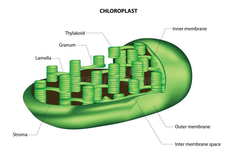
Free with trial Chloroplasts are remarkable organelles found in the cells of plants, algae, and some other photosynthetic organisms. They play a central role in the process of photosynthesis, which is crucial for sustaining life on Earth. The anatomy of chloroplasts is intricately designed to facilitate the complex series of biochemical reactions involved in photosynthesis. At first glance, chloroplasts appear as small, oval-shaped structures within the cytoplasm of plant cells. However, a closer examination reveals a highly organized internal structure that is essential for their function. The outer membrane of the chloroplast serves as a barrier, separating the interior of the organelle from the surrounding cytoplasm. This membrane is semi-permeable, allowing the passage of certain molecules in and out of the chloroplast. Enclosed within the outer membrane is the inner membrane of the chloroplast, which is also selectively permeable. Together, these membranes regulate the exchange of molecules between the chloroplast and the rest of the cell, ensuring the proper functioning of photosynthesis. The internal space of the chloroplast is filled with a fluid called the stroma. The stroma contains enzymes, DNA, ribosomes, and other components necessary for the synthesis of organic molecules during photosynthesis. Embedded within the stroma are stacks of disc-like structures called thylakoids. These interconnected membranes contain the photosynthetic pigments, including chlorophyll, which absorb light energy during the light-dependent reactions of photosynthesis. The arrangement of thylakoids in stacks, known as grana, increases the surface area available for light absorption and facilitates the efficient transfer of electrons and protons during photosynthesis. Within the thylakoid membranes, light energy is used to drive the conversion of water and carbon dioxide into oxygen and glucose. This process involves a series of complex biochemical reactions, including the photosystems, electron transport chain, and ATP synthase complexes. The intricate anatomy of chloroplasts reflects their essential role in sustaining life on Earth. Through the process of photosynthesis, chloroplasts harness the energy of sunlight to produce oxygen, a vital component of the atmosphere, and organic molecules, which serve as the basis of the food web. Understanding the anatomy and function of chloroplasts not only deepens our appreciation of the natural world but also provides insights into strategies for improving agricultural productivity and addressing global challenges such as climate change. Inner membranes vectors Chloroplast vector. Chloroplasts are remarkable organelles found in the cells of plants, algae, and some other photosynthetic organisms. They play a central role in the process of photosynthesis, which is crucial for sustaining life on Earth. The anatomy of chloroplasts is intricately designed to facilitate the complex series of biochemical reactions involved in photosynthesis. At first glance, chloroplasts appear as small, oval-shaped structures within the cytoplasm of plant cells. However, a closer examination reveals a highly organized internal structure that is essential for their function. The outer membrane of the chloroplast serves as a barrier, separating the interior of the organelle from the surrounding cytoplasm. This membrane is semi-permeable, allowing the passage of certain molecules in and out of the chloroplast. Enclosed within the outer membrane is the inner membrane of the chloroplast, which is also selectively permeable. Together, these membranes regulate the exchange of molecules between the chloroplast and the rest of the cell, ensuring the proper functioning of photosynthesis. The internal space of the chloroplast is filled with a fluid called the stroma. The stroma contains enzymes, DNA, ribosomes, and other components necessary for the synthesis of organic molecules during photosynthesis. Embedded within the stroma are stacks of disc-like structures called thylakoids. These interconnected membranes contain the photosynthetic pigments, including chlorophyll, which absorb light energy during the light-dependent reactions of photosynthesis. The arrangement of thylakoids in stacks, known as grana, increases the surface area available for light absorption and facilitates the efficient transfer of electrons and protons during photosynthesis. Within the thylakoid membranes, light energy is used to drive the conversion of water and carbon dioxide into oxygen and glucose. This process involves a series of complex biochemical reactions, including the photosystems, electron transport chain, and ATP synthase complexes. The intricate anatomy of chloroplasts reflects their essential role in sustaining life on Earth. Through the process of photosynthesis, chloroplasts harness the energy of sunlight to produce oxygen, a vital component of the atmosphere, and organic molecules, which serve as the basis of the food web. Understanding the anatomy and function of chloroplasts not only deepens our appreciation of the natural world but also provides insights into strategies for improving agricultural productivity and addressing global challenges such as climate change.
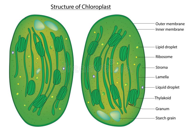
Free with trial Chloroplasts are double-membraned organelles with an outer membrane and an inner membrane. Inside the chloroplast, there is a fluid-filled space called the stroma, where various enzymes and molecules necessary for photosynthesis are located. Embedded within the stroma are stacks of membrane-bound structures called thylakoids. These thylakoid stacks are known as grana (singular: granum). Chlorophyll molecules are embedded in the thylakoid membranes and are responsible for capturing light energy during photosynthesis. Inner membranes vectors Detailed chloroplast diagram. Chloroplasts are double-membraned organelles with an outer membrane and an inner membrane. Inside the chloroplast, there is a fluid-filled space called the stroma, where various enzymes and molecules necessary for photosynthesis are located. Embedded within the stroma are stacks of membrane-bound structures called thylakoids. These thylakoid stacks are known as grana (singular: granum). Chlorophyll molecules are embedded in the thylakoid membranes and are responsible for capturing light energy during photosynthesis.
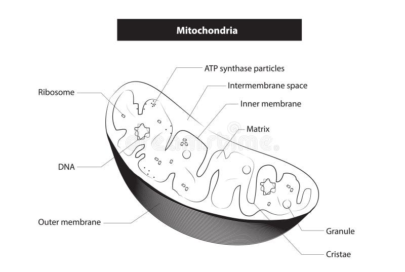
Free with trial Mitochondria have an outer membrane and an inner membrane, which is highly folded into structures called cristae. The space between the inner and outer membranes is known as the intermembrane space, while the space inside the inner membrane is called the mitochondrial matrix. Inner membranes vectors Mitochondrion anatomy. Mitochondria have an outer membrane and an inner membrane, which is highly folded into structures called cristae. The space between the inner and outer membranes is known as the intermembrane space, while the space inside the inner membrane is called the mitochondrial matrix.
Free with trial Translucent lemon slices are arranged against a bright background, revealing intricate details. The thin slices allow light to pass through, highlighting the citrus segments and seeds. The overlapping arrangement creates varying shades of yellow and green, forming natural patterns. The texture of the lemon rind and inner membranes is visible, creating a delicate, lace-like appearance. The image captures the refreshing and vibrant nature of lemons, often associated with freshness and citrus flavor. Inner membranes illustrations Translucent Lemon Slices with Sunlight Patterns. Translucent lemon slices are arranged against a bright background, revealing intricate details. The thin slices allow light to pass through, highlighting the citrus segments and seeds. The overlapping arrangement creates varying shades of yellow and green, forming natural patterns. The texture of the lemon rind and inner membranes is visible, creating a delicate, lace-like appearance. The image captures the refreshing and vibrant nature of lemons, often associated with freshness and citrus flavor.
Free with trial Detailed, cross-sectional view of a mitochondrion, showcasing its internal components such as the inner and outer membranes, cristae, matrix, ribosomes, and the location of mitochondrial DNA. Various labels highlight different sections of the organelle. Inner membranes illustrations Cross-section illustration of a mitochondrion, highlighting its internal structures. Detailed, cross-sectional view of a mitochondrion, showcasing its internal components such as the inner and outer membranes, cristae, matrix, ribosomes, and the location of mitochondrial DNA. Various labels highlight different sections of the organelle
Free with trial Close-up shot of several vibrant red bell pepper slices arranged on a clean white background. The focus is on the intricate inner structure of the peppers, showcasing the seeds and membranes. The image conveys freshness, healthy eating, and culinary ingredients. Ideal for food blogs, recipe illustrations, or health-related content. Inner membranes illustrations Sliced Red Bell Peppers on White. Close-up shot of several vibrant red bell pepper slices arranged on a clean white background. The focus is on the intricate inner structure of the peppers, showcasing the seeds and membranes. The image conveys freshness, healthy eating, and culinary ingredients. Ideal for food blogs, recipe illustrations, or health-related content.
Free with trial Cute yellow baby dragon cartoon illustration showing front, side, and back views. The dragon has large eyes, small horns, and a cheerful expression. Its wings are spread, and a row of orange spikes runs down its back. The tail ends in a star-shaped tip. The dragon's body is predominantly yellow with beige underbelly and inner wing membranes. Its limbs are short and rounded, contributing to a playful, endearing appearance. Inner membranes vectors Cute Yellow Baby Dragon Character Cartoon Illustration. Cute yellow baby dragon cartoon illustration showing front, side, and back views. The dragon has large eyes, small horns, and a cheerful expression. Its wings are spread, and a row of orange spikes runs down its back. The tail ends in a star-shaped tip. The dragon's body is predominantly yellow with beige underbelly and inner wing membranes. Its limbs are short and rounded, contributing to a playful, endearing appearance.
Free with trial Three colorful sliced bell pepper rings, red, yellow, and green, are artfully stacked and overlapping against a light blue gradient background. The peppers are fresh and vibrant, with visible seeds and inner membranes. The lighting highlights the glossy texture of the vegetables, creating a clean and appealing visual. Inner membranes illustrations Sliced Bell Peppers Red Yellow Green Stacked paprika. Three colorful sliced bell pepper rings, red, yellow, and green, are artfully stacked and overlapping against a light blue gradient background. The peppers are fresh and vibrant, with visible seeds and inner membranes. The lighting highlights the glossy texture of the vegetables, creating a clean and appealing visual
Free with trial A vibrant vector illustration featuring a stylized black bat with wide-spread wings, set against a clean white background. The bat has striking orange accents on its wing membranes, ears, and inner mouth, complemented by piercing red, glowing eyes and sharp white fangs. This cartoonish yet spooky design perfectly captures the essence of Halloween and nocturnal creatures. Ideal for holiday decorations, party invitations, web graphics, children's projects, costume designs, or any creative content requiring a classic, eerie, and easily adaptable symbol of fright and mystery. Inner membranes vectors Spooky Black and Orange Flying Bat Vector for Halloween. A vibrant vector illustration featuring a stylized black bat with wide-spread wings, set against a clean white background. The bat has striking orange accents on its wing membranes, ears, and inner mouth, complemented by piercing red, glowing eyes and sharp white fangs. This cartoonish yet spooky design perfectly captures the essence of Halloween and nocturnal creatures. Ideal for holiday decorations, party invitations, web graphics, children's projects, costume designs, or any creative content requiring a classic, eerie, and easily adaptable symbol of fright and mystery.
Free with trial A detailed close up illustration displays multiple rod shaped bacteria cells with vibrant blue outer membranes and sharp projections against a blurred purple background The cells contain groups of three circular structures inside symbolizing the inner mechanisms This image can be used in various scientific medical or educational contexts to represent microbiology infectious diseases or cellular. Inner membranes illustrations Microscopic View of Rod Shaped Bacteria Cells Floating Freely in a Purple Environment. A detailed close up illustration displays multiple rod shaped bacteria cells with vibrant blue outer membranes and sharp projections against a blurred purple background The cells contain groups of three circular structures inside symbolizing the inner mechanisms This image can be used in various scientific medical or educational contexts to represent microbiology infectious diseases or cellular
Free with trial Cute cartoon dragon with large, expressive eyes and a broad smile. Its body is light green with small beige-colored spots on the forehead. It has a round head, tiny beige horns, and matching inner wing membranes. The underbelly is a light beige, with small limbs and a tail ending in a pointed tip. The creature's wings are small and angled, resembling traditional dragon wings. This whimsical design gives the character a friendly and playful appearance. Inner membranes illustrations Baby chibi dragon swimming in magical glowing pond with fish. Cute cartoon dragon with large, expressive eyes and a broad smile. Its body is light green with small beige-colored spots on the forehead. It has a round head, tiny beige horns, and matching inner wing membranes. The underbelly is a light beige, with small limbs and a tail ending in a pointed tip. The creature's wings are small and angled, resembling traditional dragon wings. This whimsical design gives the character a friendly and playful appearance.
Free with trial Embark on a captivating journey into the microscopic realm with our interactive 3D molecular model. Unravel the intricate mechanisms of photosynthesis, witnessing the symphony of biochemical reactions that sustain life on our planet. Explore the inner workings of a plant cell, delving into the role of chloroplasts, chlorophyll, and the light- , This photo was created using generative AI. , This photo was created using generative AI. dependent and light-independent , This photo was created using generative AI. reactions , This photo was created using generative AI. Inner membranes illustrations Immerse Yourself in Photosynthesis: A 3D Molecular Exploration of Plant Cells. Embark on a captivating journey into the microscopic realm with our interactive 3D molecular model. Unravel the intricate mechanisms of photosynthesis, witnessing the symphony of biochemical reactions that sustain life on our planet. Explore the inner workings of a plant cell, delving into the role of chloroplasts, chlorophyll, and the light- , This photo was created using generative AI., This photo was created using generative AI.dependent and light-independent , This photo was created using generative AI.reactions , This photo was created using generative AI. , This photo was created using generative AI.
Free with trial A vibrant red chili pepper, meticulously cut and arranged into a heart shape, sits against a stark white background. This close-up macro shot reveals the pepper's inner seeds and membranes, creating a striking Valentine's Day image. Perfect for culinary blogs, greeting cards, or food photography projects. The image is clean and simple, suitable. Inner membranes illustrations Vibrant Red Chili Pepper Heart on White Background. A vibrant red chili pepper, meticulously cut and arranged into a heart shape, sits against a stark white background. This close-up macro shot reveals the pepper's inner seeds and membranes, creating a striking Valentine's Day image. Perfect for culinary blogs, greeting cards, or food photography projects. The image is clean and simple, suitable
Free with trial Explore intricate abstract cellular forms glowing with inner energy. A visually stunning concept for science, medical, or technology themes. Inner membranes illustrations Vibrant abstract cellular structure with glowing orange core and delicate white membranes. Explore intricate abstract cellular forms glowing with inner energy. A visually stunning concept for science, medical, or technology themes
Free with trial A striking close-up unveils the inner world of a pomegranate, its crimson arils glistening with juice. The fruit's segmented structure emphasizes its inherent beauty and the nourishing treasure it holds, a natural delicacy for healthful indulgence. Inner membranes illustrations Vibrant pomegranate section displaying juicy arils and delicate membranes on white background. A striking close-up unveils the inner world of a pomegranate, its crimson arils glistening with juice. The fruit's segmented structure emphasizes its inherent beauty and the nourishing treasure it holds, a natural delicacy for healthful indulgence.
Free with trial Delve into the intricate world of plant cells with this captivating microscopic image. Focused on a chloroplast, the vital organelle responsible for photosynthesis, this detailed view showcases the complex textures and structures within. Observe the remarkable internal architecture, revealing the inner workings of this essential component of plant anatomy. Learn about the role of chlorophyll in. Inner membranes illustrations Unveiling the Chloroplast A Microscopic Journey into Plant Cellular Structure and Photosynthesis. Delve into the intricate world of plant cells with this captivating microscopic image. Focused on a chloroplast, the vital organelle responsible for photosynthesis, this detailed view showcases the complex textures and structures within. Observe the remarkable internal architecture, revealing the inner workings of this essential component of plant anatomy. Learn about the role of chlorophyll in
Free with trial This detailed microscopic view showcases the intricate structure of a chloroplast within a plant cell. Observe the remarkable inner workings of this essential cellular organelle, responsible for photosynthesis. The image reveals the thylakoid membranes, organized into grana, and the surrounding stroma, highlighting the crucial components that facilitate the process. A close-up perspective offers. Inner membranes illustrations Unveiling the Intricate World of Chloroplasts A Detailed Microscopic Exploration of Plant Cell Structure. This detailed microscopic view showcases the intricate structure of a chloroplast within a plant cell. Observe the remarkable inner workings of this essential cellular organelle, responsible for photosynthesis. The image reveals the thylakoid membranes, organized into grana, and the surrounding stroma, highlighting the crucial components that facilitate the process. A close-up perspective offers
Free with trial Experience the vibrant burst of freshness in this captivating macro photography of juicy orange slices. The image showcases the exquisite details of the citrus fruit, from the glistening, translucent segments to the subtle texture of the peel. Each slice is a study in contrasts, with the bright orange flesh juxtaposed against the paler, almost white inner membranes. The light plays across the. Inner membranes illustrations Closeup Burst of Fresh Orange Slices Vibrant Citrus Macro Photography Showcasing Juicy Details and Texture. Experience the vibrant burst of freshness in this captivating macro photography of juicy orange slices. The image showcases the exquisite details of the citrus fruit, from the glistening, translucent segments to the subtle texture of the peel. Each slice is a study in contrasts, with the bright orange flesh juxtaposed against the paler, almost white inner membranes. The light plays across the