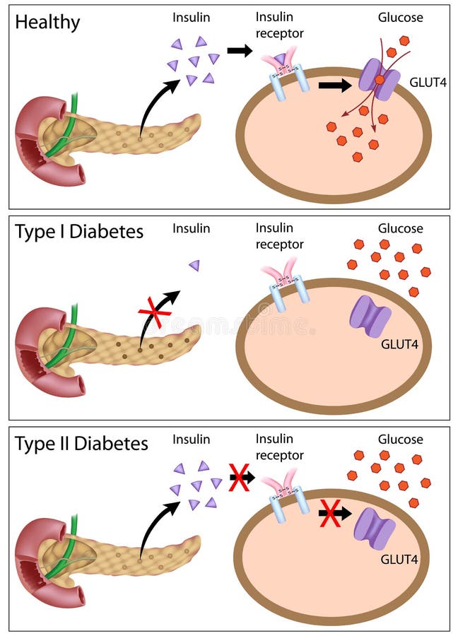
Free with trial Insulin signaling and steps affected in diabetes type 1 and type 2, eps8. Islet cells vectors Insulin action and diabetes types. Insulin signaling and steps affected in diabetes type 1 and type 2, eps8
Free with trial Glucose induces insulin exocytosis in beta cells of the pancreas, eps10. Islet cells vectors Glucose induces insulin release in beta cells. Glucose induces insulin exocytosis in beta cells of the pancreas, eps10
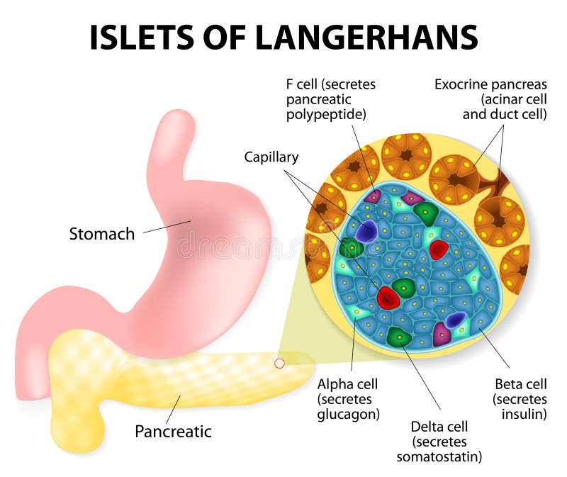
Free with trial The islets of Langerhans are responsible for the endocrine function of the pancreas. Each islet contains beta, alpha, and delta cells that are responsible for the secretion of a hormones. Islet cells vectors Islets of Langerhans
Free with trial Drawing of a pancreatic islet of Langerhans, showing the alpha, beta, and delta hormone-producing cells. Islet cells illustrations Pancreatic islet
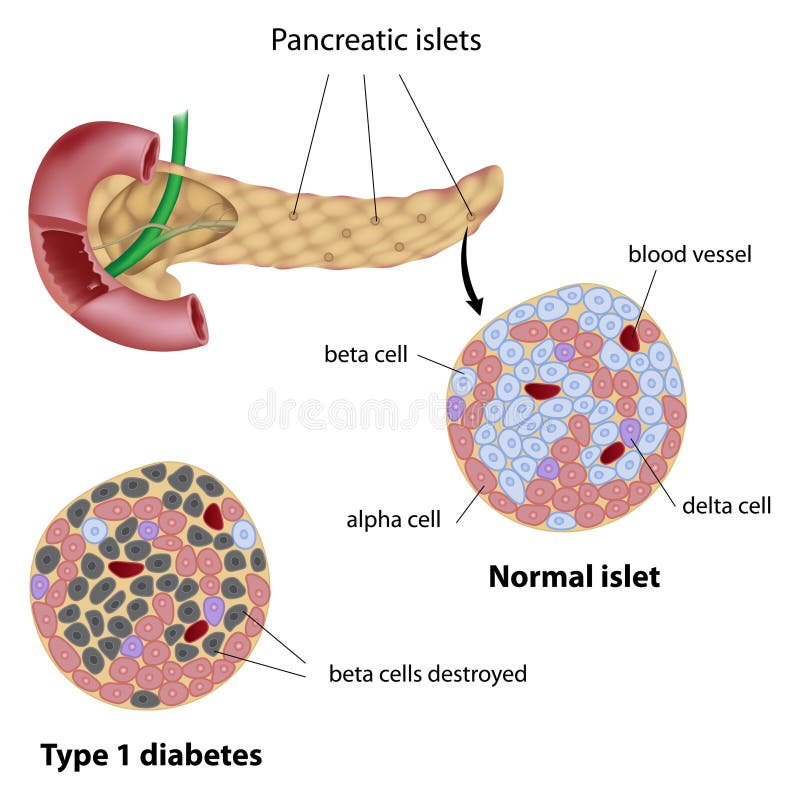
Free with trial Pancreatic islet normal and type 1 diabetic, eps8. Islet cells vectors Pancreatic islet
Free with trial Types of Diabetes. Type 1 and Type 2 Diabetes Mellitus. Insulin-Dependent Diabetes Mellitus and Non Insulin-Dependent Diabetes Mellitus. Insulin resistance and insufficient insulin production. Islet cells vectors Types of Diabetes
Free with trial Islets of Langerhans and diabetes mellitus type 1. Islet cells vectors Islets of Langerhans
Free with trial The pancreas has many islets that contain insulin-producing beta cells and glucagon-producing. Type 1 diabetes Beta cell destroyed. Islet cells vectors Type 1 diabetes Beta cell destroyed.
Free with trial A Human Figure with a Diagram Cut Out style of Moving Lipids or Fatty Acids and Red Blood Cells. Medical Image Symbolism for Hypertension, Nutrition, Diabetes and more. Raster jpg Illustration. Islet cells illustrations Body Fluids or Excretion concept. Medical and Healthcare. Editable Clip Art. A Human Figure with a Diagram Cut Out style of Moving Lipids or Fatty Acids and Red Blood Cells. Medical Image Symbolism for Hypertension, Nutrition, Diabetes and more. Raster jpg Illustration.
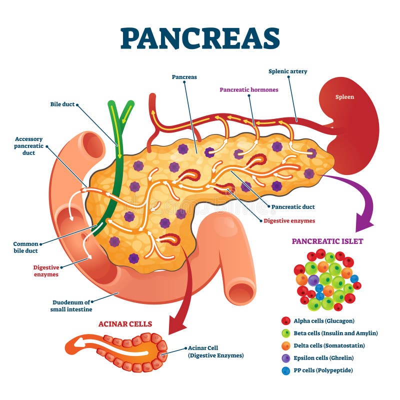
Free with trial Pancreas anatomical cross section model, vector illustration medical example. Blood flow process, cell structure and hormone functions. Digestive enzymes, pancreatic islet and other internal elements. Islet cells vectors Pancreas anatomical cross section model, vector illustration medical example
Free with trial Pancreatic islets. pancreas is an organ of the digestive system and endocrine system. silhouette of Human with highlighted internal organs. Closeup of pancreas and islets of Langerhans. Vector illustration for biological, medical, science and educational use. Islet cells vectors Pancreas. Human silhouette with highlighted internal organs. Closeup of pancreas and islets of Langerhans. pancreatic islets. pancreas is an organ of the digestive system and endocrine system. silhouette of Human with highlighted internal organs. Closeup of pancreas and islets of Langerhans. Vector illustration for biological, medical, science and educational use
Free with trial Pancreatic endocrine system anatomy, alpha, beta and delta cells secreting glucagon, insulin, and somatostatin illustration. Islet cells illustrations Pancreatic endocrine cells anatomy showing endocrine cells involved in secretion of hormones. Pancreatic endocrine system anatomy, alpha, beta and delta cells secreting glucagon, insulin, and somatostatin illustration
Free with trial Types of Diabetes. Type 1 and Type 2 Diabetes Mellitus. Insulin-Dependent Diabetes Mellitus and Non Insulin-Dependent Diabetes Mellitus. Insulin resistance and insufficient insulin production. Islet cells vectors Types of Diabetes
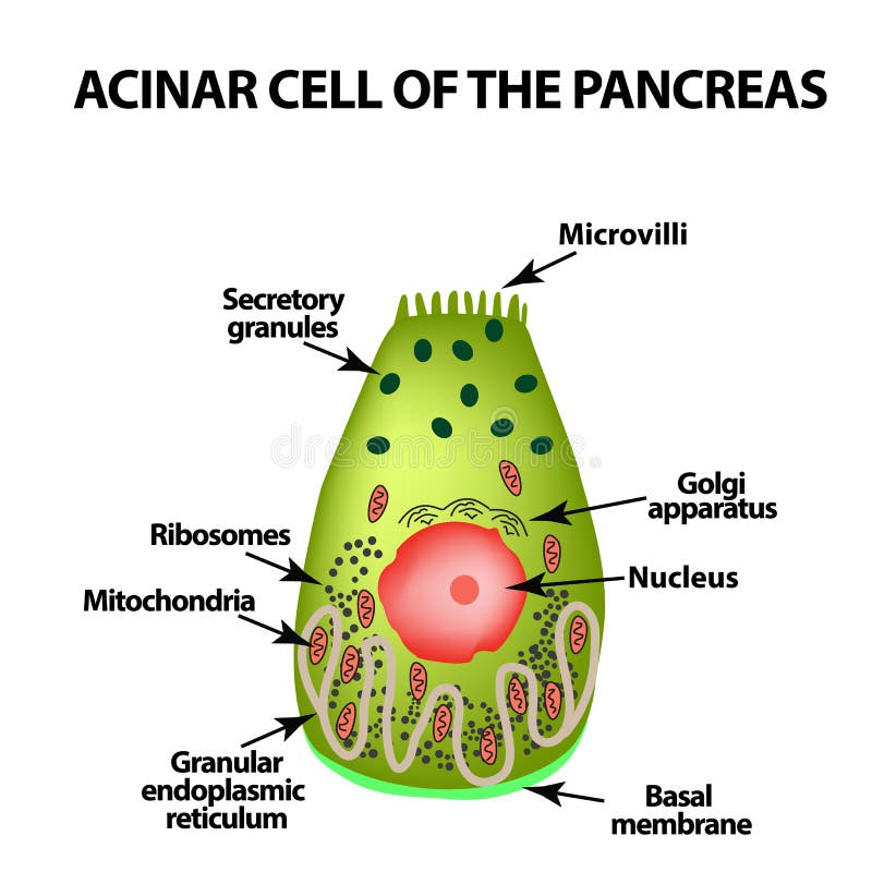
Free with trial Acinar cell of the pancreas. Acinus. Infographics. Vector illustration on isolated background. Islet cells vectors Acinar cell of the pancreas. Acinus. Infographics. Vector illustration on isolated background
Free with trial Pancreatic islet. The islets of Langerhans are responsible for endocrine function of pancreas. Each islet contains beta, alpha, and delta cells that are responsible for the secretion of a hormones. Islet cells vectors Pancreatic islet
Free with trial Vector illustration graphic diagram presentation slide of the pancreatic islet cells within the Islet of Langerhans inside the pancreas. Islet cells vectors Vector illustration slide of the pancreatic islet cells. vector illustration graphic diagram presentation slide of the pancreatic islet cells within the Islet of Langerhans inside the pancreas
Free with trial Spherical fat cells collide on black background 3d render. Islet cells illustrations Spherical fat cells collide on black background 3d
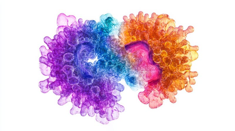
Free with trial Amyloid fibrils are formed by normally soluble proteins, which assemble to form insoluble fibers that are resistant to degradation. Their formation can accompany disease and each disease is characterized by a specific protein or peptide that aggregates. 3D cartoon model, white background. Islet cells illustrations Amyloid fibril structure of islet amyloid polypeptide. Amyloid fibrils are formed by normally soluble proteins, which assemble to form insoluble fibers that are resistant to degradation. Their formation can accompany disease and each disease is characterized by a specific protein or peptide that aggregates. 3D cartoon model, white background
Free with trial Pancreatic islets. pancreas is an organ of the digestive system and endocrine system. silhouette of Human with highlighted internal organs. Closeup of pancreas and islets of Langerhans. Islet cells vectors Pancreas. Islet of langerhans. Pancreatic islets. pancreas is an organ of the digestive system and endocrine system. silhouette of Human with highlighted internal organs. Closeup of pancreas and islets of Langerhans
Free with trial Diagram depicting the metabolic processes in diabetic ketoacidosis (DKA). The liver is central, showing glycogen conversion to glucose and ketones. Pathways indicate increased glucagon, fatty acids, glycerol, and amino acids contributing to ketone production. A blood vessel highlights increased ketone and glucose levels. Text indicates the role of insufficient or absent insulin. Arrows connect substrates, glucose, and ketones with various cells, illustrating gluconeogenesis, glycogenolysis, and ketogenesis. Islet cells vectors Diabetic Ketoacidosis (DKA) sign and symptoms. Diagram depicting the metabolic processes in diabetic ketoacidosis (DKA). The liver is central, showing glycogen conversion to glucose and ketones. Pathways indicate increased glucagon, fatty acids, glycerol, and amino acids contributing to ketone production. A blood vessel highlights increased ketone and glucose levels. Text indicates the role of insufficient or absent insulin. Arrows connect substrates, glucose, and ketones with various cells, illustrating gluconeogenesis, glycogenolysis, and ketogenesis.
Free with trial The pancreas is affected cystic tumor of the islet cells. Vector illustration on background. Islet cells vectors The pancreas is affected cystic tumor of the
Free with trial Acinar cell of the pancreas. Acinus. Infographics. Vector illustration on isolated background. Islet cells vectors Acinar cell of the pancreas. Acinus. Infographics. Vector illustration on isolated background
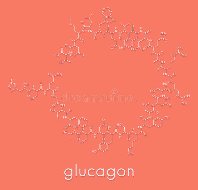
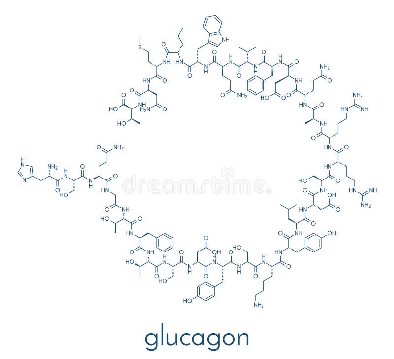
Free with trial Glucagon hypoglycemia drug molecule. Skeletal formula. Islet cells illustrations Glucagon hypoglycemia drug molecule. Skeletal formula.
Free with trial Glp 1 insulin agonis therapy diabetes type 2. Islet cells illustrations Glp-1 and insulin, diabetes type 2 theraphy. Glp 1 insulin agonis therapy diabetes type 2
Free with trial Illustration showing the progression of Diabetic Ketoacidosis (DKA) in five stages. 1) Pancreas fails to produce insulin, causing glucose buildup in the bloodstream. 2) Liver releases more glucose, worsening hyperglycemia. 3) Fat breakdown releases ketones since cells can't access glucose. 4) Kidneys attempt to filter out excess ketones and glucose, leading to frequent urination and fluid loss. 5) Dehydration and electrolyte loss occur, potentially leading to complications like shock or coma. Glucose and ketone graphics indicate blood composition changes. Islet cells vectors Stages of Diabetic Ketoacidosis (DKA). Illustration showing the progression of Diabetic Ketoacidosis (DKA) in five stages. 1) Pancreas fails to produce insulin, causing glucose buildup in the bloodstream. 2) Liver releases more glucose, worsening hyperglycemia. 3) Fat breakdown releases ketones since cells can't access glucose. 4) Kidneys attempt to filter out excess ketones and glucose, leading to frequent urination and fluid loss. 5) Dehydration and electrolyte loss occur, potentially leading to complications like shock or coma. Glucose and ketone graphics indicate blood composition changes.
Free with trial Glp 1 insulin agonis therapy diabetes type 2. Islet cells illustrations Glp-1 and insulin, diabetes type 2 theraphy. Glp 1 insulin agonis therapy diabetes type 2
Free with trial This detailed micrograph depicts a pancreatic islet within the pancreas, surrounded by darker-stained exocrine acini. The islet's lighter cells are essential for hormone secretion, crucial for regulating blood glucose levels. Islet cells illustrations Micrograph of a pancreatic islet, showing endocrine cells of Langerhans surrounded by exocrine pancreatic acini. This detailed micrograph depicts a pancreatic islet within the pancreas, surrounded by darker-stained exocrine acini. The islet's lighter cells are essential for hormone secretion, crucial for regulating blood glucose levels.
Free with trial This isometric diagram showcases islet cells in the human pancreas, highlighting their structure and function in a vibrant and detailed visualization. Perfect for educational use. Islet cells illustrations Isometric Diagram of Islet Cells in Human Pancreas Structure. This isometric diagram showcases islet cells in the human pancreas, highlighting their structure and function in a vibrant and detailed visualization. Perfect for educational use
Free with trial Explore an intricate macro photograph showcasing islet cells. The vibrant colors and textures emphasize the unique interaction and structure at a microscopic level. Islet cells illustrations Macro Photography of Islet Cells Interacting in a Unique Style. Explore an intricate macro photograph showcasing islet cells. The vibrant colors and textures emphasize the unique interaction and structure at a microscopic level
Free with trial Explore the intricate details of islet cells through macro photography, showcasing their vibrant colors and unique textures within biological structures. Islet cells illustrations Detailed Macro Photography of Islet Cells Interacting in Tissue. Explore the intricate details of islet cells through macro photography, showcasing their vibrant colors and unique textures within biological structures
Free with trial Stunning artistic visualization of pancreatic islet cells in a vibrant natural landscape, featuring golden light and abstract forms that evoke growth and harmony. Islet cells illustrations Artistic Visualization of Pancreatic Islet Cells in Natural Landscape. Stunning artistic visualization of pancreatic islet cells in a vibrant natural landscape, featuring golden light and abstract forms that evoke growth and harmony
Free with trial Explore a captivating macro image capturing the intricate details of islet cells interacting, showcasing vibrant colors and textures in a scientific context. Islet cells illustrations Macro Photography of Islet Cells Interacting in Vibrant Colors. Explore a captivating macro image capturing the intricate details of islet cells interacting, showcasing vibrant colors and textures in a scientific context
Free with trial Experience a stunning hyper-realistic 3D render showcasing pancreatic islet cells, designed for scientific illustration and research purposes, highlighting vibrant structures in microscopic detail. Islet cells illustrations Hyper-Realistic 3D Render of Pancreatic Islet Cells at Gigapixel Quality. Experience a stunning hyper-realistic 3D render showcasing pancreatic islet cells, designed for scientific illustration and research purposes, highlighting vibrant structures in microscopic detail
Free with trial Explore a hyper realistic 3D render of pancreatic islet cells, showcasing intricate details and vibrant colors, perfect for scientific and medical visualization. Islet cells illustrations Hyper Realistic 3D Render of Pancreatic Islet Cells in Gigapixel Quality. Explore a hyper realistic 3D render of pancreatic islet cells, showcasing intricate details and vibrant colors, perfect for scientific and medical visualization
Free with trial This isometric diagram illustrates islet cells in the human pancreas, showcasing their intricate structure and vital role in regulating blood sugar levels. Islet cells illustrations Isometric Diagram of Islet Cells in Human Pancreas Anatomy. This isometric diagram illustrates islet cells in the human pancreas, showcasing their intricate structure and vital role in regulating blood sugar levels
Free with trial Discover an intricate 3D render of pancreatic islet cells, showcasing vibrant colors and detailed textures in a stunning digital environment, ideal for scientific illustration. Islet cells illustrations Hyper Realistic 3D Render of Pancreatic Islet Cells in Detail. Discover an intricate 3D render of pancreatic islet cells, showcasing vibrant colors and detailed textures in a stunning digital environment, ideal for scientific illustration
Free with trial Microscopic view of pancreatic islet cells with vibrant coloration. A colorful microscopic image reveals pancreatic islet cells surrounded by connective tissue. Islet cells illustrations Microscopic view of pancreatic islet cells with vibrant coloration. A colorful microscopic image reveals pancreatic islet cells surrounded by connective tissue
Free with trial Fluorescent microscopy reveals the intricate structure of pancreatic islet cells, crucial for hormone production. Islet cells illustrations Detailed microscopic view of pancreatic islet cells with vibrant coloration. Fluorescent microscopy reveals the intricate structure of pancreatic islet cells, crucial for hormone production
Free with trial This precise illustration of islet cells offers insight into their structure and function, set against a clean white backdrop. Islet cells illustrations Explore the intricate details of pancreatic islet cells, highlighting alpha and beta cells in a clear, medical illustration. This precise illustration of islet cells offers insight into their structure and function, set against a clean white backdrop.
Free with trial This precise illustration of islet cells offers insight into their structure and function, set against a clean white backdrop. Islet cells illustrations Explore the intricate details of pancreatic islet cells, highlighting alpha and beta cells in a clear, medical illustration. This precise illustration of islet cells offers insight into their structure and function, set against a clean white backdrop.
Free with trial This precise illustration of islet cells offers insight into their structure and function, set against a clean white backdrop. Islet cells illustrations Explore the intricate details of pancreatic islet cells, highlighting alpha and beta cells in a clear, medical illustration. This precise illustration of islet cells offers insight into their structure and function, set against a clean white backdrop.
Free with trial This image showcases a hyper realistic 3D render of pancreatic islet cells, illustrating intricate details and vibrant colors, highlighting their role in health and biology. Islet cells illustrations Hyper Realistic 3D Render of Pancreatic Islet Cells Detail. This image showcases a hyper realistic 3D render of pancreatic islet cells, illustrating intricate details and vibrant colors, highlighting their role in health and biology
Free with trial Discover the intricate details of islet cells interacting in this macro photography image. Vibrant colors and unique textures create a stunning visual experience perfect for scientific and artistic needs. Islet cells illustrations Macro Photography of Islet Cells Interacting in Vibrant Colors. Discover the intricate details of islet cells interacting in this macro photography image. Vibrant colors and unique textures create a stunning visual experience perfect for scientific and artistic needs
Free with trial Rare tumors arising from the hormone-producing islet cells of the pancreas, which may be functional or nonfunctional. Islet cells vectors Islet Cell Tumors (Pancreatic Neuroendocrine Tumors). Rare tumors arising from the hormone-producing islet cells of the pancreas, which may be functional or nonfunctional.
Free with trial Dive into this artistic visualization showcasing pancreatic islet cells, blending creativity with scientific insight, perfect for educational and research purposes. Islet cells illustrations Artistic Medical Visualization of Pancreatic Islet Cells in Detail. Dive into this artistic visualization showcasing pancreatic islet cells, blending creativity with scientific insight, perfect for educational and research purposes
Free with trial Explore a vibrant and artistic visualization of pancreatic islet cells, showcasing intricate details in a colorful and abstract style, perfect for medical and biological themes. Islet cells illustrations Detailed Artistic Visualization of Pancreatic Islet Cells in Purple. Explore a vibrant and artistic visualization of pancreatic islet cells, showcasing intricate details in a colorful and abstract style, perfect for medical and biological themes
Free with trial A detailed 3D medical illustration of the Islet of Langerhans, showcasing alpha, beta, delta, PP, and epsilon cells. Ideal for scientific visualization, medical education, and research, depicting. Islet cells illustrations Detailed 3D Medical Illustration of Islet of Langerhans on White Background Depicting Alpha Beta Delta PP and Epsilon Cells for. A detailed 3D medical illustration of the Islet of Langerhans, showcasing alpha, beta, delta, PP, and epsilon cells. Ideal for scientific visualization, medical education, and research, depicting
Free with trial This breathtaking microscopic image showcases the intricate cellular architecture of pancreatic islets of Langerhans. Pancreatic islet cells, including beta cells, alpha cells, delta cells, and gamma cells, are vividly depicted, highlighting their critical roles in hormone production. The panoramic view reveals the complex interplay of these endocrine cells within the pancreatic tissue, offering. Islet cells illustrations Pancreatic Islet of Langerhans Cellular Structures Revealed in Stunning Detail via Advanced Microscopy. This breathtaking microscopic image showcases the intricate cellular architecture of pancreatic islets of Langerhans. Pancreatic islet cells, including beta cells, alpha cells, delta cells, and gamma cells, are vividly depicted, highlighting their critical roles in hormone production. The panoramic view reveals the complex interplay of these endocrine cells within the pancreatic tissue, offering
Free with trial Detailed isometric diagram illustrating islet cell structure in the human pancreas. This vibrant and educational image showcases cellular composition and functions for scientific study. Islet cells illustrations Isometric Diagram of Islet Cell Structure in Human Pancreas. Detailed isometric diagram illustrating islet cell structure in the human pancreas. This vibrant and educational image showcases cellular composition and functions for scientific study
Free with trial Explore an artistic visualization showcasing islet cell regeneration through microscopy. This vibrant illustration depicts the dynamic structure and function of cells in a scientific context. Islet cells illustrations Artistic Visualization of Islet Cell Regeneration Under Microscopy. Explore an artistic visualization showcasing islet cell regeneration through microscopy. This vibrant illustration depicts the dynamic structure and function of cells in a scientific context
Free with trial Detailed image microscopic pancreatic cells. Purple, blue colors. Science tech background. Biological cells, medical research, biology, health care, human cell scientific illustration. Islet cells illustrations Detailed image microscopic pancreatic cells. Purple, blue colors. Science tech background. Biological cells, medical research
Free with trial Explore the intricate internal structure of the pancreas, revealing blood cells and islets. Perfect for medical and scientific education, research, and healthcare communications. Islet cells vectors Detailed cross-section of human pancreas showing blood cells and islets. Explore the intricate internal structure of the pancreas, revealing blood cells and islets. Perfect for medical and scientific education, research, and healthcare communications
Free with trial This stunning microscopic image reveals the intricate cellular architecture of the pancreas, a vital organ in both the endocrine and digestive systems. The image showcases the pancreas's dual function, highlighting the specific islet cells responsible for insulin production and the acinar cells that synthesize digestive enzymes. Notice the rich detail of the beta cells, which are key in. Islet cells illustrations Detailed Microscopic View of the Pancreas Insulin Production Digestive Enzyme Synthesis and Cellular Processes. This stunning microscopic image reveals the intricate cellular architecture of the pancreas, a vital organ in both the endocrine and digestive systems. The image showcases the pancreas's dual function, highlighting the specific islet cells responsible for insulin production and the acinar cells that synthesize digestive enzymes. Notice the rich detail of the beta cells, which are key in
Free with trial Explore an artistic visualization showcasing pancreatic islets and cells in stunning detail. This captivating image illustrates cellular functions and structures. Islet cells illustrations Artistic Medical Visualization of Pancreatic Islets and Cells. Explore an artistic visualization showcasing pancreatic islets and cells in stunning detail. This captivating image illustrates cellular functions and structures
Free with trial A striking visualization of AI-assisted islet cell therapy, depicting glowing red and blue cells in a futuristic medical environment, showcasing innovation in biotechnology. Islet cells illustrations Futuristic Concept of AI-Enhanced Islet Cell Therapy in Medicine. A striking visualization of AI-assisted islet cell therapy, depicting glowing red and blue cells in a futuristic medical environment, showcasing innovation in biotechnology
Free with trial This image depicts a magnified cross-section of the human pancreas, revealing its intricate structure with visible islets of Langerhans, acinar cells, and pancreatic ducts. Islet cells illustrations This is an image of a cross section of the human pancreas. This image depicts a magnified cross-section of the human pancreas, revealing its intricate structure with visible islets of Langerhans, acinar cells, and pancreatic ducts
Free with trial Illuminated internal organ a detailed visualization of the pancreas's intricate structure and function. Aesthetic image. Generative AI. Islet cells illustrations Illuminated internal organ a detailed visualization of the pancreas\'s intricate structure and function. Generative AI
Free with trial Glp 1 insulin agonis therapy diabetes type 2. Islet cells illustrations Glp-1 and insulin, diabetes type 2 theraphy. Glp 1 insulin agonis therapy diabetes type 2
Free with trial Glp 1 insulin agonis therapy diabetes type 2. Islet cells illustrations Glp-1 and insulin, diabetes type 2 theraphy. Glp 1 insulin agonis therapy diabetes type 2
Free with trial Detailed pancreas illustration showcases its cellular structure and blood vessels for medical learning or healthcare material. Islet cells illustrations Pancreas Anatomy Illustration Showing Cells and Blood Vessels Structure. Detailed pancreas illustration showcases its cellular structure and blood vessels for medical learning or healthcare material
Free with trial Anatomy model showing the pancreas, internal cell distribution, and blood vessels for medical education or healthcare related projects. Islet cells illustrations Pancreas Anatomy Visualization Showing Cells, Vessels, and Internal Structure. Anatomy model showing the pancreas, internal cell distribution, and blood vessels for medical education or healthcare related projects
Free with trial Streptozotocin cancer drug molecule. Used in treatment of metastatic cancer of the pancreatic islet cells. Skeletal formula. Islet cells illustrations Streptozotocin cancer drug molecule. Used in treatment of metastatic cancer of the pancreatic islet cells. Skeletal formula.
Free with trial Streptozotocin cancer drug molecule. Used in treatment of metastatic cancer of the pancreatic islet cells. Skeletal formula. Islet cells vectors Streptozotocin cancer drug molecule. Used in treatment of metastatic cancer of the pancreatic islet cells. Skeletal formula.
Free with trial The pancreas is affected cystic tumor of the islet cells. Vector illustration on a black background. Pancreatitis. Islet cells vectors The pancreas is affected cystic tumor of the islet cells. Vector illustration on a black background
Free with trial The pancreas is affected cystic tumor of the islet cells. Vector illustration on a black background. Islet cells vectors The pancreas is affected cystic tumor of the islet cells. Vector illustration on a black background
Free with trial Streptozotocin cancer drug molecule. Used in treatment of metastatic cancer of the pancreatic islet cells. Atoms are represented as spheres with conventional color coding: hydrogen (white), carbon (grey), nitrogen (blue. Islet cells illustrations Streptozotocin cancer drug molecule. Used in treatment of metastatic cancer of the pancreatic islet cells. Atoms are represented
Free with trial Streptozotocin cancer drug molecule. Used in treatment of metastatic cancer of the pancreatic islet cells. Atoms are represented as spheres with conventional color coding: hydrogen (white), carbon (grey), nitrogen (blue. Islet cells illustrations Streptozotocin cancer drug molecule. Used in treatment of metastatic cancer of the pancreatic islet cells. Atoms are represented
Free with trial Streptozotocin cancer drug molecule. Used in treatment of metastatic cancer of the pancreatic islet cells. Atoms are represented as spheres with conventional color coding: hydrogen (white), carbon (grey), nitrogen (blue. Islet cells illustrations Streptozotocin cancer drug molecule. Used in treatment of metastatic cancer of the pancreatic islet cells. Atoms are represented
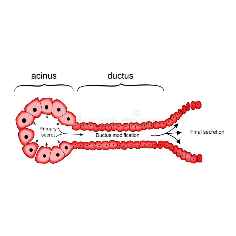
Free with trial Acinus and Ductus GI System cells anatomy and physiology vector illustration on white background. Islet cells vectors Acinus and Ductus GI System cells anatomy and physiology vector illustration
Free with trial The pancreas has many islets that contain insulin-producing beta cells and glucagon-producing. Type 1 diabetes Beta cell destroyed. Islet cells illustrations Type 1 diabetes Beta cell destroyed.
Free with trial Cancer cells, malignant cells, scientific 3D illustration. Islet cells illustrations Cancer cells, 3D illustration. Cancer cells, malignant cells, scientific 3D illustration
Free with trial Cancer cells, malignant cells, scientific 3D illustration. Islet cells illustrations Cancer cells, 3D illustration. Cancer cells, malignant cells, scientific 3D illustration
Free with trial 3D Isometric Flat Vector Conceptual Illustration of Totipotent Cells, Educational Scheme. Islet cells vectors 3D Isometric Flat Vector Conceptual Illustration of Totipotent Cells
Free with trial Cancer cells, malignant cells, scientific 3D illustration. Islet cells illustrations Cancer cells, 3D illustration. Cancer cells, malignant cells, scientific 3D illustration
Free with trial Cancer cells, malignant cells, scientific 3D illustration. Islet cells illustrations Cancer cells, 3D illustration. Cancer cells, malignant cells, scientific 3D illustration
Free with trial Cancer cells, malignant cells, scientific 3D illustration. Islet cells illustrations Cancer cells, 3D illustration. Cancer cells, malignant cells, scientific 3D illustration
Free with trial Cancer cells, malignant cells, scientific 3D illustration. Islet cells illustrations Cancer cells, 3D illustration. Cancer cells, malignant cells, scientific 3D illustration
Free with trial Diabetes stamp on white background. Islet cells illustrations Diabetes stamp on white
Free with trial The pancreas is affected cystic tumor of the islet cells. Vector illustration on background. Pancreatitis. Islet cells vectors The pancreas is affected cystic tumor of the
Free with trial Glucagon hypoglycemia drug molecule. Skeletal formula. Islet cells vectors Glucagon hypoglycemia drug molecule. Skeletal formula.