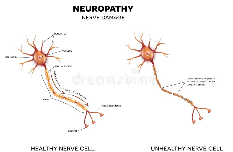
Free with trial Neuropathy, damage of peripheral nerves. Pain and loss of sensation in the extremities. This can be caused by Diabetes. Labeled synapses vectors Neuropathy, nerve damage. Neuropathy, damage of peripheral nerves. Pain and loss of sensation in the extremities. This can be caused by Diabetes.
Free with trial Neuropathy, damage of nerves. This can be caused by Diabetes. Labeled synapses vectors Nerve damage. Neuropathy, damage of nerves. This can be caused by Diabetes.
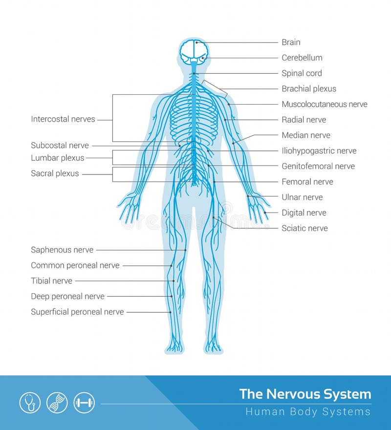
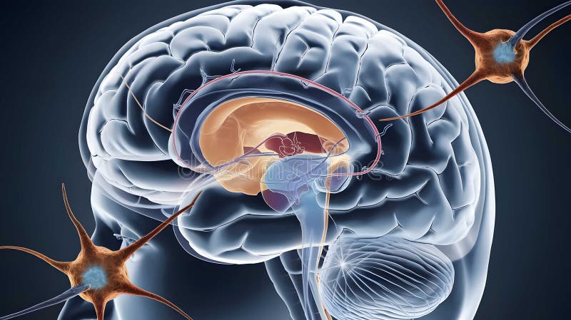
Free with trial The human nervous system vector medical illustration. Labeled synapses vectors The nervous system. The human nervous system vector medical illustration
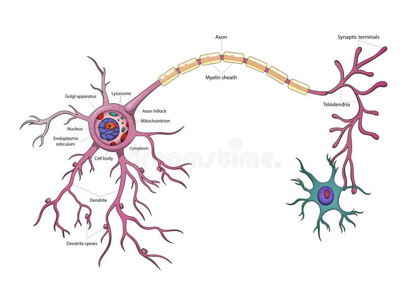
Free with trial Labeled diagram of the neuron, nerve cell that is the main part of the nervous system. Labeled synapses vectors Labeled diagram of the neuron
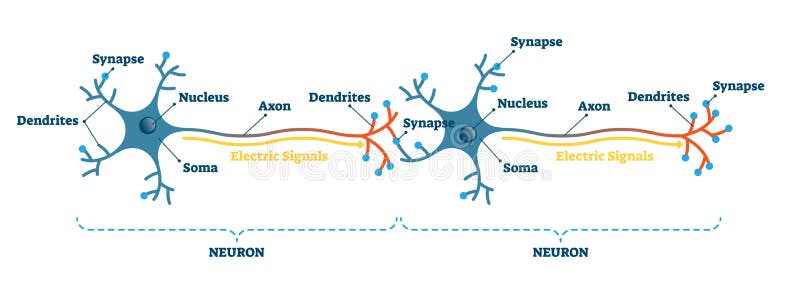
Free with trial Neuron network example diagram, vector illustration. Synapses, soma, axon and dendrites closeup scheme. Nervous system electric signal communication structure. Neurology science study information. Labeled synapses vectors Neuron network example diagram, vector illustration
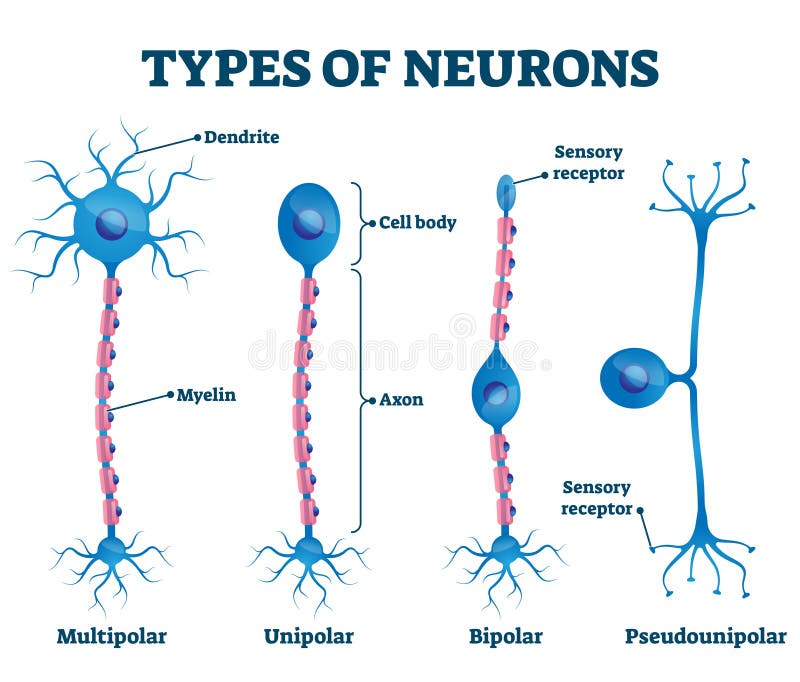
Free with trial Types of neurons vector illustration. Labeled anatomical nerve parts comparison scheme. Synapses receptors educational closeup with multipolar, unipolar, bipolar and pseudounipola sensory receptors. Labeled synapses vectors Types of neurons vector illustration. Labeled nerve parts comparison scheme. Types of neurons vector illustration. Labeled anatomical nerve parts comparison scheme. Synapses receptors educational closeup with multipolar, unipolar, bipolar and pseudounipola sensory receptors.
Free with trial Meningitis vector illustration. Medical labeled brain covering membrane inflammation scheme. Educational anatomical diagram with isolated closeup side view structure, symptoms and complications list. Labeled synapses vectors Meningitis vector illustration. Labeled brain membrane inflammation scheme. Meningitis vector illustration. Medical labeled brain covering membrane inflammation scheme. Educational anatomical diagram with isolated closeup side view structure, symptoms and complications list.
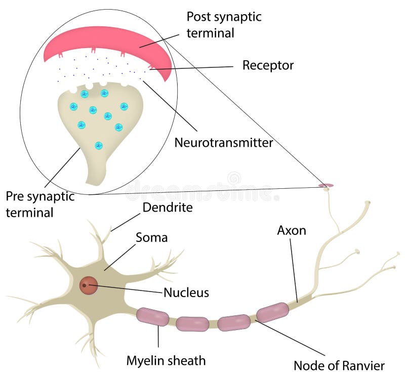
Free with trial Nerve cells and synapses are implicated in many diseases. Labeled synapses vectors Neuron and Synapse Labeled Diagram. Nerve cells and synapses are implicated in many diseases.
Free with trial Neuroscience as study of nervous system medical division outline diagram. Labeled educational field scheme with synapses, brain structures, cognitive process and neurotransmitters vector illustration. Labeled synapses vectors Neuroscience as study of nervous system medical division outline diagram
Free with trial Labeled diagram of the Neuron, nerve cell that is the main part of the nervous system. Abstract grey mesh background. Labeled synapses vectors Neuron
Free with trial Brain superfoods as educational nutrition diet products to improve memory. Diet supplement for mental benefits and boost cognition learning with right eating vector illustration. Labeled graphic list. Labeled synapses vectors Brain superfoods as educational nutrition diet products to improve memory
Free with trial Brain superfoods as nutrition diet products to improve memory outline concept. Diet supplement for mental benefits and boost learning with right eating vector illustration. Educational labeled graphic. Labeled synapses vectors Brain superfoods as nutrition diet products to improve memory outline concept
Free with trial Different types of synapses types. Neuron communication. Synaptic transmission. Labeled axosomatic, axoaxonic, axodendritic, axoextracellular, dendodentritic. Presynaptic cell and Postsynaptic cell. Labeled synapses vectors Different types of synapses types. Neuron communication. Synaptic transmission. Labeled axosomatic, axoaxonic, axodendritic
Free with trial A detailed of a human head showing the olfactory system. The a side view of a man's head with brown hair and a neutral expression. The face is labeled with various components of the olfactory system, including the olfactory bulb, axon, synapses, glomerulus, cribriform plate, dendrite, olfactory receptors, cilia, basal cells, and odorant molecules. The is set against a white background and a range. Labeled synapses illustrations Human head with labeled olfactory system components. A detailed of a human head showing the olfactory system. The a side view of a man's head with brown hair and a neutral expression. The face is labeled with various components of the olfactory system, including the olfactory bulb, axon, synapses, glomerulus, cribriform plate, dendrite, olfactory receptors, cilia, basal cells, and odorant molecules. The is set against a white background and a range
Free with trial A detailed medical of a human head showing the anatomy of the olfactory system. The a side view of a human face with a visible nose, eye, and ear. The olfactory system components are labeled and color-coded, including the olfactory bulb, axon, synapses, cribriform plate, dendrite, cilia, olfactory receptors, basal cells, and odorant molecules. The is set against a white background. Labeled synapses illustrations Human head with labeled olfactory system anatomy medical olfactory bulb. A detailed medical of a human head showing the anatomy of the olfactory system. The a side view of a human face with a visible nose, eye, and ear. The olfactory system components are labeled and color-coded, including the olfactory bulb, axon, synapses, cribriform plate, dendrite, cilia, olfactory receptors, basal cells, and odorant molecules. The is set against a white background
Free with trial Brain-shaped neural network visualization showing labeled layers including input, hidden, output neurons, synapses, weights, bias, data flow, and activation functions. Labeled synapses illustrations Neural Network Architecture Diagram. Brain-shaped neural network visualization showing labeled layers including input, hidden, output neurons, synapses, weights, bias, data flow, and activation functions.
Free with trial The image showcases a detailed visualization of neurons and their key components. It highlights the intricate network of dendrites, which receive signals from other neurons, and the axon, which transmits signals away from the neuron's cell body. The synapse, the junction where neurons communicate, is also labeled. The image emphasizes the complexity and elegance of neural connections, illustrating. Labeled synapses illustrations Neural connections: dendrites, axons, and synapses in action. The image showcases a detailed visualization of neurons and their key components. It highlights the. The image showcases a detailed visualization of neurons and their key components. It highlights the intricate network of dendrites, which receive signals from other neurons, and the axon, which transmits signals away from the neuron's cell body. The synapse, the junction where neurons communicate, is also labeled. The image emphasizes the complexity and elegance of neural connections, illustrating
Free with trial An intricate illustration of a human brain with three thought bubbles emerging from it. The bubbles are labeled 'Mind,' 'Intelligence,' and 'Psychology,' symbolizing the brain's role in these aspects. Labeled synapses illustrations Illustration of a brain with thought bubbles labeled mind, intelligence, and psychology. An intricate illustration of a human brain with three thought bubbles emerging from it. The bubbles are labeled 'Mind,' 'Intelligence,' and 'Psychology,' symbolizing the brain's role in these aspects
Free with trial A medical illustration of the human brain, showing the outer layer with labeled major lobes. The inner structures, including ventricles and meninges, are also visible. Two neurons with axons and dendrites are depicted for added detail. This AI-generated image is ideal for medical education and neuroscience studies. Labeled synapses illustrations . A medical illustration of the human brain, showing the outer layer with labeled major lobes. The inner structures, including ventricles and meninges, are also visible. Two neurons with axons and dendrites are depicted for added detail. This AI-generated image is ideal for medical education and neuroscience studies.
Free with trial Neurons with red-labeled synapses, a key component in neural communication, Neurons with red-labeled synapses, a key component in neural communication. Labeled synapses illustrations Neurons with red-labeled synapses, a key component in neural communication
Free with trial Delve into the intricate world of nerve cells with this detailed labeled diagram. Visualize the microscopic anatomy of a neuron, a fundamental unit of the nervous system. Observe the key components crucial for signal transmission, including the cell body, dendrites, axon, and myelin sheath. The diagram meticulously highlights these structures, showcasing their unique shapes and functions. Labeled synapses illustrations Unveiling the Microscopic Marvels of a Neuron A Detailed Labeled Diagram of Nerve Cell Structure. Delve into the intricate world of nerve cells with this detailed labeled diagram. Visualize the microscopic anatomy of a neuron, a fundamental unit of the nervous system. Observe the key components crucial for signal transmission, including the cell body, dendrites, axon, and myelin sheath. The diagram meticulously highlights these structures, showcasing their unique shapes and functions.
Free with trial This image is a detailed illustration of the human brain and neurons, accompanied by an EEG machine. The brain is depicted in the top left corner, showcasing its intricate structure. Three neurons are illustrated in the top center and right, highlighting their unique shapes and connections. In the bottom left, a labeled diagram of synapses is provided, explaining the connections between neurons. The EEG machine and its connection to the brain are shown in the bottom center and right, demonstrating the technology used to monitor brain activity. This image is ideal for educational materials, research papers, and medical presentations, providing a comprehensive visual representation of the brain, neurons, and EEG technology. Labeled synapses illustrations A detailed illustration of the human brain and neurons with an EEG machine. This image is a detailed illustration of the human brain and neurons, accompanied by an EEG machine. The brain is depicted in the top left corner, showcasing its intricate structure. Three neurons are illustrated in the top center and right, highlighting their unique shapes and connections. In the bottom left, a labeled diagram of synapses is provided, explaining the connections between neurons. The EEG machine and its connection to the brain are shown in the bottom center and right, demonstrating the technology used to monitor brain activity. This image is ideal for educational materials, research papers, and medical presentations, providing a comprehensive visual representation of the brain, neurons, and EEG technology.
Free with trial This detailed diagram highlights key areas like the dentate gyrus and CA regions, providing a visual guide to brain anatomy. Labeled synapses illustrations Anatomical illustration of the human brain with labeled regions and structures. This detailed diagram highlights key areas like the dentate gyrus and CA regions, providing a visual guide to brain anatomy
Free with trial This detailed illustration offers an enhanced visualization of the intricate neural connections within the human brain, showcasing the complexity of the brain's structure and function. The image highlights a network of neurons, depicted with vibrant colors to represent different pathways and types of connections. Synapses sparkle with intensity, illustrating the dynamic communication between neurons. The backdrop features a subtle gradient that emphasizes the interconnectedness of the brain regions, while key areas such as the cortex, cerebellum, and brainstem are labeled for clarity. This visualization serves as an informative tool for understanding the brain's architecture and its role in cognition, emotion, and sensory processing, making it ideal for educational materials, neuroscience research, or medical presentations. Labeled synapses illustrations Enhanced Visualization of Neural Connections in the Human Brain. This detailed illustration offers an enhanced visualization of the intricate neural connections within the human brain, showcasing the complexity of the brain's structure and function. The image highlights a network of neurons, depicted with vibrant colors to represent different pathways and types of connections. Synapses sparkle with intensity, illustrating the dynamic communication between neurons. The backdrop features a subtle gradient that emphasizes the interconnectedness of the brain regions, while key areas such as the cortex, cerebellum, and brainstem are labeled for clarity. This visualization serves as an informative tool for understanding the brain's architecture and its role in cognition, emotion, and sensory processing, making it ideal for educational materials, neuroscience research, or medical presentations.
Free with trial A luminous illustration of a human brain, showcasing intricate neural pathways and vibrant colors. Various sections of the brain are highlighted with glowing outlines, simulating electrical or energetic activity. Labels appear near different brain regions, but with nonspecific or fictional names. The image conveys a sense of complexity and connectivity within the brain's structure. The background features dynamic, radiating lines, enhancing the impression of motion and interconnectivity. Labeled synapses illustrations Glowing Human Labeled Brain with Neural Pathways and Function Labels. A luminous illustration of a human brain, showcasing intricate neural pathways and vibrant colors. Various sections of the brain are highlighted with glowing outlines, simulating electrical or energetic activity. Labels appear near different brain regions, but with nonspecific or fictional names. The image conveys a sense of complexity and connectivity within the brain's structure. The background features dynamic, radiating lines, enhancing the impression of motion and interconnectivity.
Free with trial Detailed vintage illustration of the human brain with labeled parts. Labeled synapses illustrations Vintage Anatomical Brain Illustration. Detailed vintage illustration of the human brain with labeled parts
Free with trial A detailed, scientific diagram illustrating the flow of neural signals and pathways in the human brain, with labeled sections and vibrant color-coded lines representing various neural circuits and their interconnections. Labeled synapses illustrations Neuroscience Diagram: Flowchart Representation of Brain Function. A detailed, scientific diagram illustrating the flow of neural signals and pathways in the human brain, with labeled sections and vibrant color-coded lines representing various neural circuits and their interconnections.
Free with trial A conceptual sketch illustrating the comparison between a biological human brain and an artificial neural network. The human brain on the left is detailed with synaptic links leading to the neural interface. On the right, the artificial neural network is represented with interconnected nodes, displaying inputs, data, synapses, and weights. Arrows illustrate the flow of information and connections between the brain and the network. The elements are labeled to depict their respective roles in processing and connectivity. Labeled synapses illustrations Conceptual sketch comparing a biological human brain to an artificial neural network through a neural interface. A conceptual sketch illustrating the comparison between a biological human brain and an artificial neural network. The human brain on the left is detailed with synaptic links leading to the neural interface. On the right, the artificial neural network is represented with interconnected nodes, displaying inputs, data, synapses, and weights. Arrows illustrate the flow of information and connections between the brain and the network. The elements are labeled to depict their respective roles in processing and connectivity.
Free with trial Synapse types axo-somatic, axo-axonic, axoextracellular, dendritic spine, and axo-dendritic connections diagram hand drawn schematic raster illustration. Medical science educational illustration. Labeled synapses illustrations Types of Synapses in Neural Connections diagram. synapse types axo-somatic, axo-axonic, axoextracellular, dendritic spine, and axo-dendritic connections diagram hand drawn schematic raster illustration. Medical science educational illustration
Free with trial Illustration of brain development processes with labeled diagrams. It shows various stages and sections of the brain, highlighting anatomical features like the cortex, cerebellum, and brainstem. Inset visuals detail cellular and molecular components, such as neurons and synapses, with terms like "progenitor" and "neurogenesis. " Colors differentiate functional areas and stages, while arrows indicate developmental pathways and interactions. Scientific notations and labels explain processes, providing insight into neural growth and structural formation within the brain. Labeled synapses illustrations The process of forming brain organs. Illustration of brain development processes with labeled diagrams. It shows various stages and sections of the brain, highlighting anatomical features like the cortex, cerebellum, and brainstem. Inset visuals detail cellular and molecular components, such as neurons and synapses, with terms like "progenitor" and "neurogenesis." Colors differentiate functional areas and stages, while arrows indicate developmental pathways and interactions. Scientific notations and labels explain processes, providing insight into neural growth and structural formation within the brain.
Free with trial An imaginative illustration depicting the human brain with a fishing scene incorporated. A miniature figure is fishing from a small pier built on the brain's surface, with the fishing line extending down into a stylized body of water. The brain is labeled with various anatomical regions. This image creatively blends neuroscience with a metaphor for thought processes, creativity, and the pursuit of knowledge. Suitable for educational materials, medical presentations, and creative projects. Labeled synapses illustrations Fishing for Ideas: Conceptual Brain Illustration. An imaginative illustration depicting the human brain with a fishing scene incorporated. A miniature figure is fishing from a small pier built on the brain's surface, with the fishing line extending down into a stylized body of water. The brain is labeled with various anatomical regions. This image creatively blends neuroscience with a metaphor for thought processes, creativity, and the pursuit of knowledge. Suitable for educational materials, medical presentations, and creative projects.
Free with trial This image depicts a series of intricate neural network diagrams. Each diagram showcases a dense web of interconnected nodes and edges, representing neurons and their synaptic connections. The networks are color-coded, with shades of blue, green, and gray highlighting different components or states of connectivity. The term 'Neuralty' is labeled within several diagrams, indicating a focus on. Labeled synapses illustrations Complex neural networks visualized in vibrant connectivity maps. This image depicts a series of intricate neural network diagrams. Each diagram showcases a dense web of interconnected nodes and edges, representing neurons and their synaptic connections. The networks are color-coded, with shades of blue, green, and gray highlighting different components or states of connectivity. The term 'Neuralty' is labeled within several diagrams, indicating a focus on
Free with trial A highly detailed and scientifically accurate crosssection view of the human brain showcasing intricate anatomical structures against a dark background Perfect for medical textbooks neuroscience presentations educational materials and healthcare marketing The image highlights key regions like the cerebrum cerebellum brainstem and ventricles with clear labeling potential Ideal for illustrating. Labeled synapses illustrations Detailed Cross Section of Human Brain Anatomy on Dark Background. A highly detailed and scientifically accurate crosssection view of the human brain showcasing intricate anatomical structures against a dark background Perfect for medical textbooks neuroscience presentations educational materials and healthcare marketing The image highlights key regions like the cerebrum cerebellum brainstem and ventricles with clear labeling potential Ideal for illustrating
Free with trial This conceptual illustration depicts a stylized human brain with key areas highlighted and labeled: 'Focus', 'Creativity', and 'Speed'. Arrows indicate the positive impact of 'Coffee Intake' on these cognitive functions. The blueprint-style background and technical diagrams suggest a scientific or analytical approach to understanding the effects of caffeine on the brain. Ideal for topics related to productivity, neuroscience, health, and the benefits of coffee. Labeled synapses illustrations Coffee Intake Enhancing Brain Functions: Focus, Creativity, and Speed. This conceptual illustration depicts a stylized human brain with key areas highlighted and labeled: 'Focus', 'Creativity', and 'Speed'. Arrows indicate the positive impact of 'Coffee Intake' on these cognitive functions. The blueprint-style background and technical diagrams suggest a scientific or analytical approach to understanding the effects of caffeine on the brain. Ideal for topics related to productivity, neuroscience, health, and the benefits of coffee.
Free with trial Anatomical diagram of the human brain, showcasing various regions and structures with clear labels for educational purposes. Labeled synapses vectors Detailed anatomical illustration of the human brain with labeled regions. Anatomical diagram of the human brain, showcasing various regions and structures with clear labels for educational purposes
Free with trial This image is a detailed illustration of a human brain, showcasing various parts such as the cerebrum, cerebellum, and brainstem. It also highlights the connections between different regions of the brain, with labels indicating specific areas and their functions. Labeled synapses illustrations A detailed illustration of a human brain with labeled parts and connections. This image is a detailed illustration of a human brain, showcasing various parts such as the cerebrum, cerebellum, and brainstem. It also highlights the connections between different regions of the brain, with labels indicating specific areas and their functions
Free with trial This infographic illustration depicts a neuron with a battery and a target, symbolizing the connection between mental energy and goal achievement. The battery on the left is labeled 'NOI Chehargatery' and has a green bottom and a lightning bolt, indicating a full charge. The battery on the right is labeled 'LOW Low Battery' and has a red bottom, indicating a low charge. The neuron's synapses are connected to the target, representing the relationship between mental focus and success. Labeled synapses vectors Mental health An infographic illustration of a neuron with a battery and a target. This infographic illustration depicts a neuron with a battery and a target, symbolizing the connection between mental energy and goal achievement. The battery on the left is labeled 'NOI Chehargatery' and has a green bottom and a lightning bolt, indicating a full charge. The battery on the right is labeled 'LOW Low Battery' and has a red bottom, indicating a low charge. The neuron's synapses are connected to the target, representing the relationship between mental focus and success.
Free with trial This image depicts a digital brain, formed by glowing blue circuitry, positioned above a computer chip emblazoned with 'AI'. It symbolizes the intersection of artificial intelligence and modern computing power. high-quality professional detailed elegant stylish clean crisp vibrant colorful creative. Labeled synapses illustrations A glowing blue brain constructed from circuits rests atop a microchip labeled ai. This image depicts a digital brain, formed by glowing blue circuitry, positioned above a computer chip emblazoned with 'AI'. It symbolizes the intersection of artificial intelligence and modern computing power. high-quality professional detailed elegant stylish clean crisp vibrant colorful creative
Free with trial Neurotransmitter interactions of cannabinol with neuron structure brain cell diagram schematic raster illustration. Medical science educational illustration. Labeled synapses illustrations Neurotransmitter interactions diagram science. Neurotransmitter interactions of cannabinol with neuron structure brain cell diagram schematic raster illustration. Medical science educational illustration
Free with trial This stunning digital rendering visualizes advanced artificial intelligence processing power. Rendered by Ai. A central microchip labeled XAI emits glowing blue dendritic structures that symbolize complex neural network connections and massive data propagation across a dark void. The image perfectly captures the energy and intricacy of modern machine learning algorithms and deep computing. Labeled synapses illustrations Abstract neural network flows from the XAI chip. This stunning digital rendering visualizes advanced artificial intelligence processing power. Rendered by Ai. A central microchip labeled XAI emits glowing blue dendritic structures that symbolize complex neural network connections and massive data propagation across a dark void. The image perfectly captures the energy and intricacy of modern machine learning algorithms and deep computing
Free with trial This striking image showcases a detailed 3D model of the human brain, with its various regions highlighted and labeled. The vibrant blue glow and futuristic background give it a technological feel, emphasizing the complexity of the human mind. This captivating visualization is generated by AI, offer. Labeled synapses illustrations Digital Brain: Neural Network Visualization. This striking image showcases a detailed 3D model of the human brain, with its various regions highlighted and labeled. The vibrant blue glow and futuristic background give it a technological feel, emphasizing the complexity of the human mind. This captivating visualization is generated by AI, offer
Free with trial An abstract anatomical illustration of a neural network is depicted in pastel colors. The arrangement resembles a neural structure with branching elements in shades of orange, green, and teal. It includes interconnected nodes and lines, resembling neurons and synapses. Text is scattered throughout, labeled with schematic descriptions, enhancing the scientific aesthetic. The illustration emphasizes the connectivity and complexity reminiscent of neural pathways in the brain, using simplified, colorful designs. Labeled synapses vectors Anatomical Illustration of a Neural Network
Free with trial Informative illustration depicting various aspects of brain development and disorders. The image uses a pixel art style with a muted color palette, predominantly featuring beige, light brown, and muted teal. Multiple illustrations of the human brain are shown, sectioned and labeled to highlight different areas and potential disorders. Charts and. Labeled synapses illustrations Detailed Pixel Art Illustration of Brain Development and Disorders: Anatomy Charts and Data Visualizations. Informative illustration depicting various aspects of brain development and disorders. The image uses a pixel art style with a muted color palette, predominantly featuring beige, light brown, and muted teal. Multiple illustrations of the human brain are shown, sectioned and labeled to highlight different areas and potential disorders. Charts and
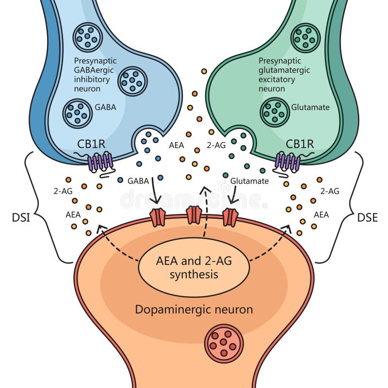
Free with trial Neurotransmitter interactions of cannabinol with neuron structure brain cell diagram schematic vector illustration. Medical science educational illustration. Labeled synapses vectors Neurotransmitter interactions diagram science. Neurotransmitter interactions of cannabinol with neuron structure brain cell diagram schematic vector illustration. Medical science educational illustration
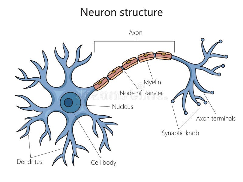
Free with trial Neuron structure brain cell diagram schematic raster illustration. Medical science educational illustration. Labeled synapses illustrations Neuron structure diagram medical science. Neuron structure brain cell diagram schematic raster illustration. Medical science educational illustration
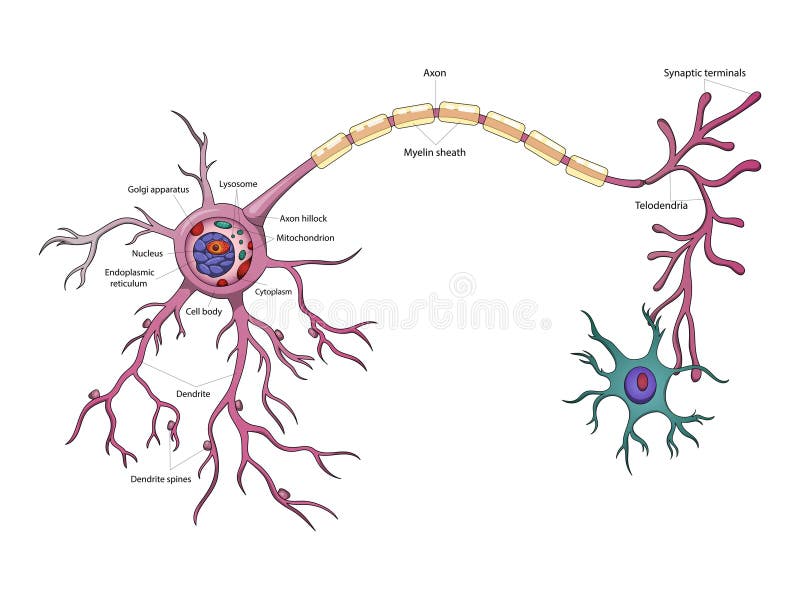
Free with trial Neuron structure brain cell diagram schematic vector illustration. Medical science educational illustration. Labeled synapses vectors Neuron structure diagram medical science. Neuron structure brain cell diagram schematic vector illustration. Medical science educational illustration
Free with trial Neuron structure brain cell diagram schematic vector illustration. Medical science educational illustration. Labeled synapses vectors Neuron structure diagram medical science. Neuron structure brain cell diagram schematic vector illustration. Medical science educational illustration
Free with trial Neuron structure brain cell diagram schematic raster illustration. Medical science educational illustration. Labeled synapses illustrations Neuron structure diagram medical science. Neuron structure brain cell diagram schematic raster illustration. Medical science educational illustration