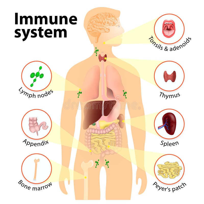
Free with trial Immune system. Human anatomy. Human silhouette with internal organs. Lymph vectors Immune system
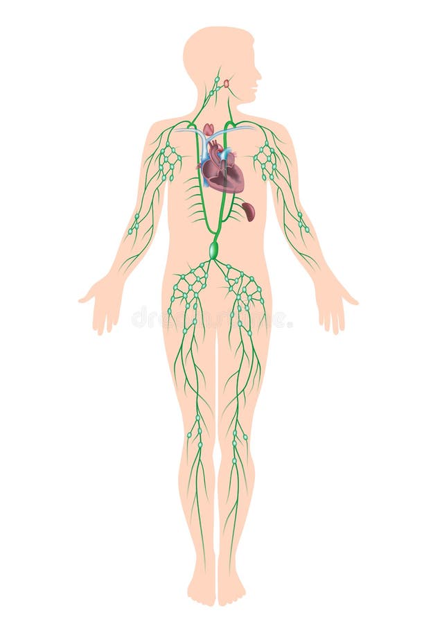
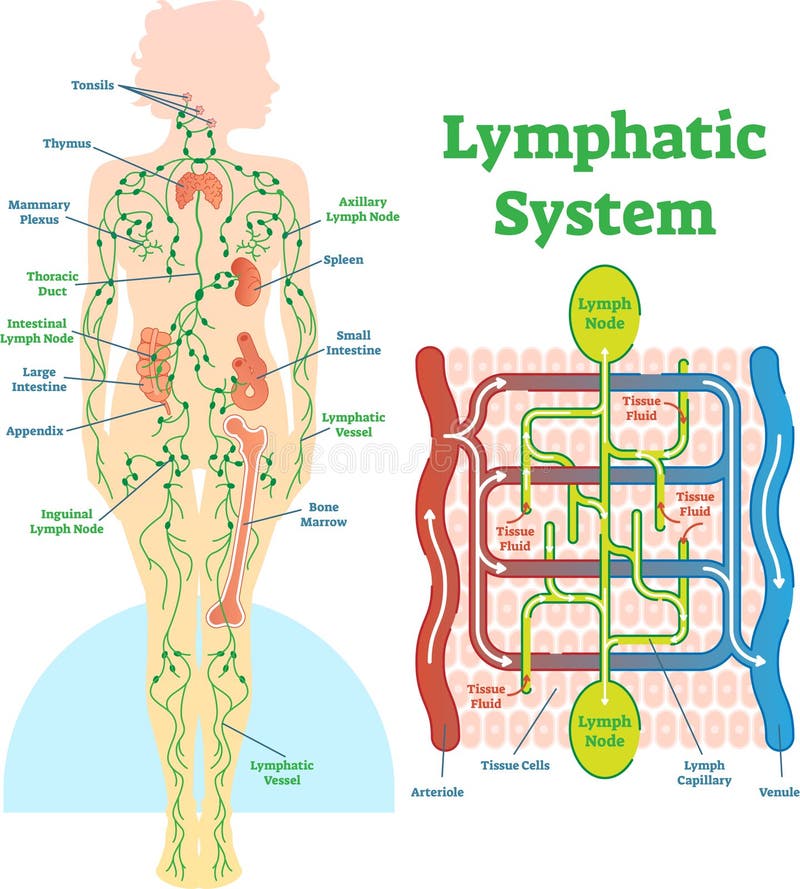
Free with trial All the organs of human lymphatic system, eps10. Lymph vectors The lymphatic system. All the organs of human lymphatic system, eps10
Free with trial Medical illustration of the different stages of colon cancer. Lymph vectors Colon cancer
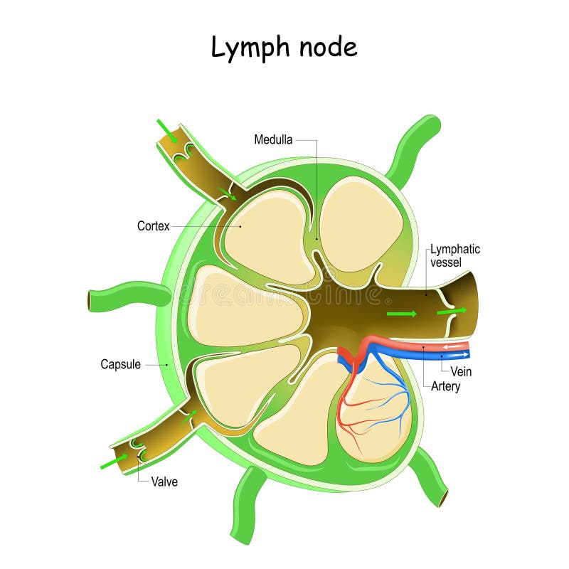
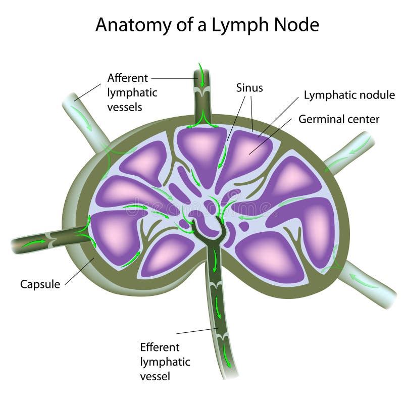
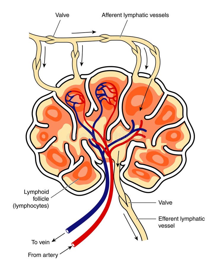
Free with trial Cross section of a lymph node, eps10. Lymph vectors Structure of a lymph node. Cross section of a lymph node, eps10
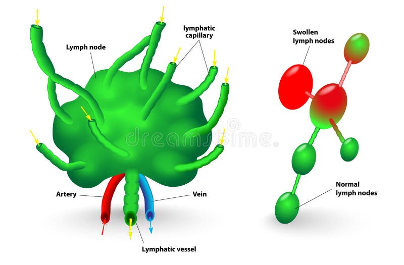
Free with trial Lymph node, lymph gland. Schematic diagram of lymph node showing the flow of lymph. Swollen lymph nodes and Normal lymph nodes. Lymph vectors Lymph node
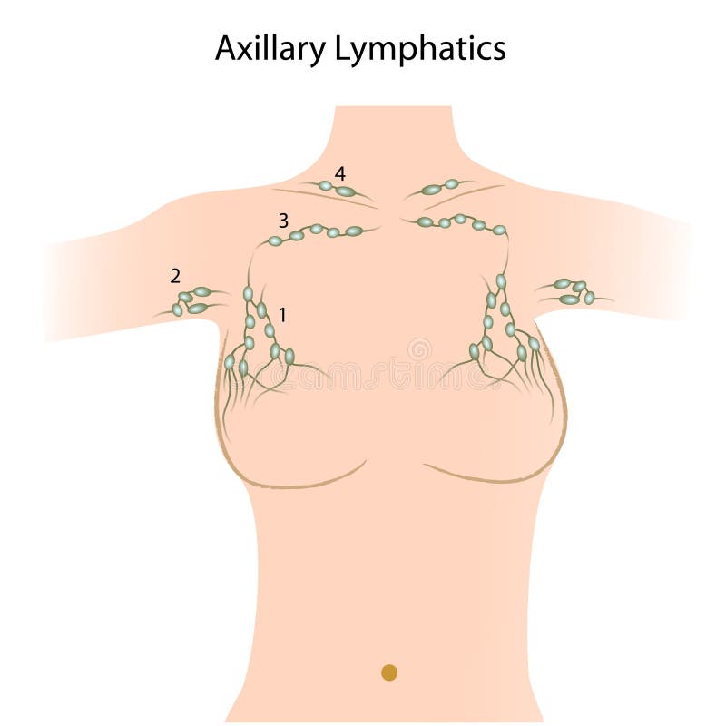
Free with trial Lymph nodes of the axillary and mammary regions, eps8. Lymph vectors Axillary lymph nodes. lymph nodes of the axillary and mammary regions, eps8
Free with trial Drawing of the axilla of a woman showing the axillary lymph nodes and lymph plexus around the breast. Lymph vectors Axillary lymph nodes
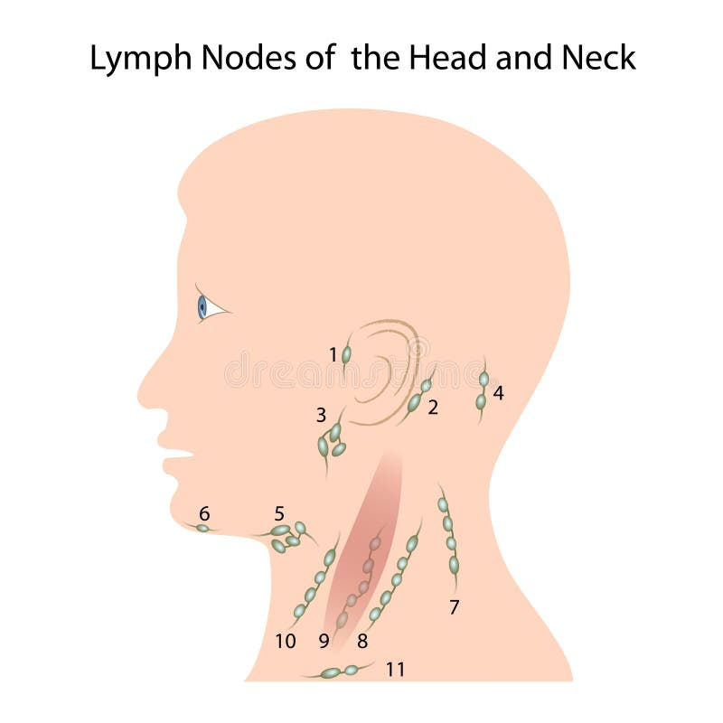
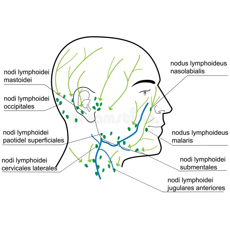
Free with trial Illustration of Lymph nodes of the head and neck, eps10. Lymph vectors Lymph nodes of the head and neck
Free with trial Cross section of a lymph node showing blood supply, follicles and lymphatic vessels. Lymph vectors Lymph node
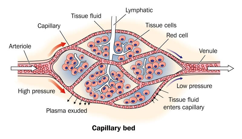
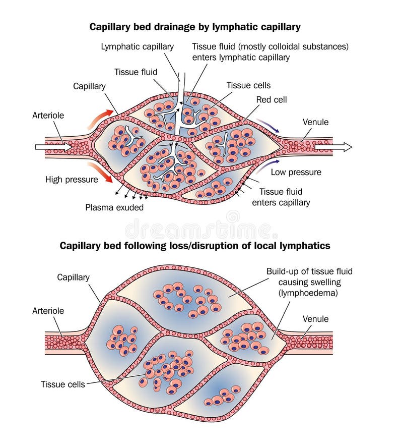
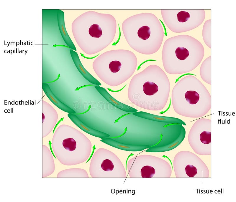
Free with trial Capillary bed showing lymph drainage. Lymph vectors Capillary bed labeled. Capillary bed showing lymph drainage
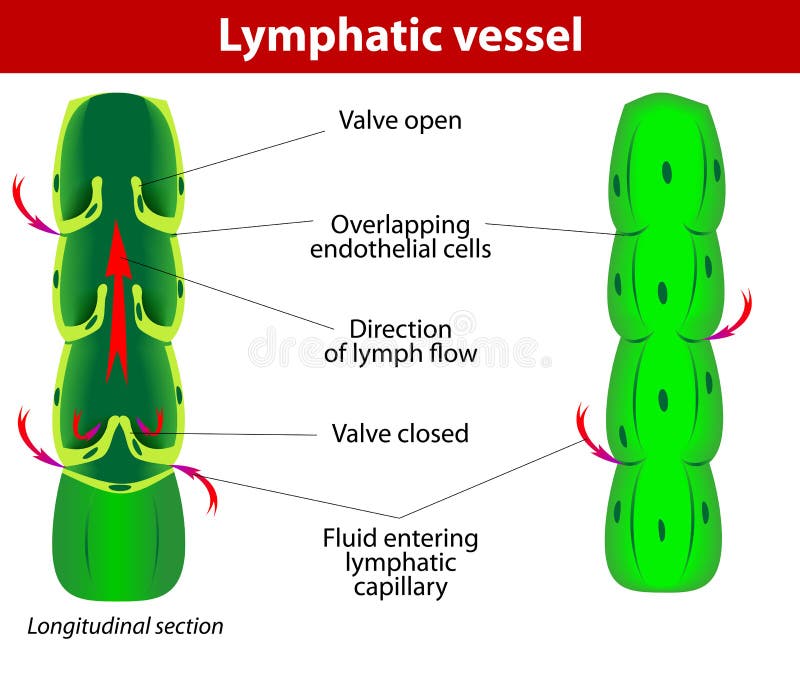
Free with trial The internal structure of a lymph vessel. Lymph vectors Lymphatic vessel. The internal structure of a lymph vessel
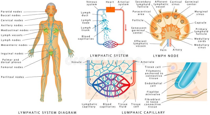
Free with trial Lymphatic system - Lymphatic diagram in human. Structure of a Lymph Node - organ of the lymphatic system. Fluid exchange between the circulatory and the lymphatic systems. Lymph vectors Lymphatic system
Free with trial Drawing of normal capillary bed with lymph vessel and oedema caused by disruption of lymphatic vessel. Lymph vectors Capillary bed lymphoedema. Drawing of normal capillary bed with lymph vessel and oedema caused by disruption of lymphatic vessel
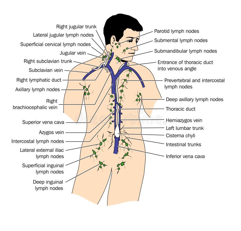
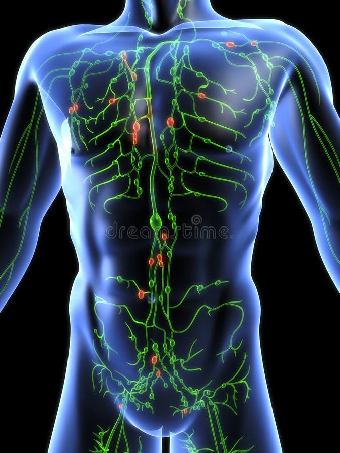
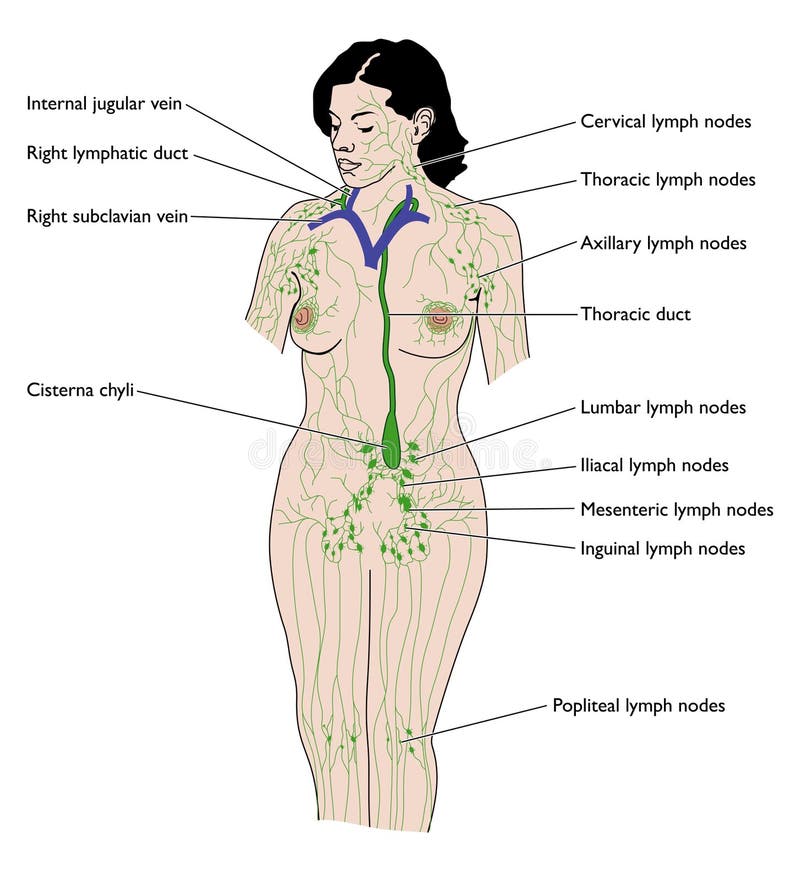
Free with trial Overview of the lymphatic system showing nodes, vessels and the thoracic duct. Lymph vectors The lymph system. Overview of the lymphatic system showing nodes, vessels and the thoracic duct
Free with trial Lymphatic system anatomical vector illustration diagram, educational medical scheme with lymph nodes and tissue fluid circulation flow. Lymph vectors Lymphatic system anatomical vector illustration diagram, educational medical scheme. Lymphatic system anatomical vector illustration diagram, educational medical scheme with lymph nodes and tissue fluid circulation flow.
Free with trial The lymph system, relationship of lymphatic capillaries to tissue cells and blood capillaries, vector medical illustration. Lymph vectors Lymphatic and Blood Capillaries. The lymph system, relationship of lymphatic capillaries to tissue cells and blood capillaries, vector medical illustration
Free with trial Human anatomy illustration of the lymphatic system. Lymph illustrations Lymphatic system
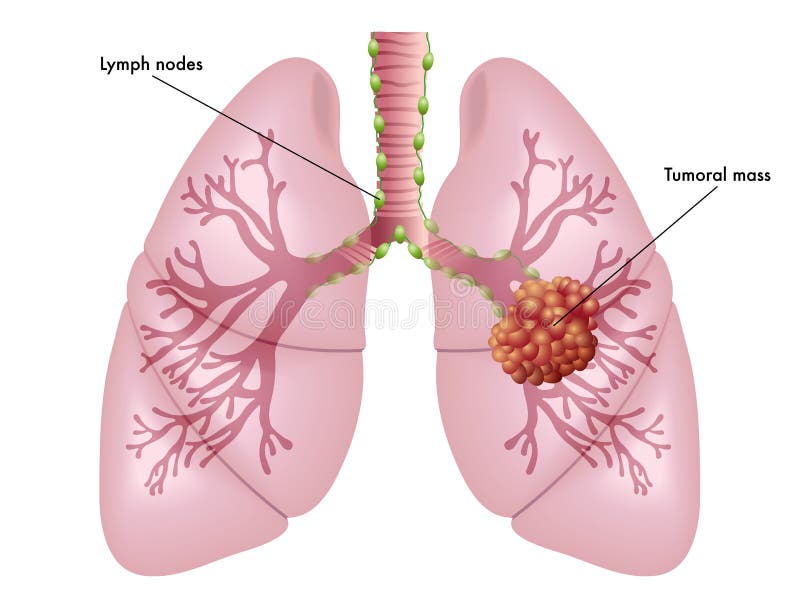
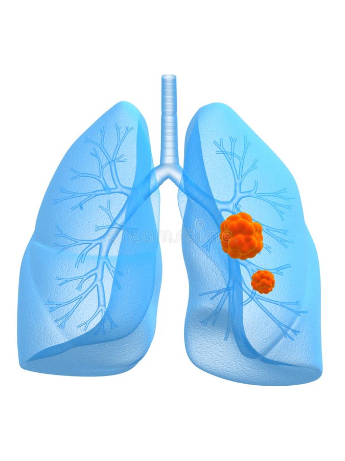
Free with trial Simple medical illustration of the symptoms of lung cancer. Lymph vectors Lung cancer
Free with trial The skin is the largest organ of the body, with a total area of about 20 square feet. The skin protects us from microbes and the elements, helps regulate body temperature, and permits the sensations of touch, heat, and cold. Skin has three layers: The epidermis, the outermost layer of skin, provides a waterproof barrier and creates our skin tone. The dermis, beneath the epidermis, contains tough connective tissue, hair follicles, and sweat glands. The deeper subcutaneous tissue hypodermis is made of fat and connective tissue. The skin’s color is created by special cells called melanocytes, which produce the pigment melanin. Melanocytes are located in the epidermis. Lymph illustrations Skin Anatomy. The skin is the largest organ of the body, with a total area of about 20 square feet. The skin protects us from microbes and the elements, helps regulate body temperature, and permits the sensations of touch, heat, and cold. Skin has three layers: The epidermis, the outermost layer of skin, provides a waterproof barrier and creates our skin tone. The dermis, beneath the epidermis, contains tough connective tissue, hair follicles, and sweat glands. The deeper subcutaneous tissue hypodermis is made of fat and connective tissue. The skin’s color is created by special cells called melanocytes, which produce the pigment melanin. Melanocytes are located in the epidermis.
Free with trial Human anatomy illustration of the lymphatic system. Lymph illustrations Lymphatic system
Free with trial Vestibular system. inner ear and its relationship to balance and equilibrum. Schematic representation of the membranous labyrinth and ross-section of the cochlea. Human Biology. Lymph vectors Vestibular system
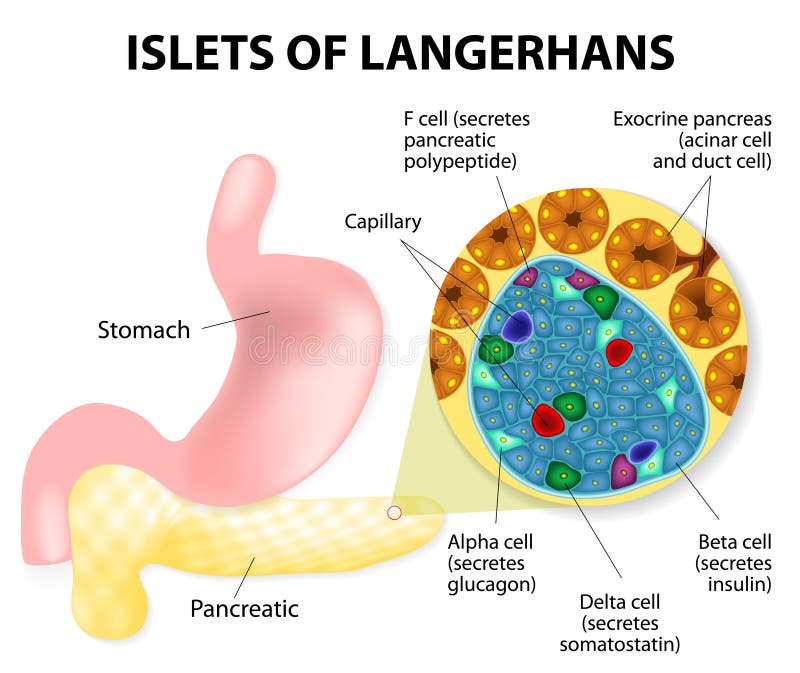
Free with trial The islets of Langerhans are responsible for the endocrine function of the pancreas. Each islet contains beta, alpha, and delta cells that are responsible for the secretion of a hormones. Lymph vectors Islets of Langerhans
Free with trial Showing the arteries, veins and lymphatic vessel with enlarged sections. Lymph vectors Arterial-venous-lymphatic. Showing the arteries, veins and lymphatic vessel with enlarged sections.
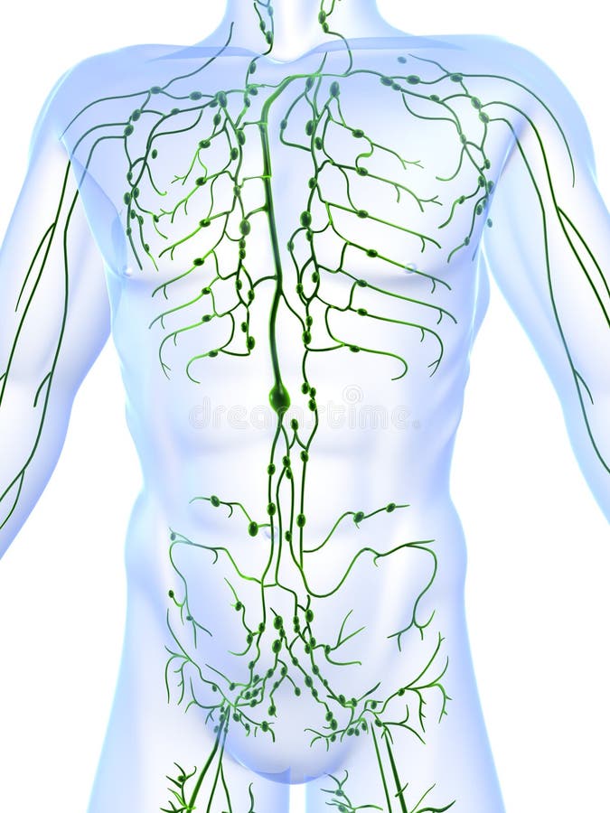
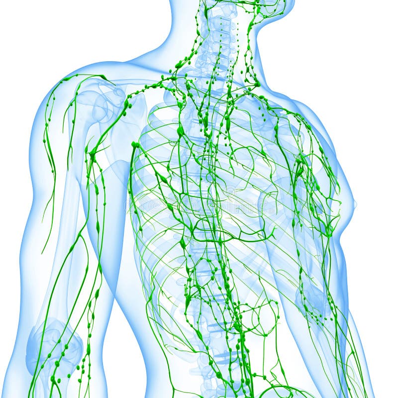
Free with trial 3d art illustration of transparent lymphatic system of man. Lymph illustrations Transparent lymphatic system of man
Free with trial Diagram of Effects of Sleep deprivation. Illustration about disease diagnosis. Lymph vectors Diagram of Effects of Sleep deprivation.
Free with trial Lymphocytes are white blood cells or leucocytes in the human immune system consisting of B and T cells which form antibodies for immunity and natural killer cells which fight viruses and tumours. Lymph illustrations Lymphocyte
Free with trial Immune system scheme on a girl body. For children education. Lymph vectors Immune system
Free with trial 3d rendered anatomy illustration of human lung and bronchi. Lymph illustrations Lung and bronchi
Free with trial Human anatomy illustration of the lymphatic system. Lymph illustrations Lymphatic system
Free with trial 3d rendered anatomy illustration of human lung with bronchi. Lymph illustrations Lung and bronchi. 3d rendered anatomy illustration of human lung with bronchi
Free with trial Uptake of tissue fluid by lymphatic capillary, eps8. Lymph vectors Lymphatic capillary
Free with trial Overview of the lymphatic system, including ducts, nodes cisterna chyli and thoracic duct. Lymph vectors The lymphatic system
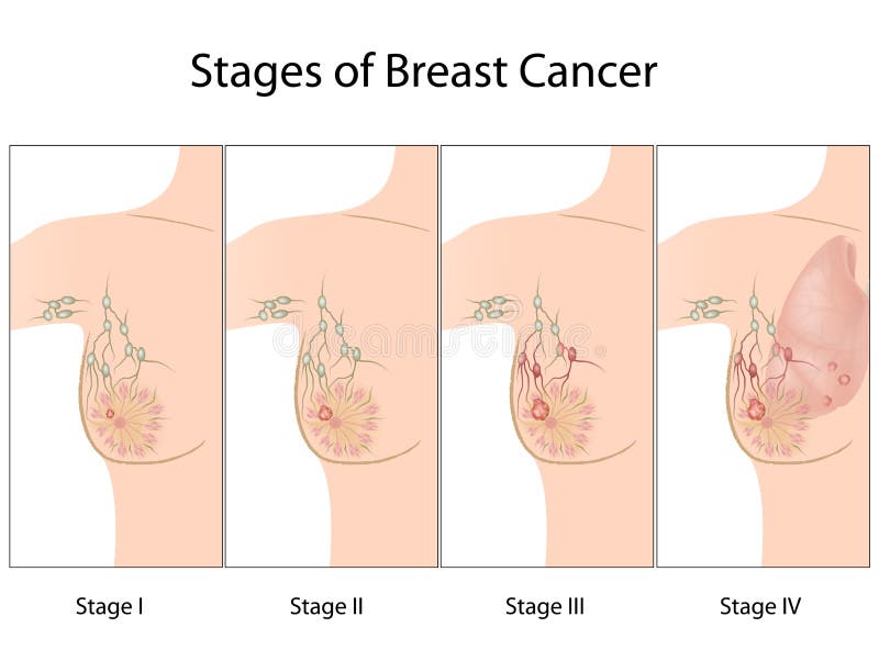
Free with trial Drawing to show the various stages of breast cancer, from early onset disease through local spread to advanced metastatic disease. Lymph vectors Stages of breast cancer
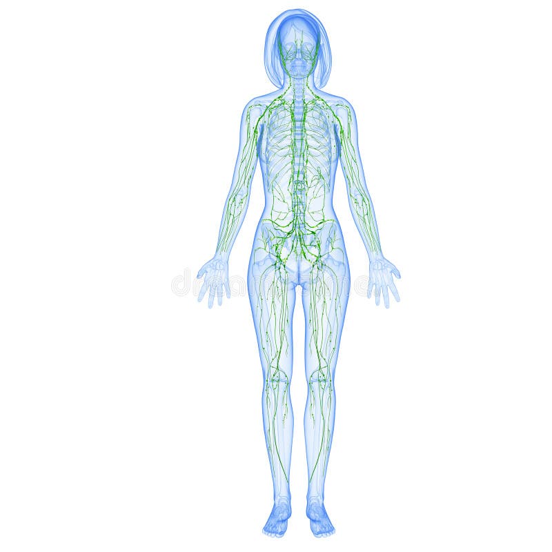
Free with trial Female anatomy illustration of the Lymphatic system isolated. Lymph illustrations Female Lymphatic system. Female anatomy illustration of the Lymphatic system isolated
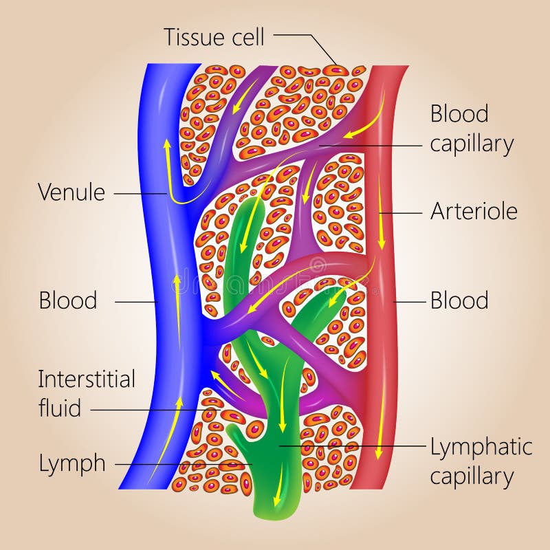
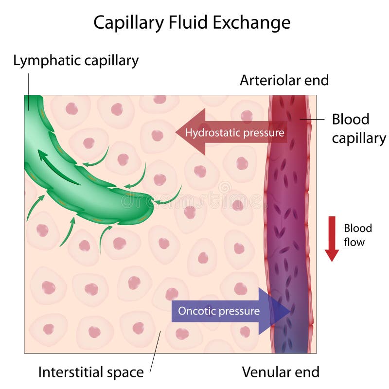
Free with trial Mechanism of capillary fluid exchange between the blood and body tissues, eps10. Lymph vectors Capillary Fluid Exchange
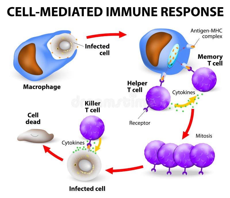
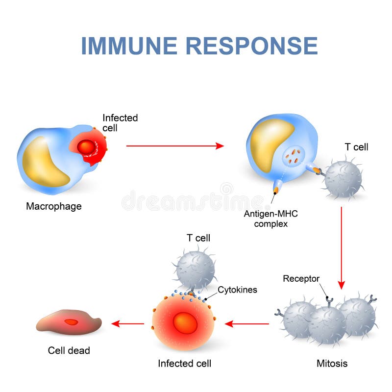
Free with trial Cell-mediated immunity. T lymphocytes do not secrete antibodies. this response incorporates activated macrophages, natural killer cells, antigen-specific cytotoxic T-lymphocytes as well as release of cytokines. Lymph vectors Cell-mediated immune response. Cell-mediated immunity. T lymphocytes do not secrete antibodies. this response incorporates activated macrophages, natural killer cells, antigen-specific cytotoxic T-lymphocytes as well as release of cytokines.
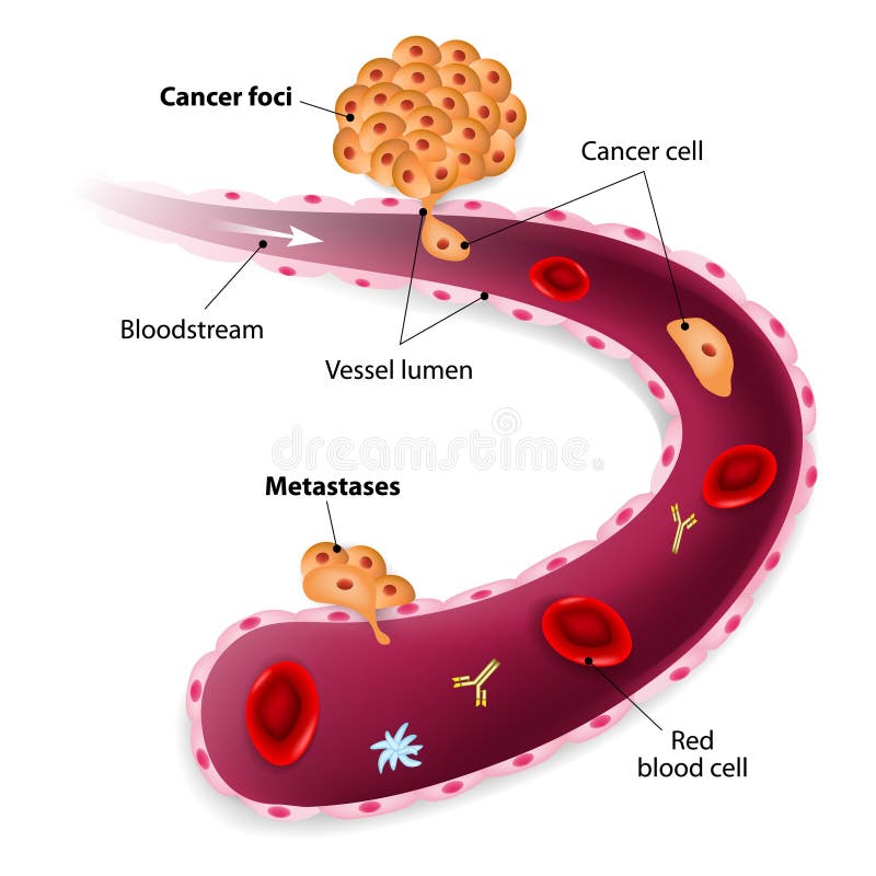
Free with trial Cancer cell squeezes through blood vessel during Metastases. Lymph vectors Cancer cells, cancer foci and Metastases. Cancer cell squeezes through blood vessel during Metastases
Free with trial Anatomy of human respiratory system, 3D image. Lymph illustrations Anatomy of human respiratory system
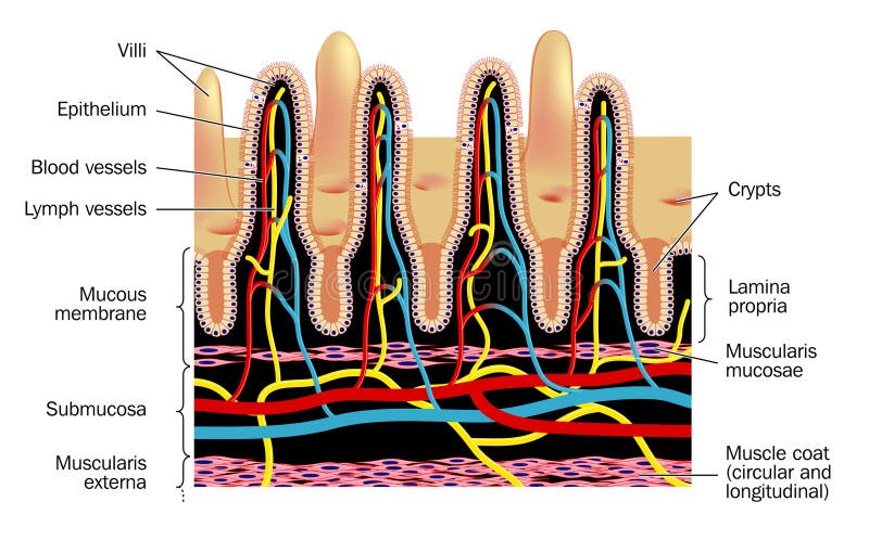
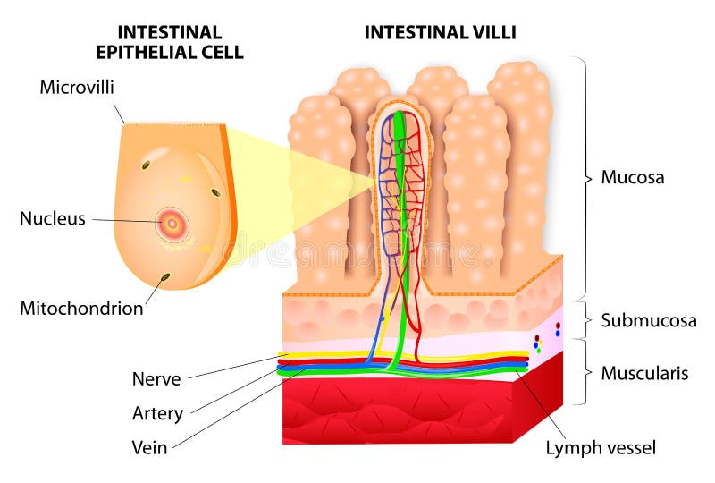
Free with trial Detail of small intestinal villim, showing the villi and the crypts, blood supply and lymph vessels. Lymph vectors Microvilli. Detail of small intestinal villim, showing the villi and the crypts, blood supply and lymph vessels
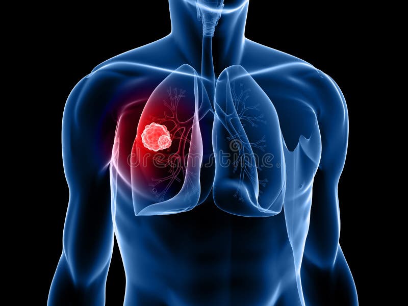
Free with trial 3d rendered x-ray illustration of a human torso with tumor in lung. Lymph illustrations Lung tumor. 3d rendered x-ray illustration of a human torso with tumor in lung
Free with trial 3d rendered illustration of human lung with carzinoma. Lymph illustrations Lung cancer. 3d rendered illustration of human lung with carzinoma
Free with trial 3d rendered illustration of a body shape with tumor in lung. Lymph illustrations Lung cancer. 3d rendered illustration of a body shape with tumor in lung
Free with trial 3d rendered anatomy illustration of a human lung wit carzinoma. Lymph illustrations Lung cancer. 3d rendered anatomy illustration of a human lung wit carzinoma
Free with trial Structure of Villi and microvilli showing arteries, veins, nerve and lymph vessel. Lymph vectors Microvilli. Detail of the small intestine. Vector. Structure of Villi and microvilli showing arteries, veins, nerve and lymph vessel.
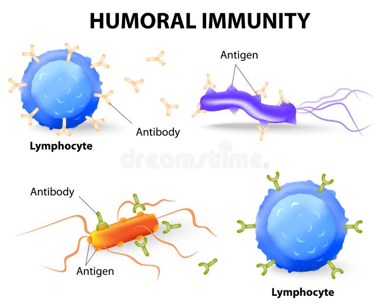
Free with trial Humoral immunity. Lymphocyte, antibody and antigen. Vector diagram. Lymph vectors Humoral immunity. Lymphocyte, antibody and antigen
Free with trial 3d rendered anatomy illustration of human lung with bronchi. Lymph illustrations Lung and bronchi. 3d rendered anatomy illustration of human lung with bronchi
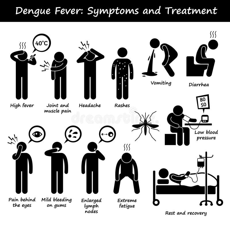
Free with trial A set of human pictogram representing the symptoms of dengue fever by aedes mosquito. This include high fever, joint and muscle pain, headache, skin rashes, vomiting, diarrhea, bleeding gum, enlarged lymph node, fatigue, and low blood pressure. Lymph vectors Dengue Fever Symptoms and Treatment Aedes Mosquito. A set of human pictogram representing the symptoms of dengue fever by aedes mosquito. This include high fever, joint and muscle pain, headache, skin rashes, vomiting, diarrhea, bleeding gum, enlarged lymph node, fatigue, and low blood pressure.
Free with trial 3d rendered illustration from a profile of a bronch. Lymph illustrations Asthma bronchiales. 3d rendered illustration from a profile of a bronch
Free with trial Medical illustration of the effects of the Rheumatoid Arthritis. Lymph vectors Rheumatoid Arthritis
Free with trial Cell-mediated immunity. T lymphocytes do not secrete antibodies. this response incorporates activated macrophages, natural killer cells, antigen-specific cytotoxic T-lymphocytes as well as release of cytokines. Lymph vectors Cell-mediated immunity.
Free with trial Female anatomy illustration of the Lymphatic system. Lymph illustrations Female Lymphatic system. Female anatomy illustration of the Lymphatic system
Free with trial Steps of phagocytosis. antigen presenting cell in human immune response. Antigen-presenting cell engulf antigens and process them so that they can be recognized by lymphocytes. Lymph vectors Antigen-presenting cell
Free with trial 3D medical illustration - lungs with visible bronchi. Lymph illustrations Lungs with visible bronchi
Free with trial Main white blood cell leukocytes type overview. Lymph illustrations White blood cells overview. Main white blood cell leukocytes type overview .
Free with trial White Blood cell formation from differentiation of hematopoietic stem cell. Lymph illustrations White Blood cell formation
Free with trial Cancer cell with in lymphocytes in the surrounding trying to eliminate the tumor cell. Lymph illustrations Cancer cell with lymphocytes. Cancer cell with in lymphocytes in the surrounding trying to eliminate the tumor cell
Free with trial Autoimmune disease infographic. Medical Infographic set elements and symbols for design. Lymph vectors Autoimmune disease infographic
Free with trial Female anatomy illustration of the lymphatic system of male half body. Lymph illustrations Female Lymphatic system of half body. Female anatomy illustration of the lymphatic system of male half body
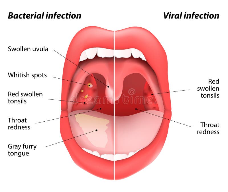
Free with trial The differences between viral and bacterial infections. Open mouth view of tonsils. Vector illustration of a disease inflammation of the tonsils caused by viral or bacterial infection. Lymph vectors The differences between viral and bacterial infections
Free with trial Lymphocytes are white blood cells or leucocytes in the human immune system consisting of B and T cells which form antibodies for immunity and natural killer cells which fight viruses and tumours. Lymph illustrations Lymphocytes are white blood cells or leucocytes in the human immune system consisting of B and T cells which form antibodies for
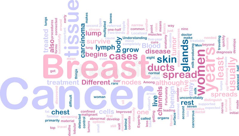
Free with trial Word cloud tags concept illustration of breast cancer. Lymph illustrations Breast cancer wordcloud. Word cloud tags concept illustration of breast cancer
Free with trial Illustration of lymph nodes of the head and neck. Lymph vectors Lymph nodes of the head and neck.
Free with trial Lymph nodes each of a number of small swellings in the lymphatic system where lymph is filtered and lymphocytes are formed. Lymph illustrations Lymph nodes
Free with trial Lymph nodes each of a number of small swellings in the lymphatic system where lymph is filtered and lymphocytes are formed. Lymph illustrations Lymph nodes
Free with trial Drawing of the lymph nodes of the chest and axilla, showing the relations to the underlying muscle and ribs. Lymph vectors Lymph nodes of the chest and axilla
Free with trial Lymph is the fluid that circulates throughout the lymphatic system. The lymph is formed when the interstitial fluid (the fluid which lies in the interstices of all body tissues)[1] is collected through lymph capillaries. It is then transported through lymph vessels to lymph nodes before emptying ultimately into the right or the left subclavian vein, where it mixes back with blood. Lymph illustrations Lymph
Free with trial Lymph nodes are small, bean-shaped glands throughout the body. They are part of the lymph system, which carries fluid (lymph fluid), nutrients, and waste material between the body tissues and the bloodstream. The lymph system is an important part of the immune system, the body's defense system against disease. The lymph nodes filter lymph fluid as it flows through them, trapping bacteria, viruses, and other foreign substances, which are then destroyed by special white blood cells called lymphocytes. Lymph illustrations Lymph nodes
Free with trial Lymph Node Anatomy. Labeled diagram showing the flow of lymph. Afferent and efferent vessels. Vector illustration. Lymph vectors Lymph Node Anatomy
Free with trial Illustration inflammation of the submandibular lymph nodes. Lymph vectors Lymphadenitis . inflammation of the lymph nodes. Illustration inflammation of the submandibular lymph nodes
Free with trial Lymphatic circulation system with lymph transportation to organs an heart vector illustration. Labeled anatomical scheme with part of cardiovascular network. Pulmonary circuit medical explanation. Lymph vectors Lymphatic circulation system with lymph transportation vector illustration. Lymphatic circulation system with lymph transportation to organs an heart vector. Lymphatic circulation system with lymph transportation to organs an heart vector illustration. Labeled anatomical scheme with part of cardiovascular network. Pulmonary circuit medical explanation.


![Lymph is the fluid that circulates throughout the lymphatic system. The lymph is formed when the interstitial fluid (the fluid which lies in the interstices of all body tissues)[1] is collected through lymph capillaries. It is then transported through lymph vessels to lymph nodes before emptying ultimately into the right or the left subclavian vein, where it mixes back with blood. Lymph illustrations](https://thumbs.dreamstime.com/b/lymph-fluid-circulates-throughout-lymphatic-system-formed-interstitial-fluid-fluid-which-55400876.jpg)