Free with trial Signal transduction pathway by insulin receptor, usually defective in type 2 diabetes, eps8. Membrane receptor vectors Effect of Insulin on glucose uptake. Signal transduction pathway by insulin receptor, usually defective in type 2 diabetes, eps8
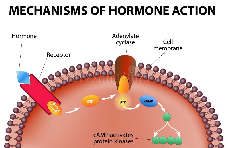
Free with trial Hormones bind to receptors on the plasma membrane. The hormone itself is the first messenger. Binding to the receptors activates a second messenger inside the cell. The second messenger causes intracellular effects. Membrane receptor vectors Mechanisms of hormone action. Hormones bind to receptors on the plasma membrane. The hormone itself is the first messenger. Binding to the receptors activates a second messenger inside the cell. The second messenger causes intracellular effects
Free with trial When a hormone outside of a capillary, it can act on a target cell. A steroid hormone is capable of crossing through the cell membrane of the target cell. A protein hormone attaches to the cells membrane and activates a receptor that releases, in turn, a messenger within the cell. Membrane receptor vectors Hormones work. Vector. When a hormone outside of a capillary, it can act on a target cell. A steroid hormone is capable of crossing through the cell membrane of the target cell. A protein hormone attaches to the cells membrane and activates a receptor that releases, in turn, a messenger within the cell.
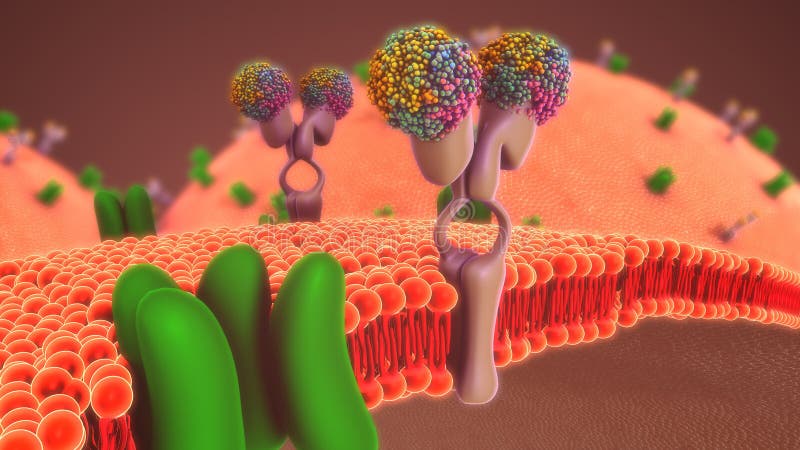
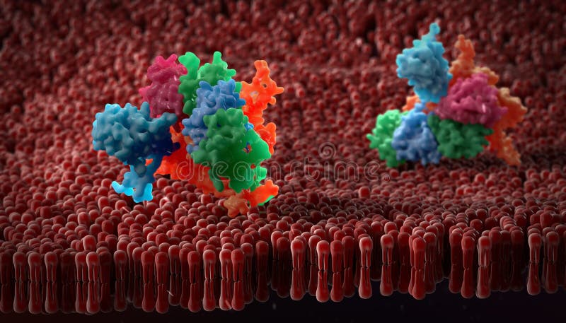
Free with trial Artistic impression of a plasma membrane of a human cell. The plasma membrane is a bilayer composed of phopholipids in which lots of transmembrane and surface proteins reside. Its function is to separate the intracellular content. Membrane receptor illustrations Plasma membrane of a cell with associated proteins. Artistic impression of a plasma membrane of a human cell. The plasma membrane is a bilayer composed of phopholipids in which lots of transmembrane and surface proteins reside. Its function is to separate the intracellular content
Free with trial Plasma Membrane Of A Cell With other molecules. Membrane receptor illustrations Plasma Membrane Of A Cell
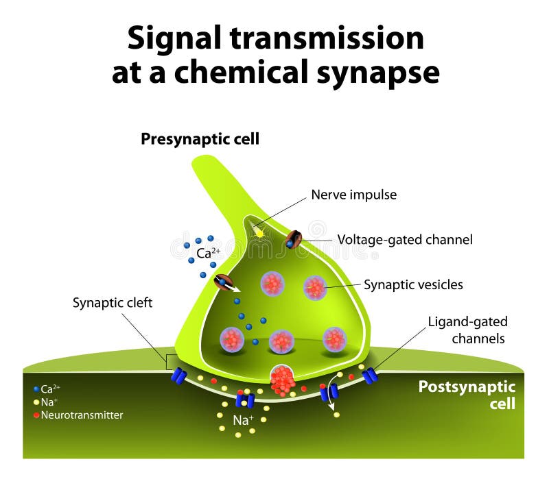
Free with trial Signal transmission at a chemical synapse. one neuron releases neurotransmitter molecules into a synaptic cleft that is adjacent to another neuron. Membrane receptor vectors Chemical synapse
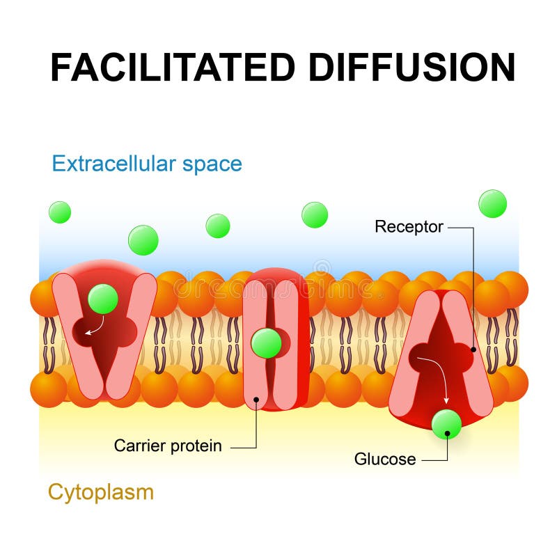
Free with trial Facilitated diffusion or facilitated transport or passive-mediated transport. Carrier protein. Membrane receptor vectors Facilitated diffusion or passive-mediated transport. Facilitated diffusion or facilitated transport or passive-mediated transport. Carrier protein
Free with trial Human cell, anatomy image. 2 D illustration, on white background. Membrane receptor illustrations Human cell, anatomy image.
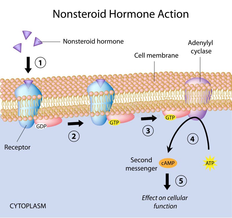
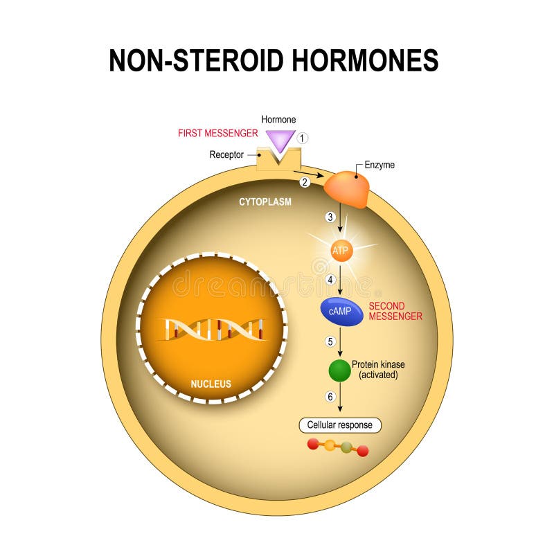
Free with trial Signal transduction pathway triggered by nonsteroid hormones, eps10. Membrane receptor vectors Nonsteroid hormones action. Signal transduction pathway triggered by nonsteroid hormones, eps10
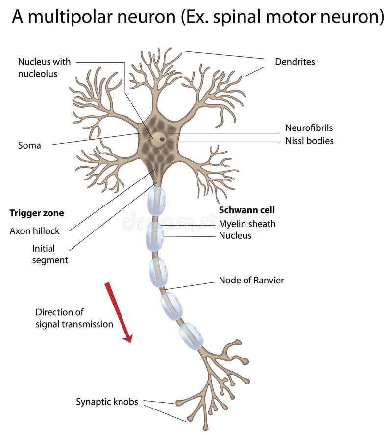
Free with trial Eps10, gradient and mesh printing compatible, used transparency. Membrane receptor vectors Motor neuron, detailed and accurate, labeled vers. Eps10, gradient and mesh printing compatible, used transparency
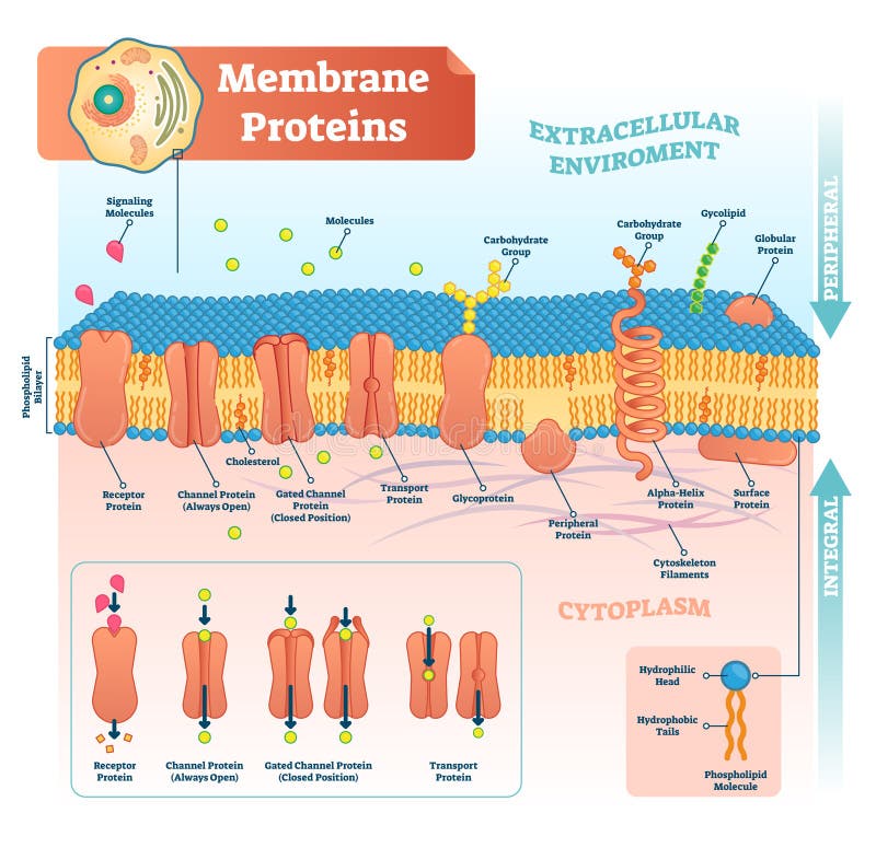
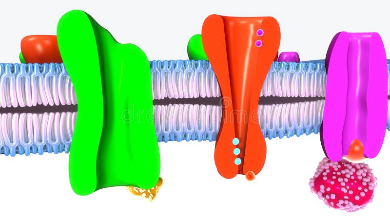
Free with trial Membrane proteins labeled vector illustration. Detailed microscopic structure scheme. Anatomical diagram with receptor, open channel, closed gated and transport protein. Membrane receptor vectors Membrane proteins labeled vector illustration. Detailed structure scheme. Membrane proteins labeled vector illustration. Detailed microscopic structure scheme. Anatomical diagram with receptor, open channel, closed gated and transport protein.
Free with trial Dendritic cells vector illustration. Anatomical labeled scheme with progenitor, immature, nucleus and membrane extensions. Antigen and receptor diagram. Microscopic closeup with biological structure. Membrane receptor vectors Dendritic cells vector illustration. Anatomical labeled closeup scheme with progenitor, immature, nucleus, antigen and receptor. Dendritic cells vector illustration. Anatomical labeled scheme with progenitor, immature, nucleus and membrane extensions. Antigen and receptor diagram. Microscopic closeup with biological structure.
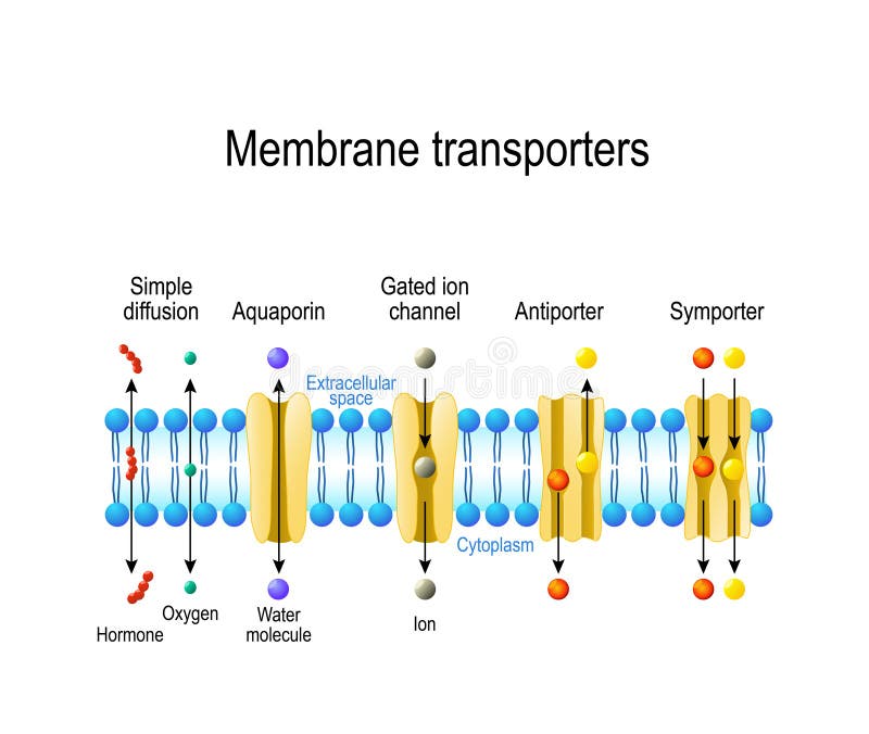
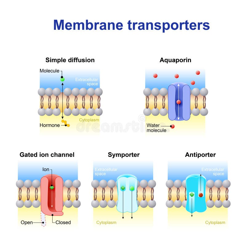
Free with trial Mechanisms for the transport of ions and molecules across cell membranes. Types of a channel in the cell membrane: simple diffusion, Aquaporin, Gated ion channel, Symporter and Antiporter. Membrane receptor vectors Types of a channel in the cell membrane
Free with trial Plasma Membrane Of A Cell With other molecules. Membrane receptor illustrations Plasma Membrane Of A Cell
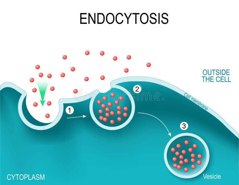
Free with trial Endocytosis. Type of vesicle transport that carry very large molecules across the cell membrane. Vector illustration. Membrane receptor vectors Endocytosis Vesicle Transport Cell Membrane. Endocytosis. Type of vesicle transport that carry very large molecules across the cell membrane. Vector illustration
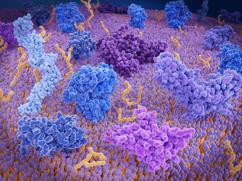
Free with trial Cell-surface receptors, also known as transmembrane receptors, are cell surface, membrane-anchored, or integral proteins that bind to external ligand molecules. This type of receptor spans the plasma membrane and performs signal transduction, converting an extracellular signal into an intracellular signal. Membrane receptor illustrations Cell membrane receptors. Cell-surface receptors, also known as transmembrane receptors, are cell surface, membrane-anchored, or integral proteins that bind to external ligand molecules. This type of receptor spans the plasma membrane and performs signal transduction, converting an extracellular signal into an intracellular signal.
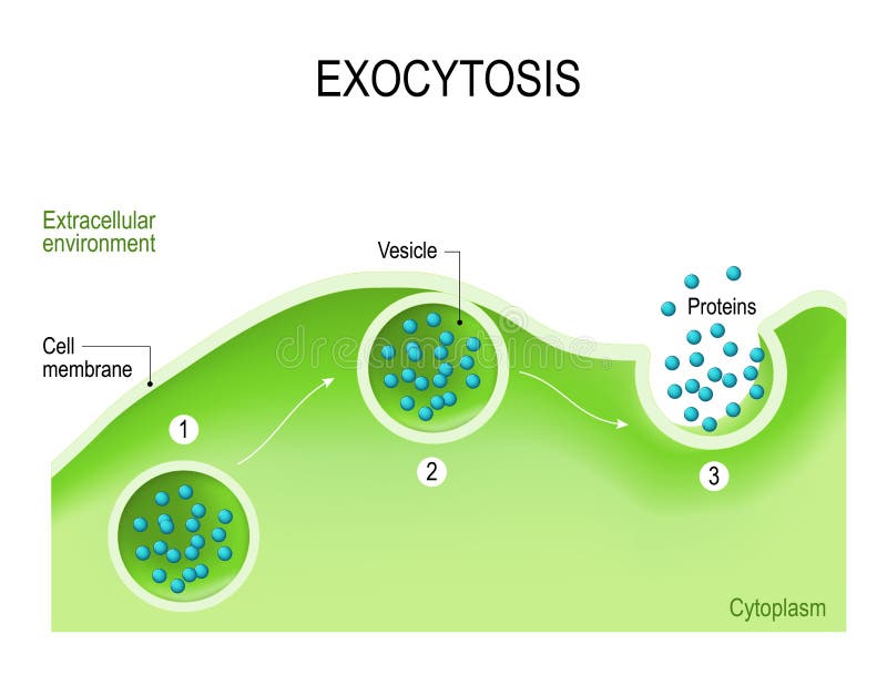
Free with trial Exocytosis - vesicle transport that carry very large molecules across the cell membrane. Vector illustration. Membrane receptor vectors Exocytosis Vesicle Transport Cell Membrane. Exocytosis - vesicle transport that carry very large molecules across the cell membrane. Vector illustration
Free with trial Chimeric antigen receptor T cell and Antibody molecule. IgE and CAR. Artificial T cell receptors are proteins that have been engineered for cancer therapy. genetically engineered. Vector diagram for medical, educational and science use. Membrane receptor vectors Chimeric antigen receptor T cell and Antibody molecule. IgE and CAR
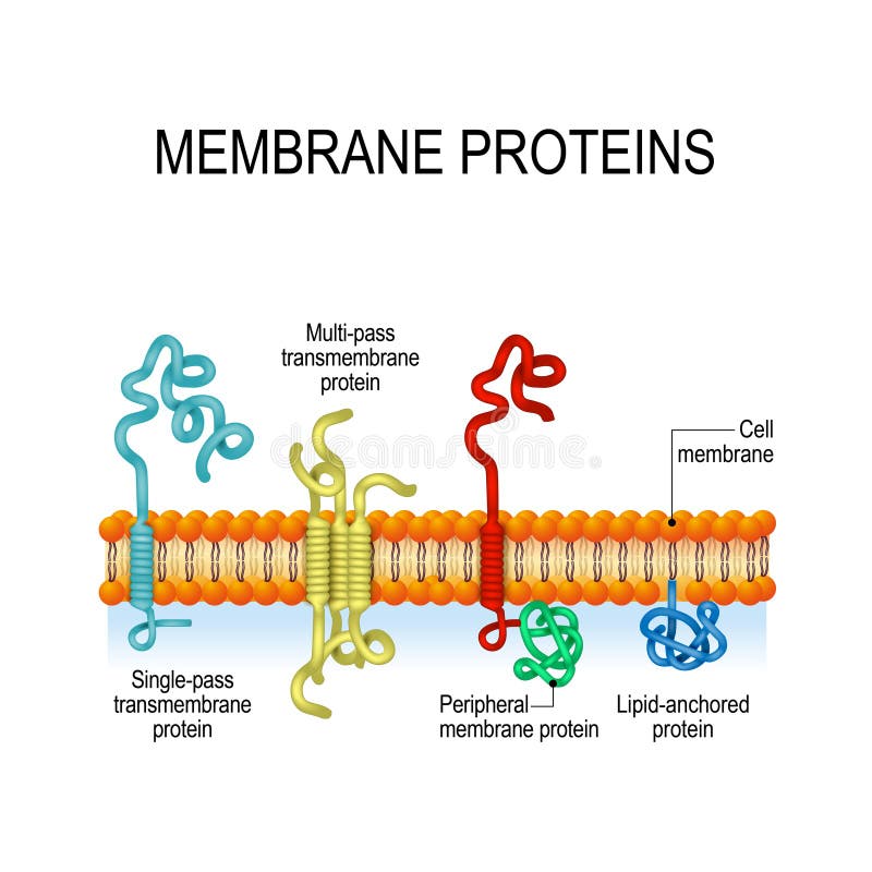
Free with trial Membrane proteins. integral, and Peripheral membrane proteins, Single-pass, and Multi-pass transmembrane α-helix, Lipid-anchored protein. Vector illustration for biological, science and educational use. Membrane receptor vectors Membrane proteins. Vector illustration for biological, science and educational use. Membrane proteins. integral, and Peripheral membrane proteins, Single-pass, and Multi-pass transmembrane α-helix, Lipid-anchored protein. Vector illustration for biological, science and educational use
Free with trial Symport and antiport - are an integral membrane protein. cell membrane transport system. How it works. Membrane receptor vectors Symport and antiport. cell membrane transport systems. Symport and antiport - are an integral membrane protein. cell membrane transport system. How it works
Free with trial Cell-surface receptors, also known as transmembrane receptors, are cell surface, membrane-anchored, or integral proteins that bind to external ligand molecules. This type of receptor spans the plasma membrane and performs signal transduction, converting an extracellular signal into an intracellular signal. Membrane receptor illustrations Cell membrane receptors. Cell-surface receptors, also known as transmembrane receptors, are cell surface, membrane-anchored, or integral proteins that bind to external ligand molecules. This type of receptor spans the plasma membrane and performs signal transduction, converting an extracellular signal into an intracellular signal.
Free with trial Anatomy of the Lysosome: Hydrolytic enzymes, Membrane and transport proteins. This organelle use the enzymes to break down and digest food particles, engulfed viruses or bacteria in the cell. Vector diagram for medical use. Membrane receptor vectors Anatomy of the Lysosome: Hydrolytic enzymes, Membrane and transport proteins.
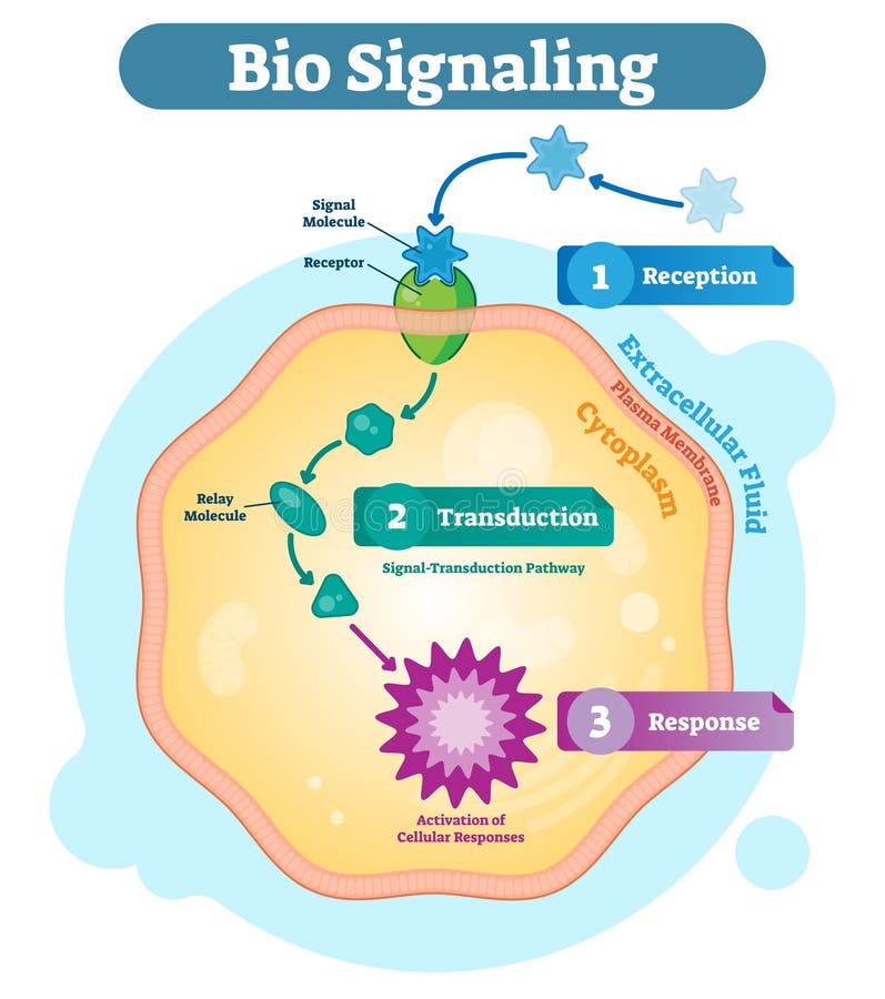
Free with trial Bio signaling cell communication network system, micro biological anatomy labeled diagram vector illustration with receptor, transduction and response activity. Cell cross section scheme. Membrane receptor vectors Bio signaling cell communication system, biological anatomy diagram vector illustration with receptor, transduction and response. Bio signaling cell communication network system, micro biological anatomy labeled diagram vector illustration with receptor, transduction and response activity. Cell cross section scheme.
Free with trial Endocytosis and exocytosis in the cell. endocytosis: transports proteins into the cell. exocytosis: transports molecules out of the cytoplasm. vesicles fuses with membrane, contents are secreted into the extracellular environment. Membrane receptor vectors Endocytosis and exocytosis in the cell
Free with trial Exocytosis. Cell transports molecules out of the cell. vesicles are carried to the cell membrane, fuses with membrane, contents are secreted into the extracellular environment. Membrane receptor vectors Exocytosis
Free with trial Events at the plasma membrane of a neuron when stimulated by a chemical, eps10. Membrane receptor vectors Neuron and local potential. Events at the plasma membrane of a neuron when stimulated by a chemical, eps10
Free with trial Animal cell with nucleus, cytoplasm, DNA, enzime, protein kinase, receptor, and hormone. How non-steroid hormones work. Non-steroid hormones interact with receptors on the cell membrane and activate secondary messenger systems that carry out their effects within the cell. Human endocrine system. Membrane receptor vectors Animal cell with nucleus, cytoplasm, DNA, enzime, protein kinase
Free with trial The different types of endocytosis: receptor-mediated endocytosis, pinocytosis cell drinking and phagocytosis cell eating. vesicle, coated vesicle, and phagosome. vector illustration for medical, educational and science use. Membrane receptor vectors The different types of endocytosis
Free with trial Mechanisms for the transport of ions and molecules across cell membranes. Types of a channel in the cell membrane: simple diffusion, Aquaporin, Gated ion channel, Symporter and Antiporter. Membrane receptor vectors Mechanisms for the transport of ions and molecules across cell m
Free with trial Anatomy of the Lysosome: Hydrolytic enzymes, Membrane and transport proteins. This organelle use the enzymes to break down and digest food particles, engulfed viruses or bacteria in the cell. Vector diagram for medical use. Membrane receptor vectors Anatomy of the Lysosome. Vector diagram for medical use. Anatomy of the Lysosome: Hydrolytic enzymes, Membrane and transport proteins. This organelle use the enzymes to break down and digest food particles, engulfed viruses or bacteria in the cell. Vector diagram for medical use
Free with trial Dendritic cells present antigens green to lymphocytes through their membran bound MHC-molecules violet. CD4 molecules light blue bind to other portions of the MHC, strengthening the interaction. After binding to the MHC-antigen complex, The T-cell receptor blue sends a signal cascade through an attached G-protein into the T-lymphocyte cell, that activates an immune response. Membrane receptor illustrations Activation of the immune response: antigen presenting cell activates T-lymphocytes (smaller c. Dendritic cells present antigens green to lymphocytes through their membran bound MHC-molecules violet. CD4 molecules light blue bind to other portions of the MHC, strengthening the interaction. After binding to the MHC-antigen complex, The T-cell receptor blue sends a signal cascade through an attached G-protein into the T-lymphocyte cell, that activates an immune response.
Free with trial 3d computer illustration of an antigen presenting cell. The antigen is a peptide from a tumor cell, bacteria or virus. Dendritic cells present antigens to lymphocytes through their membran bound MHC-molecules. After binding to the MHC-antigen complex, the T-cell receptor sends a a signal cascade into the T-lymphocyte cell, that activates an immune response. Membrane receptor illustrations Dendritic cell presenting an antigen to T-lymphocytes. 3d computer illustration of an antigen presenting cell. The antigen is a peptide from a tumor cell, bacteria or virus. Dendritic cells present antigens to lymphocytes through their membran bound MHC-molecules . After binding to the MHC-antigen complex, the T-cell receptor sends a a signal cascade into the T-lymphocyte cell, that activates an immune response.
Free with trial The binding of LDL particles to the LDL receptors mediates the endocytosis of the particles through clathrin coated vesicles, which are then processed in the inner cell. Membrane receptor illustrations LDL particles binding to LDL receptors on the cell membrane. The binding of LDL particles to the LDL receptors mediates the endocytosis of the particles through clathrin coated vesicles, which are then processed in the inner cell.
Free with trial Illustration of a neuron or nerve cell from the central nervous sytem showing the cell body or soma, dendrites and axon which act as conductors and receptors in transmission of signals. Membrane receptor illustrations Neuron or nerve cell
Free with trial Properties of cytokines. signal transduction between cells. endocrine, paracrine and autocrine secretion. Membrane receptor vectors Properties of cytokines
Free with trial Cancer and cytotoxic T-cells. T lymphocyte kills cancer cells. T-cell immune responses, release the perforin and granzymes, and attack cancerous cells. Through the action of perforin, granzymes enter the cytoplasm of the target cell, and lead to apoptosis cell death. Membrane receptor vectors Cancer and cytotoxic T-cells
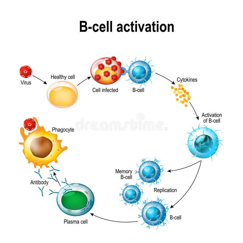
Free with trial Activation of B-cell leukocytes: lymphoblast, activation, memory B-leukocyte, virus, plasma cell, antibody, antigen, and naive lymphocyte. Membrane receptor vectors Activation of B-cell leukocytes
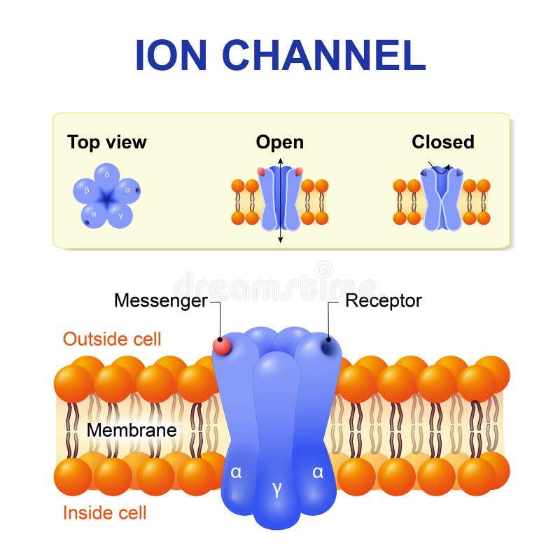
Free with trial Ion channel. structure of the channel. Vector diagram. Membrane receptor vectors Ion channel
Free with trial Molecular model of the protein, rhodopsin, which is involved in vision. Membrane receptor illustrations Membrane protein. Molecular model of the protein, rhodopsin, which is involved in vision
Free with trial Energy is required for the normal functioning of the organs in the body. Many tissues can also use fat or protein as an energy source but others, such as the brain and red blood cells, can only use glucose. Glucose is stored in the body as glycogen. Membrane receptor illustrations Glucose Metabolism. Energy is required for the normal functioning of the organs in the body. Many tissues can also use fat or protein as an energy source but others, such as the brain and red blood cells, can only use glucose. Glucose is stored in the body as glycogen.
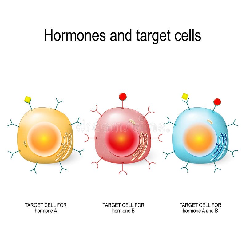
Free with trial Hormones, Receptors and Target Cells. each type of hormone is designed only certain cells. These cells will have receptors on them that are specific for a certain hormone. Vector illustratio. Membrane receptor vectors Hormones, Receptors and Target Cells
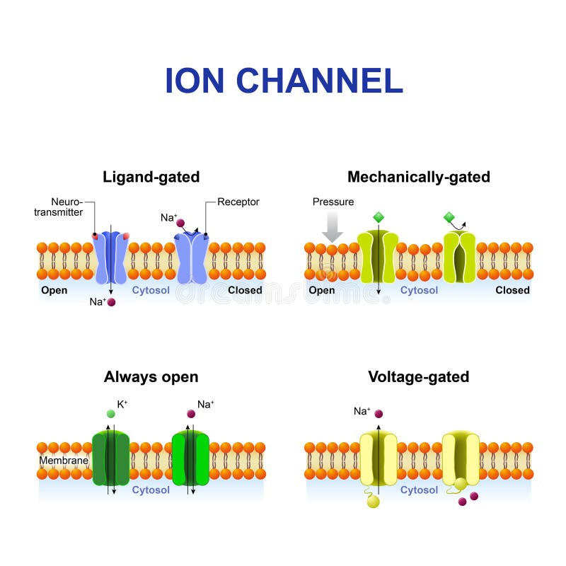
Free with trial Types of ion channel. Classification by gating. mechanism of action. Voltage-Gated, Ligand-gated, Mechanically-gated and Always open ion channels. Membrane receptor vectors Types of ion channel
Free with trial Illustration of a neuron or nerve cell from the central nervous sytem showing the cell body or soma, dendrites and axon which act as conductors and receptors in transmission of signals. Membrane receptor illustrations Neuron or nerve cell
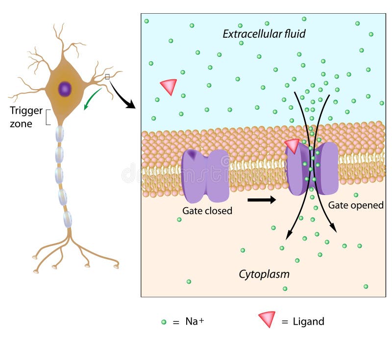
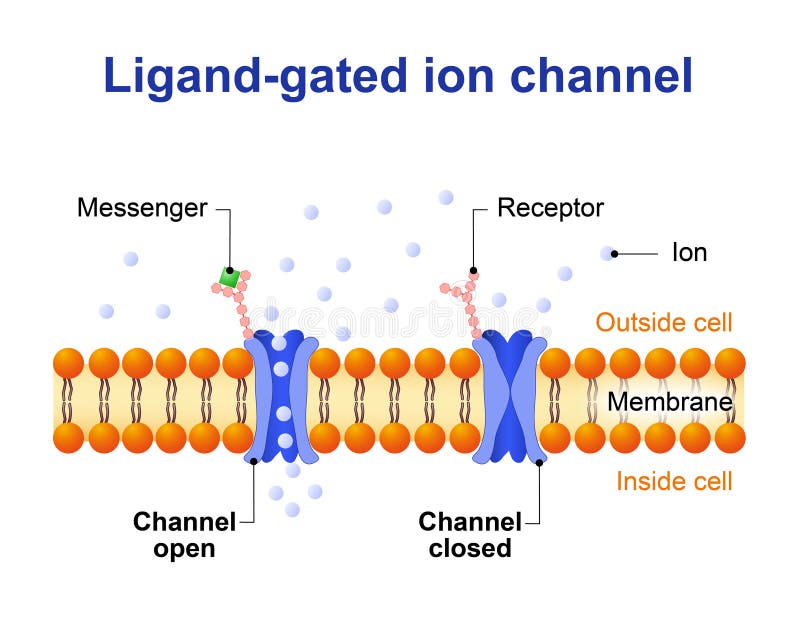
Free with trial Ligand-gated ion channel. channel proteins which open to ions Na, K, Ca, or Cl. Membrane receptor vectors Ligand-gated ion channel
Free with trial Different types of synapses. Neuron to Neuron Transmission. Membrane receptor vectors Different types of synapses
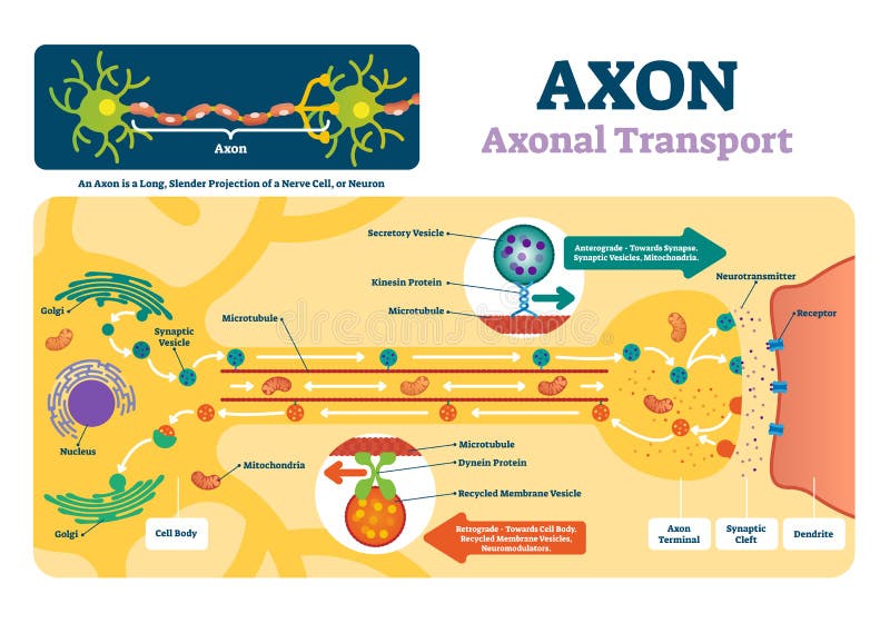
Free with trial Axon vector illustration. Labeled diagram with explanation and structure. Closeup with cell body, terminal, synaptic cleft and dendrite. Nerve cell projection or neuron. Membrane receptor vectors Axon vector illustration. Labeled diagram with explanation and structure.
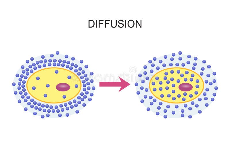
Free with trial Diffusion Across Cell Membranes. Vector illustration design. Membrane receptor vectors Diffusion Across Cell Membranes
Free with trial Eps10, gradient and mesh printing compatible, used transparency. Membrane receptor vectors Motor neuron, detail and accurate,non-labeled vs. Eps10, gradient and mesh printing compatible, used transparency
Free with trial Energy is required for the normal functioning of the organs in the body. Many tissues can also use fat or protein as an energy source but others, such as the brain and red blood cells, can only use glucose. Glucose is stored in the body as glycogen. Membrane receptor illustrations Glucose Metabolism. Energy is required for the normal functioning of the organs in the body. Many tissues can also use fat or protein as an energy source but others, such as the brain and red blood cells, can only use glucose. Glucose is stored in the body as glycogen.
Free with trial Acetylcholine vector illustration. Labeled scheme with structure of neurotransmitter, neuromuscular junction, synaptic vesicle, axon and cleft. Anatomical closeup diagram. Membrane receptor vectors Acetylcholine vector illustration. Labeled scheme with neurotransmitter. Acetylcholine vector illustration. Labeled scheme with structure of neurotransmitter, neuromuscular junction, synaptic vesicle, axon and cleft. Anatomical closeup diagram
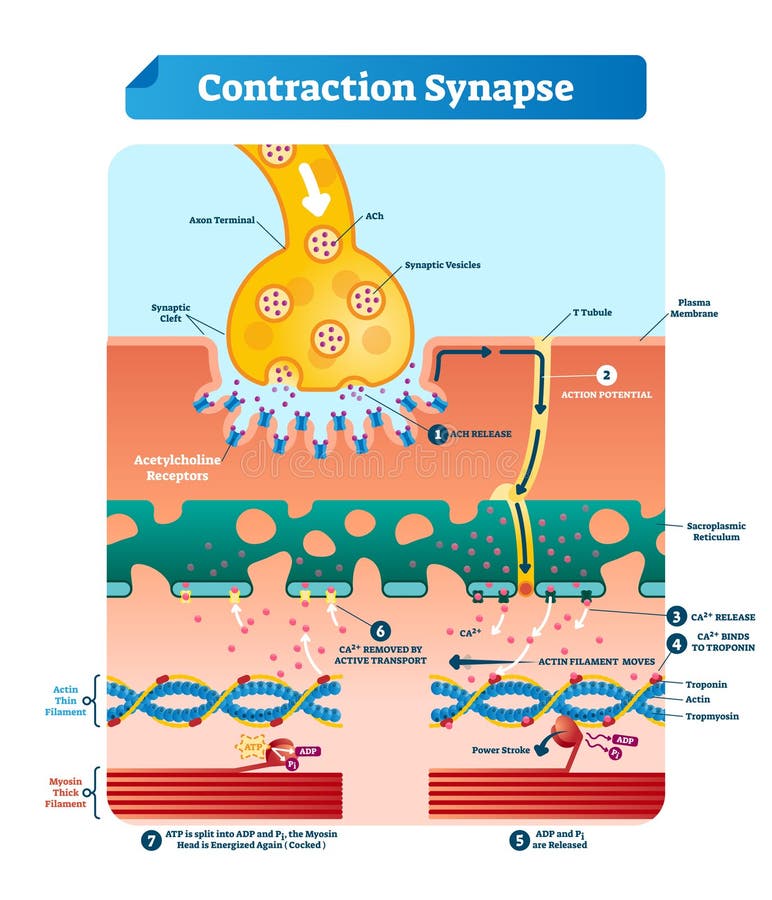
Free with trial Contraction synapse vector illustration. Labeled closeup medical structure scheme. Diagram with full cycle of ACH release, action potential, troponin bonding and filament. Membrane receptor vectors Contraction synapse vector illustration. Labeled medical structure scheme. Contraction synapse vector illustration. Labeled closeup medical structure scheme. Diagram with full cycle of ACH release, action potential, troponin bonding and filament
Free with trial The spike protein red mediates the coronavirus entry into host cells. It binds to the angiotensin converting enzyme 2 blue through its S1 subunit and then fuses viral and host membranes through the S2 subunits. Membrane receptor illustrations The coronavirus spike protein bound to the ACE2 protein on the surface of a human cell. The spike protein red mediates the coronavirus entry into host cells. It binds to the angiotensin converting enzyme 2 blue through its S1 subunit and then fuses viral and host membranes through the S2 subunits
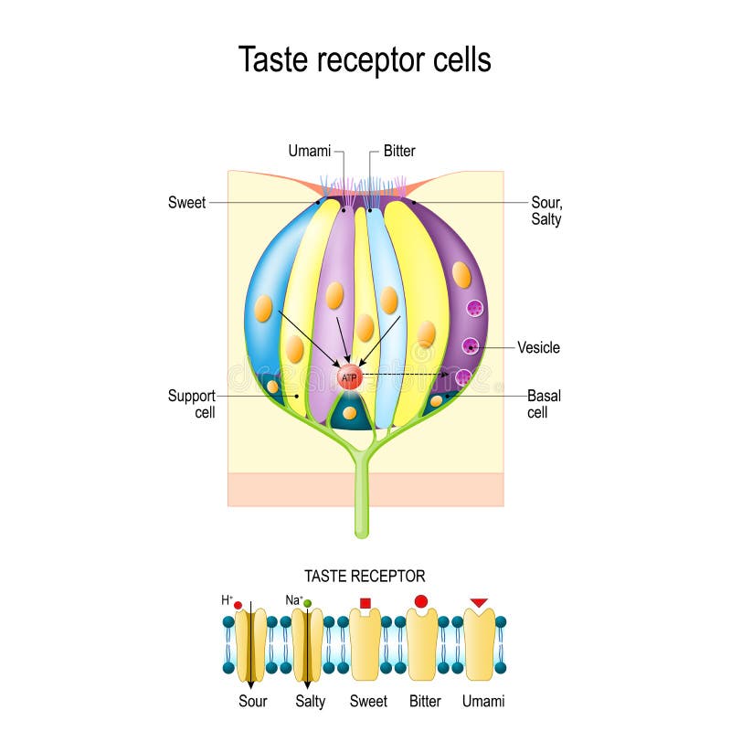
Free with trial Taste bud with receptor cells. Types of Taste receptors. Cell membrane and ion channels for sour, salty, sweet, umami. This diagram above depicts the signal transduction pathway of the different taste. Membrane receptor vectors Taste bud with receptor cells. Types of Taste receptors. Cell membrane and ion channels for sour, salty, sweet, umami
Free with trial Cell membrane. GABA receptor and various sita for ligands bind. Top view of ion channel which illustrates the five combined subunits that form Cl ion channel pore. Membrane receptor vectors Cell membrane. GABA receptor. Top view of ion channel. Cell membrane. GABA receptor and various sita for ligands bind. Top view of ion channel which illustrates the five combined subunits that form Cl ion channel pore
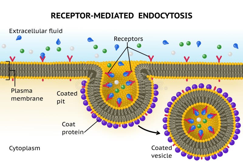
Free with trial Receptor-mediated endocytosis includes membrane proteins called receptors that bind specific molecules ligands. Membrane receptor illustrations Receptor-mediated endocytosis
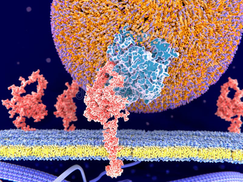
Free with trial 3d computer illustration of a LDL particle binding to its receptor on the surface of a cell. The LDL-receptor is a membrane protein found in almost every human cell. It binds the Apoprotein B100 from LDL-particles and mediates their internalization endocytosis in the cell. This occurs in almost all cells but mainly in the liver where it removes about 70 % of LDL from the circulation. Membrane receptor illustrations LDL particle binding to the LDL-receptor. 3d computer illustration of a LDL particle binding to its receptor on the surface of a cell. The LDL-receptor is a membrane protein found in almost every human cell. It binds the Apoprotein B100 from LDL-particles and mediates their internalization endocytosis in the cell. This occurs in almost all cells but mainly in the liver where it removes about 70 % of LDL from the circulation.
Free with trial 5 membrane proteins with their ligands: left to right Potassium channel, delta-opioid receptor, LDL receptor, acetylcholine receptor, histamine receptor,. Membrane receptor illustrations Structure variety of membrane proteins:. 5 membrane proteins with their ligands: left to right Potassium channel, delta-opioid receptor, LDL receptor, acetylcholine receptor, histamine receptor,
Free with trial Cell with membrane proteins. Ion channels: ligand-gated, voltage-gated, antiporter, symporter, always open, and aquaporin. Neuro transmitter. Vector illustration for medical, educational, and science use. Membrane receptor vectors Cell membrane with ion channels. Cell with membrane proteins. Ion channels: ligand-gated, voltage-gated, antiporter, symporter, always open, and aquaporin. Neuro transmitter. Vector illustration for medical, educational, and science use
Free with trial Glucose transport through cell membrane via transporters activated by insulin affecting its receptors. Membrane receptor vectors Glucose transport through cell membrane via
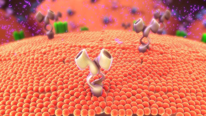
Free with trial Cell-surface receptors, also known as transmembrane receptors, are cell surface, membrane-anchored, or integral proteins that bind to external ligand molecules. This type of receptor spans the plasma membrane and performs signal transduction, converting an extracellular signal into an intracellular signal. Membrane receptor illustrations Cell membrane. Cell-surface receptors, also known as transmembrane receptors, are cell surface, membrane-anchored, or integral proteins that bind to external ligand molecules. This type of receptor spans the plasma membrane and performs signal transduction, converting an extracellular signal into an intracellular signal.
Free with trial Plasma membrane of a human cell. The plasma membrane is a bilayer composed of phopholipids in which lots of transmembrane and surface proteins reside. Membrane receptor illustrations Plasma membrane of a human cell.
Free with trial Immunologically active proteins on a T-cell. TCR blue, CD-4 light blue, CD-28 dark blue, PD-1 magenta, CTLA-4 violet, Ca-channel dark violet. The T-cell receptor, CD-4 and CD-28 activate T-cells, while PD-1 and CTLA-4 inhibit the activation of T-cells. Membrane receptor illustrations T-cell receptor, CD-4, CD-28, PD-1 and CTLA-4 and a calcium chan. Immunologically active proteins on a T-cell. TCR blue, CD-4 light blue, CD-28 dark blue, PD-1 magenta, CTLA-4 violet, Ca-channel dark violet. The T-cell receptor, CD-4 and CD-28 activate T-cells, while PD-1 and CTLA-4 inhibit the activation of T-cells.
Free with trial 3d computer illustration of a chimeric antigen receptor. CARs are engineered cell receptors that allow T cells to recognize and attack cancer cells in a specific way. They are built by connecting several functional parts from different proteins. In this image a signal protein ZAP70 is attached to the intracellular domain. Membrane receptor illustrations Chimeric antigen receptor CAR. 3d computer illustration of a chimeric antigen receptor. CARs are engineered cell receptors that allow T cells to recognize and attack cancer cells in a specific way. They are built by connecting several functional parts from different proteins. In this image a signal protein ZAP70 is attached to the intracellular domain.
Free with trial Glucose transport through cell membrane via transporters activated by insulin. Membrane receptor vectors Glucose transport through cell membrane via
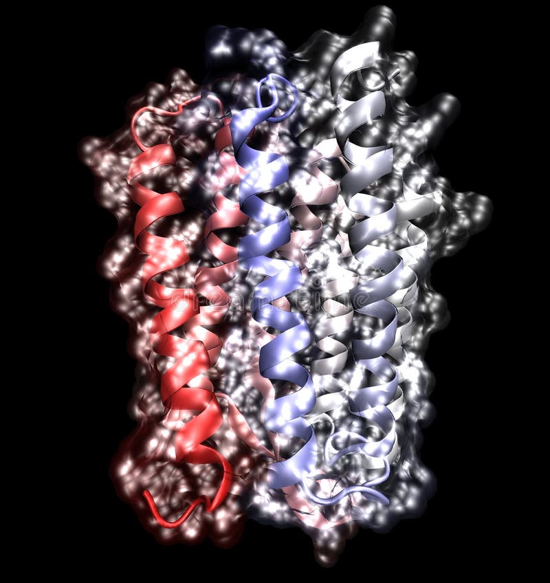
Free with trial Toll-like receptor TLR4 bound to MD2 orange and LPS lipopolysaccharide; grey. Proteins shown as cartoon, space-filling model of LPS. Membrane receptor illustrations TLR4 toll-like receptor, cartoon model. Toll-like receptor TLR4 bound to MD2 orange and LPS lipopolysaccharide; grey. Proteins shown as cartoon, space-filling model of LPS.
Free with trial Lysosome Hydrolytic enzymes, Membrane and transport proteins, science and medical use, Vector Illustration. Membrane receptor vectors Lysosome Hydrolytic enzymes, Membrane and transport proteins
Free with trial Nasal mucosa anatomy. Nasal mucous membrane lining the respiratory tract. Ciliated, basal and goblet cells. Flat vector illustration. Membrane receptor vectors Nasal mucosa anatomy. Nasal mucous membrane lining the respiratory tract. Ciliated, basal and goblet cells. Flat vector illustration
Free with trial Molecular model of the protein, rhodopsin, which is involved in vision. A faulty version of this protein can lead to blindness. Membrane receptor illustrations Membrane protein on white. Molecular model of the protein, rhodopsin, which is involved in vision. A faulty version of this protein can lead to blindness
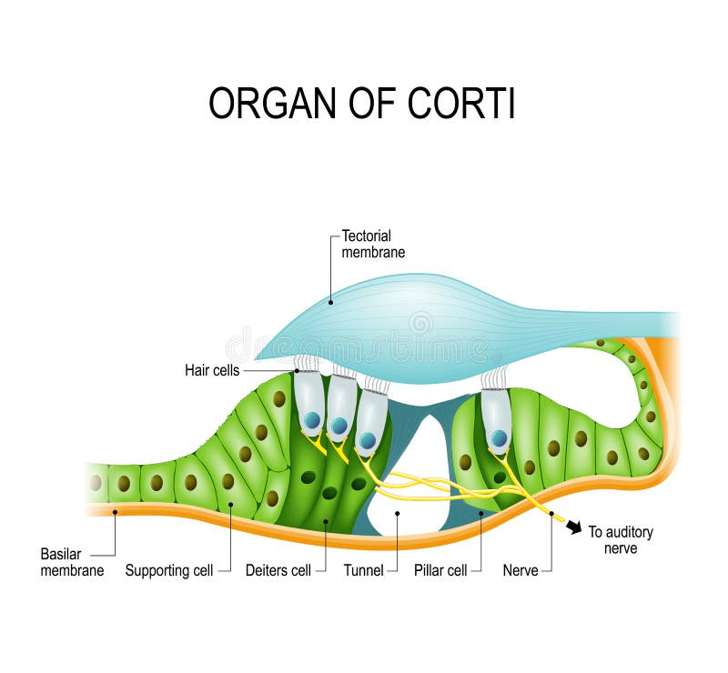
Free with trial The organ of Corti spiral organ in a cross-section. Organ of Corti is the receptor organ for hearing, a neuron located in the cochlea of the inner ear. Hair Cell. Membrane receptor vectors The organ of Corti in a cross-section. The organ of Corti spiral organ in a cross-section. Organ of Corti is the receptor organ for hearing, a neuron located in the cochlea of the inner ear. Hair Cell
Free with trial CARs are engineered cell receptors that allow T cells to recognize and attack cancer cells in a specific way. They are built by connecting several functional parts from different proteins. In this image a signal protein ZAP70 is attached to the intracellular domain. Membrane receptor illustrations Chimeric antigen receptor CAR. CARs are engineered cell receptors that allow T cells to recognize and attack cancer cells in a specific way. They are built by connecting several functional parts from different proteins. In this image a signal protein ZAP70 is attached to the intracellular domain.
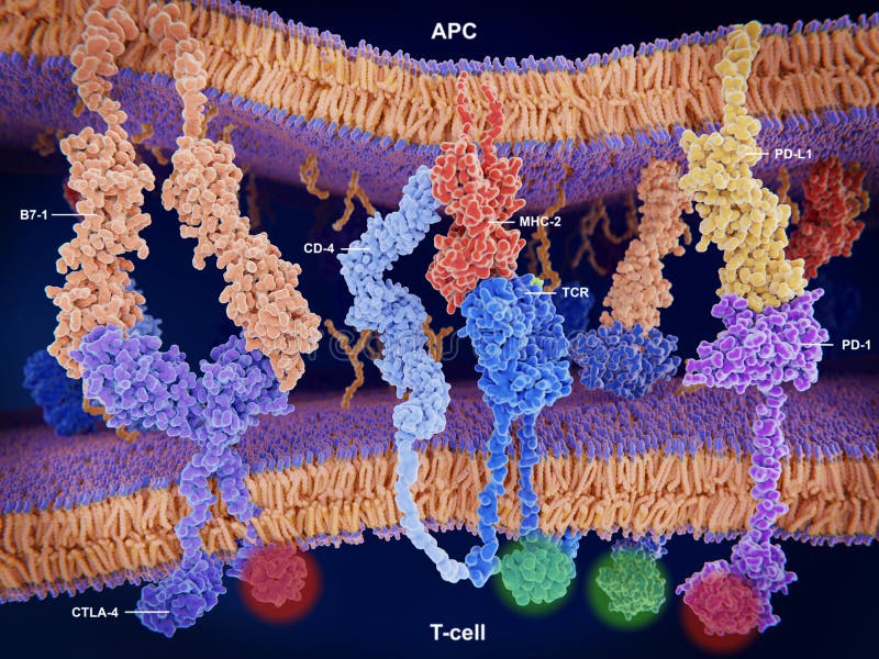
Free with trial Interactions of MHC-II with the T-cell receptor and CD4 and B7-1 with CD-28 activates T-cells while the interactions of P7-1 with CTLA-4 and PD-L1 with PD-1 deactivates T-cells. Membrane receptor illustrations Activation and inhibition of the immune response on T-cells. Interactions of MHC-II with the T-cell receptor and CD4 and B7-1 with CD-28 activates T-cells while the interactions of P7-1 with CTLA-4 and PD-L1 with PD-1 deactivates T-cells.
Free with trial T-cell receptors activate in T-lymphocytes the immune response to antigens presented by antigen presenting cells. Source: PDB entry 6jxr. Membrane receptor illustrations Structure of a T-cell receptor bound to a cell membrane. T-cell receptors activate in T-lymphocytes the immune response to antigens presented by antigen presenting cells. Source: PDB entry 6jxr
Free with trial Endocytosis and exocytosis are types of vesicle transport that carry very large molecules across the cell membrane. Vector illustration. Membrane receptor vectors Endocytosis and Exocytosis Diagram. Endocytosis and exocytosis are types of vesicle transport that carry very large molecules across the cell membrane. Vector illustration
Free with trial G protein coupled receptors gated ion channel. Structure of a G protein-coupled receptor (GPCR). Mechanism for the transport of ions. Cell membrane receptors for ligands bind. vector illustration. Membrane receptor vectors G protein coupled receptors gated ion channel. Vector illustration. G protein coupled receptors gated ion channel. Structure of a G protein-coupled receptor (GPCR). Mechanism for the transport of ions. Cell membrane receptors for ligands bind. vector illustration
Free with trial Endocytosis process with closeup cell side view in anatomical outline diagram. Phagocytosis, pinocytosis or receptor mediated stages explanation vector illustration. Labeled intracellular invagination. Membrane receptor vectors Endocytosis process with closeup cell side view in anatomical outline diagram
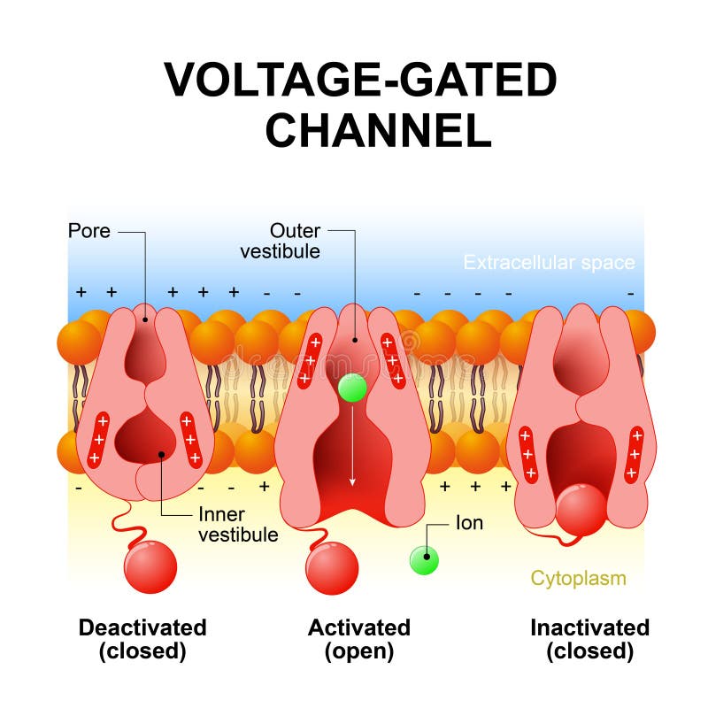
Free with trial Voltage-gated channels. inactivation gate, deactivation and activation ion channel. Open and close gate. Interior of the cell is negatively charged and the exterior is positively charged and vice versa. Membrane receptor vectors Voltage-gated channels
Free with trial Active transport is usually associated with accumulating high concentrations of molecules that the cell needs, such as ions, glucose and amino acids. ... Examples of active transport include the uptake of glucose in the intestines in humans and the uptake of mineral ions into root hair cells of plants. Membrane receptor illustrations Active Transport human cell. Active transport is usually associated with accumulating high concentrations of molecules that the cell needs, such as ions, glucose and amino acids. ... Examples of active transport include the uptake of glucose in the intestines in humans and the uptake of mineral ions into root hair cells of plants.
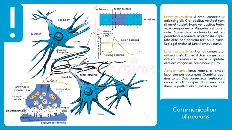
Free with trial Communication of neurons. Infographic diagram. Membrane receptor vectors Communication of neurons.
Free with trial Endocytosis. The cell transports proteins into the cell. Membrane receptor vectors Endocytosis.