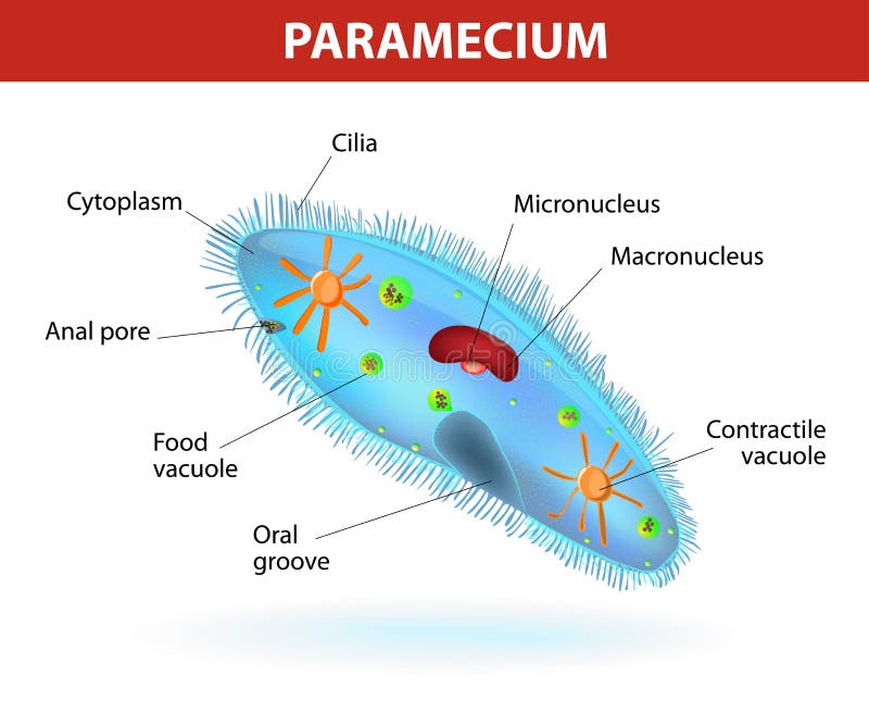
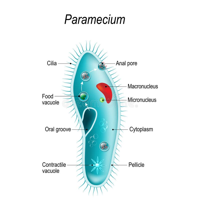
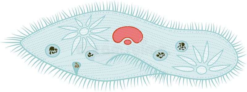
Free with trial Anatomy of a paramecium. Vector diagram. Ciliate protozoan that lives in stagnant freshwater. Paramecium covered with cilia, which allow it to move about and to feed on bacteria. Micro cilia vectors Structure of a paramecium. Anatomy of a paramecium. Vector diagram. Ciliate protozoan that lives in stagnant freshwater. Paramecium covered with cilia, which allow it to move about and to feed on bacteria.
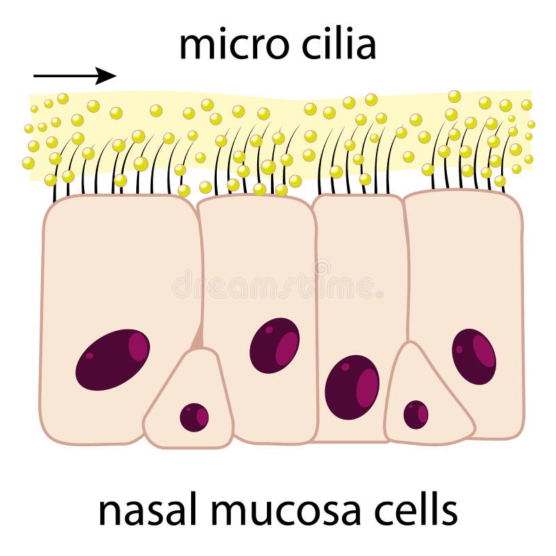
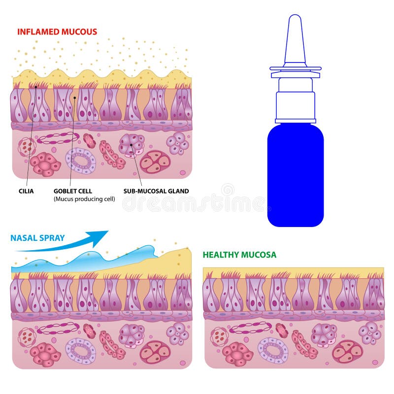
Free with trial Inflamed and normal nasal mucosa cells and micro cilia vector scheme with nasal spray effect and bottle. Micro cilia vectors Nasal mucosa cells and micro cilia vector scheme
Free with trial Eyebrow perfectly shaped. permanent make-up and tattooing. Cosmetic for eyebrows. Beauty salon. Eyelash extension. A beautiful make-up. Thick fuzzy cilia. Mascara for volume and length. Micro cilia illustrations Eyebrow perfectly shaped. permanent make-up and tattooing. Cosmetic for eyebrows. Eyelash extension. A beautiful make-up. Thick fu. eyebrow perfectly shaped. permanent make-up and tattooing. Cosmetic for eyebrows. Beauty salon. Eyelash extension. A beautiful make-up. Thick fuzzy cilia. Mascara for volume and length.
Free with trial Paramecium is a genus of unicellular Ciliate protozoa, commonly studied as a representative of the Ciliate group. Paramecia are widespread in freshwater, brackish and marine environments, and are often very abundant in stagnant basins and ponds. Micro cilia illustrations Paramecium
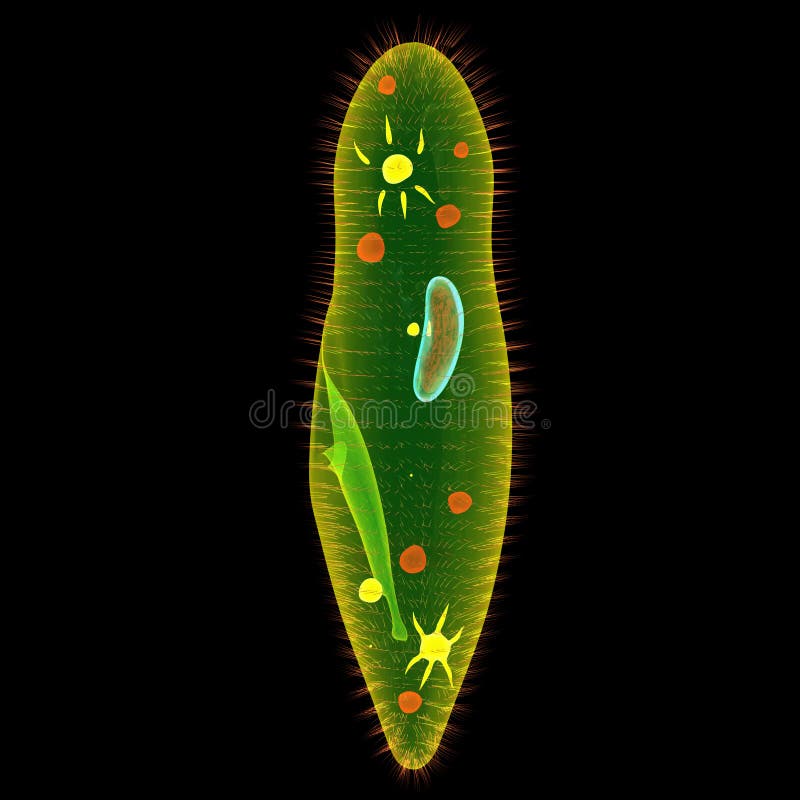
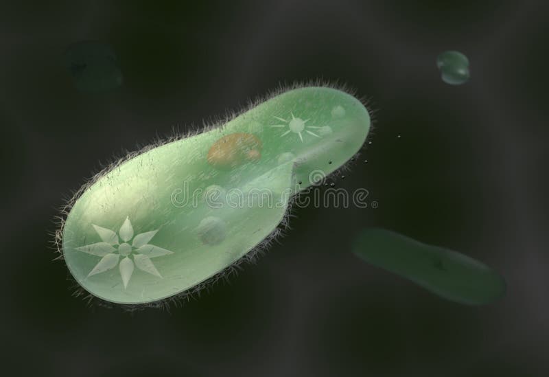
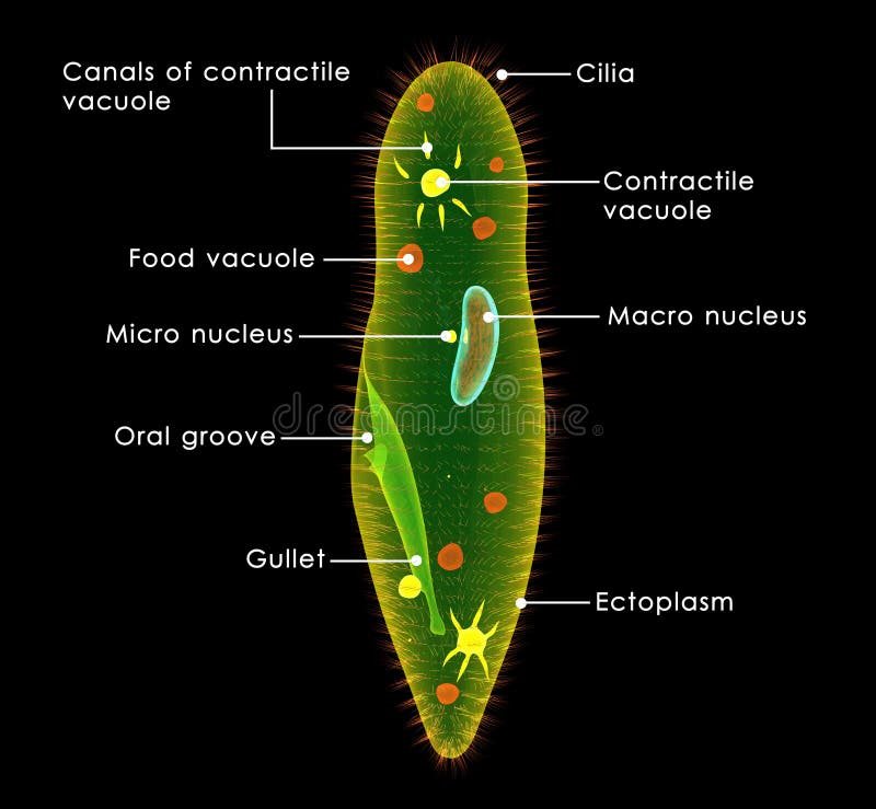
Free with trial Structure of a paramecium caudatum on green backround. Micro cilia illustrations Infusoria paramecium. Structure of a paramecium caudatum on green backround
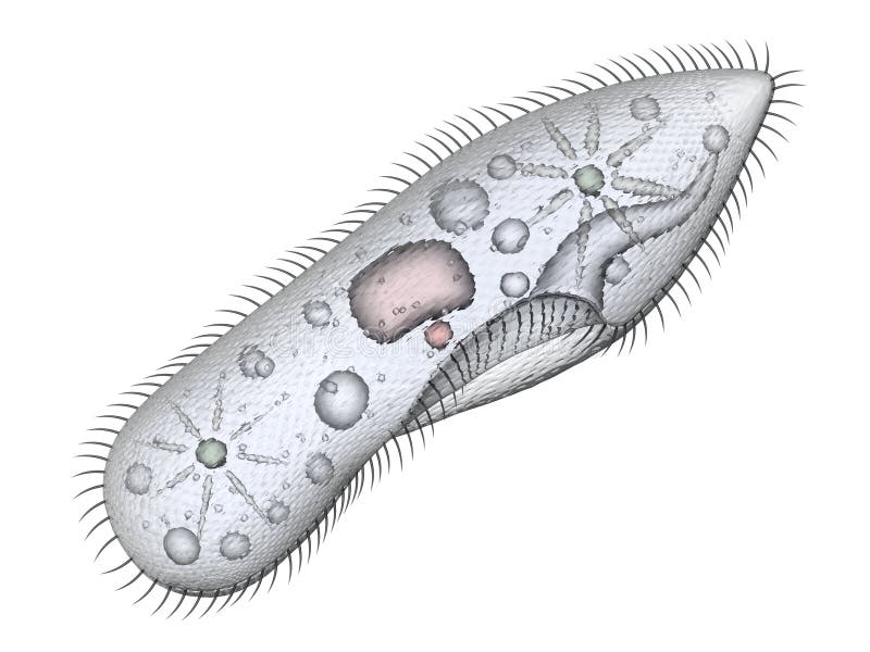
Free with trial Structure of a paramecium caudatum isolated on white. Micro cilia illustrations Infusoria paramecium. Structure of a paramecium caudatum isolated on white
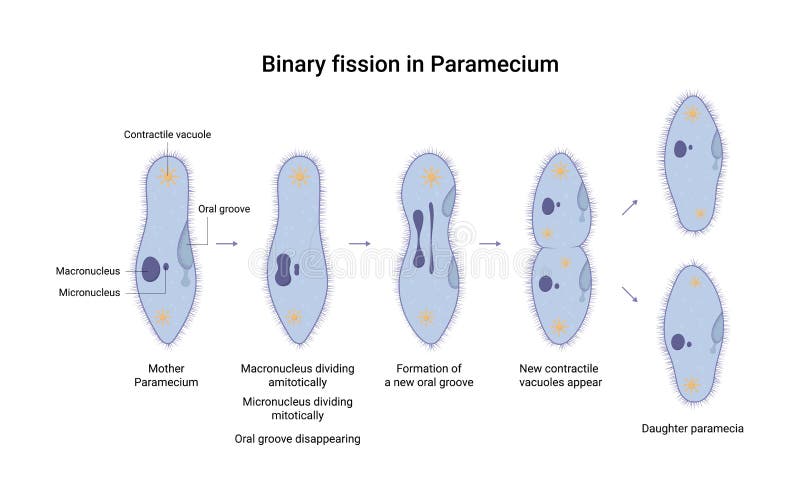
Free with trial Asexual reproduction in Protozoa. Paramecia division. Micro cilia vectors Asexual reproduction in Protozoa. Paramecia division
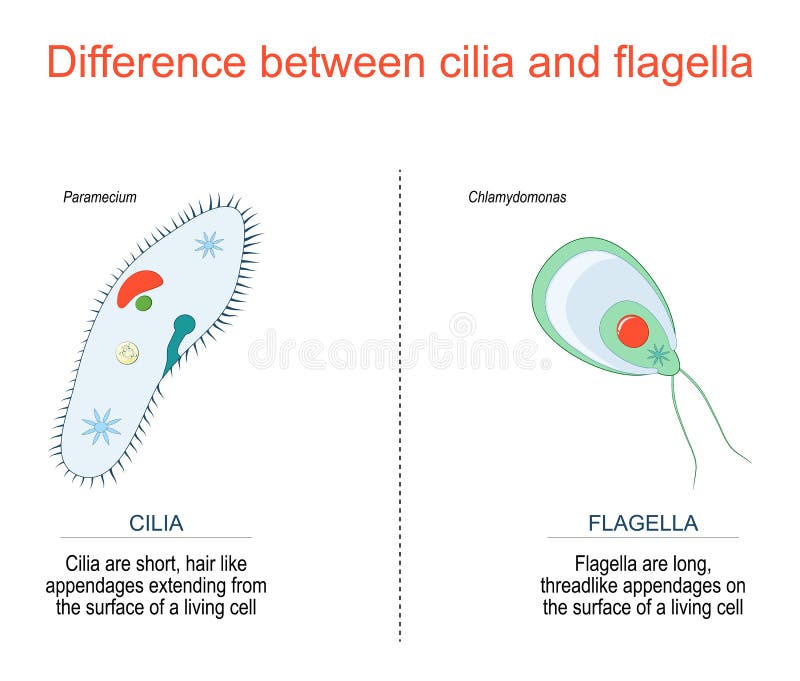
Free with trial Difference between cilia and flagella for example Paramecium and Chlamydomonas. Micro cilia vectors Cilia and flagella. Paramecium and Chlamydomonas. Difference between cilia and flagella for example Paramecium and Chlamydomonas
Free with trial Cilia and flagella biological structure difference comparison outline diagram. Labeled educational microorganisms closeup view with basal body and bacteria motion types explanation vector illustration. Micro cilia vectors Cilia and flagella biological structure difference comparison outline diagram
Free with trial Aspergillosis lung infection caused by Aspergillus, vector outline diagram. Irritated airway, excess mucus and damaged cilia caused by common mold fungus spores. Microbiological danger for human health. Micro cilia vectors Aspergillosis lung infection caused by Aspergillus, vector outline diagram
Free with trial Structure of a paramecium caudatum isolated on white. Micro cilia illustrations Paramecium
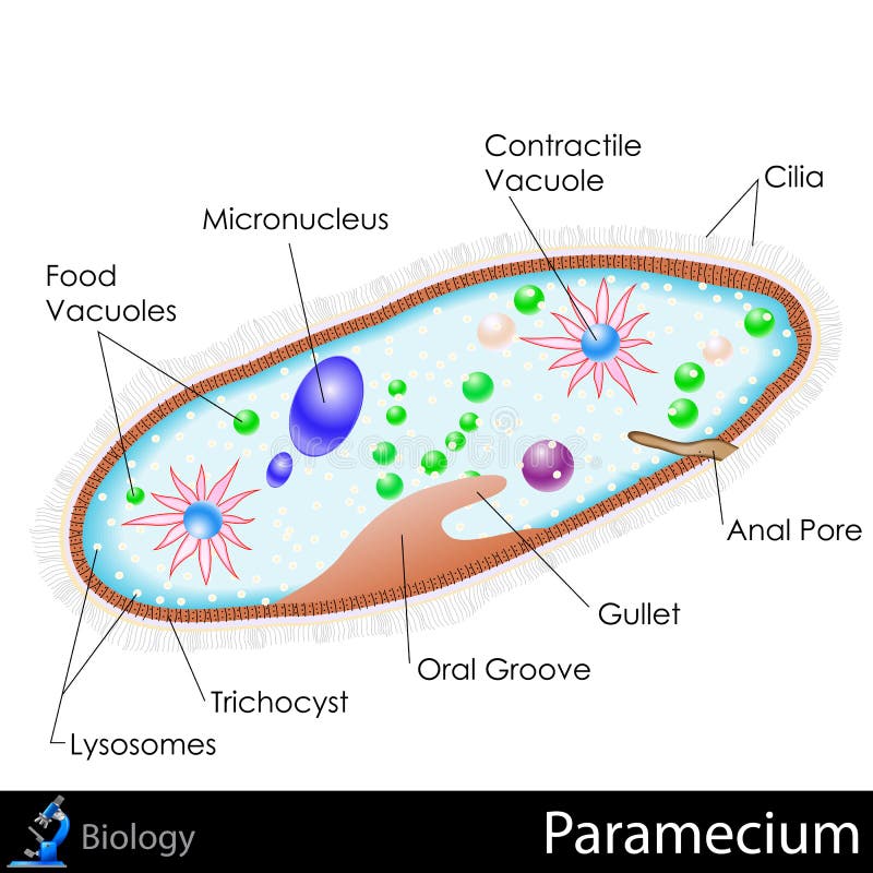
Free with trial Easy to edit vector illustration of diagram of paramecium. Micro cilia vectors Paramecium Diagram. Easy to edit vector illustration of diagram of paramecium
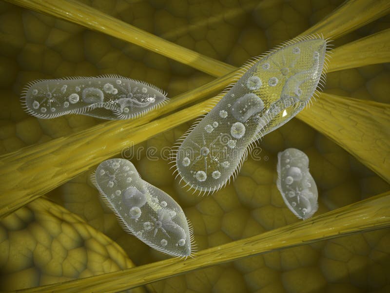
Free with trial Structure of a paramecium caudatum on yellow backround. Micro cilia illustrations Paramecium
Free with trial Structure of a paramecium caudatum. Vector illustration for educational and science use. Micro cilia vectors Structure of a paramecium caudatum
Free with trial Bacteria Staphylococcus in nose. 3D illustration shows spherical bacteria Staphylococcus aureus adhering to hair-like cilia on nasal epithelial cells. Micro cilia illustrations Bacteria Staphylococcus in nose
Free with trial Biological micro organism paramecium caudatum. 3d image. Micro cilia illustrations Biological micro organism paramecium caudatum
Free with trial Model biological micro organism paramecium caudatum 3d render. Micro cilia illustrations Model biological micro organism paramecium caudatum 3d illustration. Model biological micro organism paramecium caudatum 3d render
Free with trial Model biological micro organism paramecium caudatum 3d render. Micro cilia illustrations Model biological micro organism paramecium caudatum 3d illustration. Model biological micro organism paramecium caudatum 3d render
Free with trial 3d model biological micro organism paramecium caudatum. Micro cilia illustrations Paramecium caudatum
Free with trial Eyebrow perfectly shaped. permanent make-up and tattooing. Cosmetic for eyebrows. Beauty salon. Eyelash extension. A beautiful make-up. Thick fuzzy cilia. Mascara for volume and length. Micro cilia illustrations Eyebrow perfectly shaped. permanent make-up and tattooing. Cosmetic for eyebrows. Eyelash extension. A beautiful make-up. Thick. eyebrow perfectly shaped. permanent make-up and tattooing. Cosmetic for eyebrows. Beauty salon. Eyelash extension. A beautiful make-up. Thick fuzzy cilia. Mascara for volume and length
Free with trial Anatomy of Paramecium caudatum. Vector diagram for educational, science, and biological use. Micro cilia vectors Anatomy of Paramecium
Free with trial Infusoria paramecium protozoa and amoeba. Micro cilia illustrations Infusoria paramecium protozoa
Free with trial Paramecium is a genus of unicellular ciliated protozoa, commonly studied as a representative of the ciliate group. Paramecia are widespread in freshwater, brackish and marine environments, and are often very abundant in stagnant basins and ponds. Micro cilia illustrations Paramecium
Free with trial Vorticella is a genus of protozoa, with over 16 known species. They are stalked, inverted bell-shaped ciliates, placed among the peritrichs. Micro cilia illustrations Vorticella
Free with trial This illustration is the structure and diagram of paramecium. use to study for student and interested. Micro cilia vectors Structure of Paramecium. This illustration is the structure and diagram of paramecium. use to study for student and interested.
Free with trial Vector illustration of binary fission of Paramecium. Educational illustration. Micro cilia vectors Vector illustration of binary fission of Paramecium. Educational illustration
Free with trial Cross-section diagram of a Paramecium caudatum, showing its internal structure. Digital illustration. Micro cilia illustrations Paramecium internal structure. Cross-section diagram of a Paramecium caudatum, showing its internal structure. Digital illustration
Free with trial Infusoria paramecium protozoa and amoeba. Micro cilia illustrations Infusoria paramecium protozoa
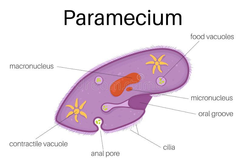
Free with trial Anatomy of a paramecium. Vector illustration. Micro cilia vectors Anatomy of a paramecium. Vector
Free with trial Infusoria paramecium protozoa set isolated on white. Micro cilia illustrations Infusoria paramecium protozoa set
Free with trial This illustration is the structure and diagram of paramecium. use to study for student and interested. Micro cilia vectors The structure of paramecium. This illustration is the structure and diagram of paramecium. use to study for student and interested.
Free with trial Paramecium microscopic closeup structure with anatomical outline diagram. Educational labeled scheme with bacteria inner parts as zoology study vector illustration. Biology or microscope basic example. Micro cilia vectors Paramecium microscopic closeup structure with anatomical outline diagram
Free with trial Structure Infusorian of the shoeshoe type or Paramecium caudatum. Paramecium caudatum is a species of unicellular protist in the phylum Ciliophora. Micro cilia vectors Structure Infusorian of the shoeshoe type or Paramecium caudatum
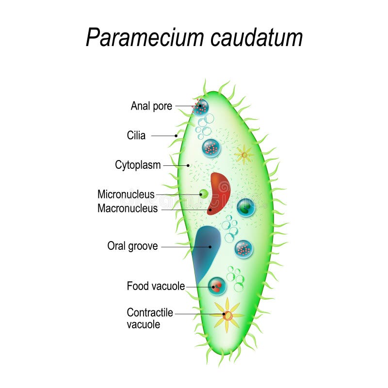
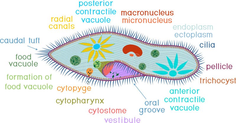
Free with trial Structure of Paramecium caudatum on white background with titles. Micro cilia vectors Structure of Paramecium caudatum with titles. Structure of Paramecium caudatum on white background with titles
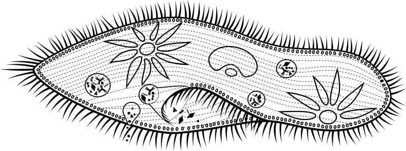
Free with trial Coloring page with structure of Paramecium caudatum with titles. Micro cilia vectors Coloring page. Structure of Paramecium caudatum with titles
Free with trial Mucociliary Transport System black and white simple vector icon, medical illustration. Micro cilia vectors Mucociliary Transport System icon, vector medical illustration. Mucociliary Transport System black and white simple vector icon, medical illustration
Free with trial Infusoria structure hand-drawn image. Paramecium caudatum. Editable vector illustration isolated on dark grey blackboard background. Biological concept in unique style. Micro cilia vectors Infusoria Hand drawn Image. Infusoria structure hand-drawn image. Paramecium caudatum. Editable vector illustration isolated on dark grey blackboard background. Biological concept in unique style.
Free with trial Coloring page. Structure of Paramecium caudatum on white background. Micro cilia vectors Coloring page. Structure of Paramecium caudatum
Free with trial Structure of Paramecium caudatum isolated on white background. Micro cilia vectors Structure of Paramecium caudatum
Free with trial Structure of Paramecium caudatum on white background with titles. Micro cilia vectors Structure of Paramecium caudatum with titles. Structure of Paramecium caudatum on white background with titles
Free with trial Mucociliary Transport System color simple vector icon, medical illustration. Micro cilia vectors Mucociliary Transport System icon, vector medical illustration. Mucociliary Transport System color simple vector icon, medical illustration
Free with trial Bacteria Staphylococcus in nose. 3D illustration shows spherical bacteria Staphylococcus aureus adhering to hair-like cilia on nasal epithelial cells. Micro cilia illustrations Bacteria Staphylococcus in nose
Free with trial Bacteria Staphylococcus in nose. 3D illustration shows spherical bacteria Staphylococcus aureus adhering to hair-like cilia on nasal epithelial cells. Micro cilia illustrations Bacteria Staphylococcus in nose
Free with trial This dramatic biomedical illustration showcases a high-resolution view of rod-shaped organisms interacting with cellular structures. These luminous entities are surrounded by fine, hair-like projections possibly representing cilia or microvilli of an epithelial surface. Rendered by Ai, the deep monochromatic blue tones and sharp focus emphasize the tension and detail of this microscopic. Micro cilia illustrations Microscopic bacteria trapped in blue cell cilia. This dramatic biomedical illustration showcases a high-resolution view of rod-shaped organisms interacting with cellular structures. These luminous entities are surrounded by fine, hair-like projections possibly representing cilia or microvilli of an epithelial surface. Rendered by Ai, the deep monochromatic blue tones and sharp focus emphasize the tension and detail of this microscopic
Free with trial Detailed microscopic view of cilia and pathogens on mucosal tissue, ideal for medical education. Features blue substrate with pink outgrowths and pathogens. Perfect for illustrating cellular biology. Micro cilia illustrations Detailed Microscopic Visualization of Cilia Cells and Pathogens on Mucosal Tissue Surface with Blue Substrate and Pink Outgrowths. Detailed microscopic view of cilia and pathogens on mucosal tissue, ideal for medical education. Features blue substrate with pink outgrowths and pathogens. Perfect for illustrating cellular biology
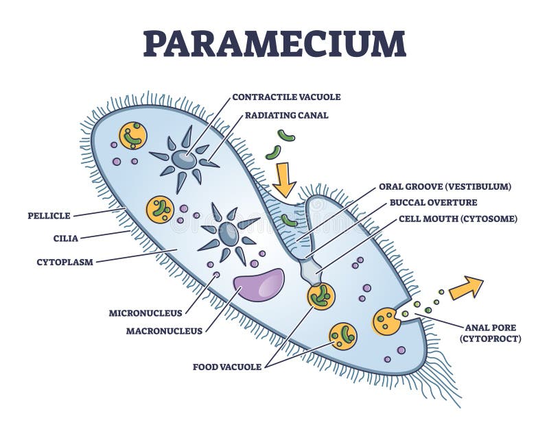
Free with trial Paramecium structure anatomy diagram poster chart, medical illustration vector. Science biology study. Labeled cilia, vacuole, radiation canal, macronucleus, vestibulum, cytoplasm, pellicle. Micro cilia vectors Paramecium structure anatomy diagram poster chart, medical illustration vector. Science biology study. Labeled cilia, vacuole
Free with trial Close-up macro shot of the edge of a poppy leaf, showcasing the fine details of its cilia. Micro cilia illustrations Macro of Poppy Leaf with Cilia. Close-up macro shot of the edge of a poppy leaf, showcasing the fine details of its cilia.
Free with trial A high-resolution microscopic image of a Paramecium, an oval-shaped single-celled ciliate protozoan. The organism is covered in cilia used for movement and feeding. Internal structures such as vacuoles, macronucleus, and micronucleus are visible, highlighting its cellular complexity. Micro cilia illustrations Microscopic view of Paramecium showing cilia, vacuoles, and internal structures in high-resolution detail. A high-resolution microscopic image of a Paramecium, an oval-shaped single-celled ciliate protozoan. The organism is covered in cilia used for movement and feeding. Internal structures such as vacuoles, macronucleus, and micronucleus are visible, highlighting its cellular complexity.
Free with trial A vibrant, close-up microscopic image of a paramecium, highlighting its detailed cellular form and the fine cilia used for movement and feeding. The background is a soft, abstract blur, emphasizing the organism. Generative AI. Micro cilia illustrations Microscopic view of a paramecium a singlecelled ciliate protozoan showcasing its intricate cellular structure and cilia. A vibrant, close-up microscopic image of a paramecium, highlighting its detailed cellular form and the fine cilia used for movement and feeding. The background is a soft, abstract blur, emphasizing the organism. Generative AI
Free with trial A fascinating glimpse into the world of ciliates with an upclose view of their complex and beautiful patterns of cilia and cytoplasmic. Micro cilia illustrations A fascinating glimpse into the world of ciliates with an upclose view of their complex and beautiful patterns of cilia
Free with trial A of Rotifers swimming in a drop of water their translucent bodies and beating cilia creating mesmerizing patterns under the microscope. Micro cilia illustrations A of Rotifers swimming in a drop of water their translucent bodies and beating cilia creating mesmerizing patterns under
Free with trial A digital illustration of microscopic life forms, showcasing various cellular structures. The image includes a variety of organelles and cells, depicted in blue tones with a glowing effect. The scene features circular and elongated shapes, some with spiky or hairy textures resembling cilia or flagella, suggesting motion and life. The background color shifts subtly, adding depth to the representation of a watery environment, emphasizing the complexity and diversity of these micro-organisms. Micro cilia illustrations Cell, organelles, tissues, the birth of new life. A digital illustration of microscopic life forms, showcasing various cellular structures. The image includes a variety of organelles and cells, depicted in blue tones with a glowing effect. The scene features circular and elongated shapes, some with spiky or hairy textures resembling cilia or flagella, suggesting motion and life. The background color shifts subtly, adding depth to the representation of a watery environment, emphasizing the complexity and diversity of these micro-organisms.
Free with trial This captivating image showcases a single-celled paramecium, a fascinating microorganism teeming with biological activity. Captured using brightfield microscopy with minimal light, the image highlights the intricate details of this protozoan. Notice the characteristic slipper shape and the intricate cilia, tiny hair-like structures that propel the paramecium through its aquatic environment. Micro cilia illustrations Unveiling the Microscopic World A Detailed Look at a Paramecium Under LowLight Brightfield Microscopy. This captivating image showcases a single-celled paramecium, a fascinating microorganism teeming with biological activity. Captured using brightfield microscopy with minimal light, the image highlights the intricate details of this protozoan. Notice the characteristic slipper shape and the intricate cilia, tiny hair-like structures that propel the paramecium through its aquatic environment.
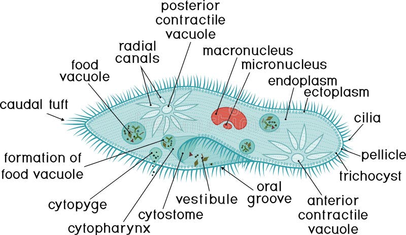
Free with trial Paramecium, nucleus, contractile vacuole, other organelles. Anatomy of Paramecium caudatum with titles. Vector illustration. Labeled diagram of Eukaryotic cell. unicellular animals. Science Biology. Micro cilia vectors Anatomy of Paramecium caudatum with titles
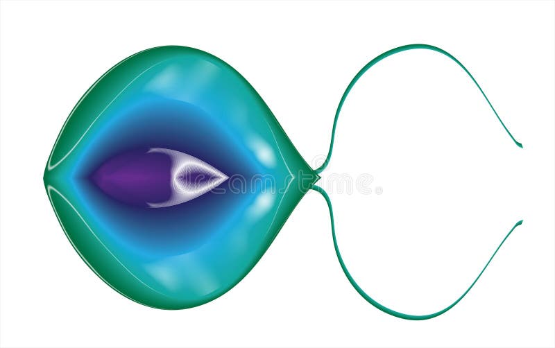
Free with trial This stunning scientific illustration depicts a transparent, protective cyst housing internal fibrous structures, likely representing the cytoskeleton or flagella of a dormant microorganism. Rendered by Ai, the image uses vibrant green illumination against a deep teal background to highlight the intricate biological detail of the enclosed form. This visualization is perfect for educational. Micro cilia illustrations Vivid 3D rendering of an encapsulated protist. This stunning scientific illustration depicts a transparent, protective cyst housing internal fibrous structures, likely representing the cytoskeleton or flagella of a dormant microorganism. Rendered by Ai, the image uses vibrant green illumination against a deep teal background to highlight the intricate biological detail of the enclosed form. This visualization is perfect for educational
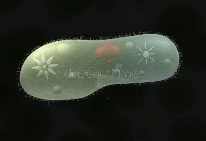
Free with trial Paramecium caudatum. Close-up of Unicellular organism on dark background. Protozoa anatomy. Realistic vector illustration. Micro cilia vectors Paramecium caudatum. Anatomy of Unicellular. Paramecium caudatum. Close-up of Unicellular organism on dark background. Protozoa anatomy. Realistic vector illustration
Free with trial A fascinating contrast between the delicacy of a two-tone corolla and the silent threat of the jaw-like traps at its base. Emerging from a carpet of damp moss, this carnivorous plant combines floral elegance with a formidable hunting strategy. Micro cilia illustrations Venus flytrap flower : Carnivorous Beauty. A fascinating contrast between the delicacy of a two-tone corolla and the silent threat of the jaw-like traps at its base. Emerging from a carpet of damp moss, this carnivorous plant combines floral elegance with a formidable hunting strategy.
Free with trial A macro 3D visualization of a biological process, showing numerous golden, liquid-filled receptors or cilia growing from a textured cellular surface. Concept: Biological processes. Micro cilia vectors Abstract Field of Golden Receptors on a Cellular Surface. A macro 3D visualization of a biological process, showing numerous golden, liquid-filled receptors or cilia growing from a textured cellular surface. Concept: Biological processes
Free with trial Different types of bacteria flagella types. Flagellar Arrangement in Bacteria. Various forms of flagellation with corresponding designations. Virus microbiology illustration vector. Micro cilia vectors Different types of bacteria flagella types. Flagellar Arrangement in Bacteria. Various forms of flagellation with corresponding
Free with trial Many bacterias under the microscope. Micro cilia illustrations Bacterias under the microscope
Free with trial Abstract oval shape tiny protist amoeba organelle pellicle parasite element. Line black hand drawn lab microbe icon sign symbol pictogram diagram sketch Art doodle cartoon style design. Closeup view. Micro cilia illustrations Paramecium caudatum. Vector drawing icon. Abstract oval shape tiny protist amoeba organelle pellicle parasite element. Line black hand drawn lab microbe icon sign symbol pictogram diagram sketch Art doodle cartoon style design. Closeup view
Free with trial Abstract oval shape tiny protist amoeba organelle pellicle parasite element. Line black hand drawn lab microbe icon sign symbol pictogram diagram sketch Art doodle cartoon style design. Closeup view. Micro cilia vectors Paramecium caudatum. Vector drawing icon. Abstract oval shape tiny protist amoeba organelle pellicle parasite element. Line black hand drawn lab microbe icon sign symbol pictogram diagram sketch Art doodle cartoon style design. Closeup view
Free with trial A detailed 3D rendering of a spiral-shaped bacterium, representative of spirochetes like Borrelia burgdorferi or Treponema pallidum, showing fine surface structures against a black background. Micro cilia illustrations Microscopic view of a spiral bacterium with flagella. A detailed 3D rendering of a spiral-shaped bacterium, representative of spirochetes like Borrelia. A detailed 3D rendering of a spiral-shaped bacterium, representative of spirochetes like Borrelia burgdorferi or Treponema pallidum, showing fine surface structures against a black background
Free with trial A bunch of bubbles with a few white pills in the middle. The bubbles are all different sizes and are scattered throughout the image. Micro cilia illustrations A bunch of bubbles with a few white pills in the middle
Free with trial Microscopic paramecium glides water other microorganisms. Underwater aquatic ballet, natural life spectacle. Microbe world, vibrant details, focus cells structure scientific study. Micro cilia illustrations Microscopic paramecium glides water other microorganisms. Underwater aquatic ballet, natural life spectacle. Microbe world
Free with trial Close up 3D art render of probiotics bacteria biology. Microscopic medicine research, medicals. Scientific bacterial epidemic background with microorganism, virus, cells elements. Micro cilia illustrations Close up 3D art render of probiotics bacteria biology. Microscopic medicine research, medicals. Scientific bacterial epidemic
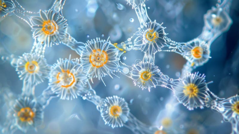
Free with trial Explore the intricate world of marine ciliates, fascinating single-celled organisms that play a vital role in aquatic ecosystems. This captivating image, captured using light microscopy at a 400x magnification, reveals the remarkable detail of a ciliate in its natural environment. Ciliates are protists, a diverse group of eukaryotic microorganisms. Their defining characteristic is the presence. Micro cilia illustrations Unveiling the Microscopic Wonders of Marine Ciliates A Detailed Look at a Singlecelled Plankton Protist Under 400x. Explore the intricate world of marine ciliates, fascinating single-celled organisms that play a vital role in aquatic ecosystems. This captivating image, captured using light microscopy at a 400x magnification, reveals the remarkable detail of a ciliate in its natural environment. Ciliates are protists, a diverse group of eukaryotic microorganisms. Their defining characteristic is the presence
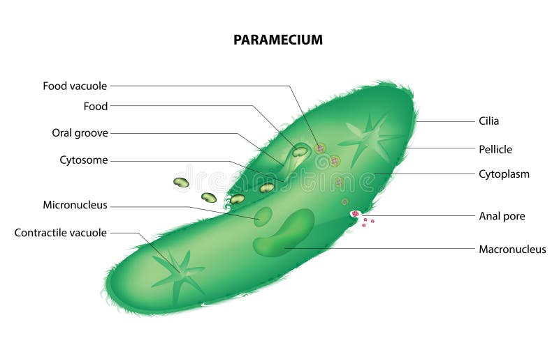
Free with trial Paramecium is a genus of single-celled, ciliate protists belonging to the phylum Ciliophora. These organisms are commonly found in freshwater environments, such as ponds, lakes, and slow-moving streams, where they feed on bacteria, algae, and other small microorganisms. Paramecium has a distinctive shape and is well-known for its ciliated surface, which it uses for movement and feeding. Micro cilia vectors Paramecium
Free with trial Flagella are long, whip-like structures used by various organisms for movement. They are composed of protein filaments and are typically found in bacteria, archaea, and eukaryotic cells such as sperm cells. Flagella can exhibit different arrangements and types, which are important for understanding the locomotion mechanisms of different organisms. Micro cilia vectors Flagella types. Flagella are long, whip-like structures used by various organisms for movement. They are composed of protein filaments and are typically found in bacteria, archaea, and eukaryotic cells such as sperm cells. Flagella can exhibit different arrangements and types, which are important for understanding the locomotion mechanisms of different organisms.
Free with trial Protozoa exhibit a remarkable diversity of forms, lifestyles, and feeding strategies. They can be classified into several groups based on their mode of movement, such as flagellates (which move using whip-like structures called flagella), ciliates (which use hair-like structures called cilia for locomotion), amoebae (which move by extending and retracting pseudopods or "false feet"), and sporozoans (which are typically non-motile and often parasitic). Micro cilia vectors Protozoa anatomy. Protozoa exhibit a remarkable diversity of forms, lifestyles, and feeding strategies. They can be classified into several groups based on their mode of movement, such as flagellates (which move using whip-like structures called flagella), ciliates (which use hair-like structures called cilia for locomotion), amoebae (which move by extending and retracting pseudopods or "false feet"), and sporozoans (which are typically non-motile and often parasitic).
Free with trial 3d rendering of Giardia, is a microscopic parasite that lives in the intestines. The parasite can cause a bowel infection called giardiasis. Micro cilia illustrations 3d rendering of Giardia inside of intestine. 3d rendering of Giardia, is a microscopic parasite that lives in the intestines. The parasite can cause a bowel infection called giardiasis
Free with trial 3d rendering of Giardia, is a microscopic parasite that lives in the intestines. The parasite can cause a bowel infection called giardiasis. Micro cilia illustrations 3d rendering of Giardia inside of intestine. 3d rendering of Giardia, is a microscopic parasite that lives in the intestines. The parasite can cause a bowel infection called giardiasis
Free with trial 3d rendering of Giardia, is a microscopic parasite that lives in the intestines. The parasite can cause a bowel infection called giardiasis. Micro cilia illustrations 3d rendering of Giardia inside of intestine. 3d rendering of Giardia, is a microscopic parasite that lives in the intestines. The parasite can cause a bowel infection called giardiasis
Free with trial 3d rendering of Giardia, is a microscopic parasite that lives in the intestines. The parasite can cause a bowel infection called giardiasis. Micro cilia illustrations 3d rendering of Giardia inside of intestine. 3d rendering of Giardia, is a microscopic parasite that lives in the intestines. The parasite can cause a bowel infection called giardiasis
Free with trial 3d rendering of Giardia, is a microscopic parasite that lives in the intestines. The parasite can cause a bowel infection called giardiasis. Micro cilia illustrations 3d rendering of Giardia inside of intestine. 3d rendering of Giardia, is a microscopic parasite that lives in the intestines. The parasite can cause a bowel infection called giardiasis