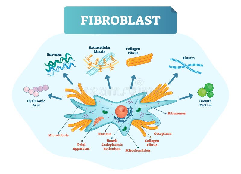
Free with trial Fibroblast vector illustration. Microscopic close-up with extracellular, collagen fibrils, elastin, hyaluronic acid. Scheme with microtubule, golgi apparatus, nucleus, mitochondrion and ribosomes. Microscopy visualization vectors Fibroblast vector illustration. Scheme with extracellular, elastin, hyaluronic acid, microtubule, golgi apparatus and ribosomes. Fibroblast vector illustration. Fibroblast vector illustration. Microscopic close-up with extracellular, collagen fibrils, elastin, hyaluronic acid. Scheme with microtubule, golgi apparatus, nucleus, mitochondrion and ribosomes.
Free with trial Microscope scientific research molecular structure colorful abstract shapes paper cut style. Vector illustration. Microscopy visualization vectors Microscope scientific research molecular structure colorful abstract shapes paper cut style. Vector illustration
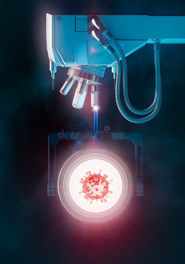
Free with trial 3D illustration depicting a red coronavirus pathogen with characteristic spike proteins viewed through an optical electron scanning microscope against glowing white illuminated background. The scientific visualization shows the microscope's cylindrical objective lenses with flexible turquoise cables positioned above the magnified viral specimen in clinical laboratory setting, representing medical research, virology study, pandemic diagnosis, or infectious disease detection through advanced microscopy technology. Microscopy visualization illustrations Red coronavirus under electron microscope scientific analysis. 3D illustration depicting a red coronavirus pathogen with characteristic spike proteins viewed. 3D illustration depicting a red coronavirus pathogen with characteristic spike proteins viewed through an optical electron scanning microscope against glowing white illuminated background. The scientific visualization shows the microscope's cylindrical objective lenses with flexible turquoise cables positioned above the magnified viral specimen in clinical laboratory setting, representing medical research, virology study, pandemic diagnosis, or infectious disease detection through advanced microscopy technology.
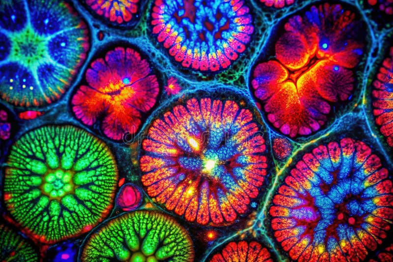
Free with trial This detailed immunofluorescence microscopy image showcases the intricate network of tight junctions within the intestinal mucosa. The image, captured under optimized studio lighting, presents a clear visualization of protein expression within the epithelial cells lining the intestine. The precise focus and depth of field highlight the structural components of the tissue, including the cellular. Microscopy visualization illustrations Visualizing Intestinal Mucosa Tight Junctions Immunofluorescence Microscopy Reveals Protein Expression and Tissue. This detailed immunofluorescence microscopy. This detailed immunofluorescence microscopy image showcases the intricate network of tight junctions within the intestinal mucosa. The image, captured under optimized studio lighting, presents a clear visualization of protein expression within the epithelial cells lining the intestine. The precise focus and depth of field highlight the structural components of the tissue, including the cellular
Free with trial This stunning image captures the intricate vascular system of the retina using specialized bio-imaging techniques. The dominant red network represents major blood vessels, while the striking green lines highlight finer capillaries near the central optic disc. Rendered by Ai, this visualization offers a deep look into ophthalmic anatomy, providing valuable data for medical research and diagnosis. Microscopy visualization illustrations Retinal vascular network under fluorescence microscopy. This stunning image captures the intricate vascular system of the retina using specialized bio-imaging. This stunning image captures the intricate vascular system of the retina using specialized bio-imaging techniques. The dominant red network represents major blood vessels, while the striking green lines highlight finer capillaries near the central optic disc. Rendered by Ai, this visualization offers a deep look into ophthalmic anatomy, providing valuable data for medical research and diagnosis
Free with trial Futuristic visualization of freeze dried exosome particles with glowing spheres and micro particles, Generated by AI. Microscopy visualization illustrations Futuristic visualization of freeze dried exosome particles with glowing spheres and micro particles. Generated by AI
Free with trial False color light microscopy image of Fusobacterium spp. in mutualistic interaction with colorectal cancer cells, rod-shaped bacteria in red or orange, cancer cells with irregular nuclei and purple-stained cytoplasm. Generative AI. Microscopy visualization illustrations False color light microscopy image of Fusobacterium spp. in mutualistic interaction with colorectal cancer cells. Generative AI. False color light microscopy. False color light microscopy image of Fusobacterium spp. in mutualistic interaction with colorectal cancer cells, rod-shaped bacteria in red or orange, cancer cells with irregular nuclei and purple-stained cytoplasm. Generative AI
Free with trial This exceptionally detailed fluorescent microscopy image showcases the intricate structure of blue asbestos fibers. Under intense magnification, the individual fibers' morphology becomes strikingly apparent, revealing their unique characteristics. The vibrant blue fluorescence highlights the fibers, clearly differentiating them from surrounding material. This enhanced visualization allows for. Microscopy visualization illustrations Detailed Fluorescent Microscopy Image of Blue Asbestos Fibers Enhanced Magnification for Scientific Analysis. This exceptionally detailed fluorescent microscopy. This exceptionally detailed fluorescent microscopy image showcases the intricate structure of blue asbestos fibers. Under intense magnification, the individual fibers' morphology becomes strikingly apparent, revealing their unique characteristics. The vibrant blue fluorescence highlights the fibers, clearly differentiating them from surrounding material. This enhanced visualization allows for
Free with trial Close up of blue exosome particles suspended in transparent spheres with advanced 3d visualization, Generated by AI. Microscopy visualization illustrations Close up of blue exosome particles suspended in transparent spheres with advanced 3d visualization. Generated by AI
Free with trial Close up of blue exosome particles in transparent spheres 3d visualization for biotechnology. Generative AI. Microscopy visualization illustrations Close up of blue exosome particles in transparent spheres 3d visualization for biotechnology. Generative AI
Free with trial Advanced visualization of freeze dried exosome particles with soft glowing spheres and depth effects, Generated by AI. Microscopy visualization illustrations Advanced visualization of freeze dried exosome particles with soft glowing spheres and depth effects. Generated by AI
Free with trial High-resolution visualization of cancer cells in the human body, emphasizing their complex structure and growth dynamics in a dark. Background ,Generative AI. Microscopy visualization illustrations High-resolution visualization of cancer cells in the human body, emphasizing their complex structure and growth dynamics in a dark. Background ,Generative AI
Free with trial Photorealistic visualization of advanced healing peptides in tissue reconstruction and repair, Generated by AI. Microscopy visualization illustrations Photorealistic visualization of advanced healing peptides in tissue reconstruction and repair. Generated by AI
Free with trial This vibrant image showcases the intricate network of cells, their cytoskeletons glowing in red and nuclei in blue, with additional green elements. The detailed view was generated by AI for enhanced visualization, highlighting cellular structures for scientific study or educational purposes. Microscopy visualization illustrations Microscopic Cell Structures. This vibrant image showcases the intricate network of cells, their cytoskeletons glowing in red and nuclei in blue, with additional. This vibrant image showcases the intricate network of cells, their cytoskeletons glowing in red and nuclei in blue, with additional green elements. The detailed view was generated by AI for enhanced visualization, highlighting cellular structures for scientific study or educational purposes.
Free with trial An intricate display of Salmonella enterica reveals its complex shape and vibrant colors. This portrayal highlights the microscopic world of bacteria critical to understanding public health. Microscopy visualization illustrations Detailed Visualization of Salmonella Enterica Showcasing Its Structure and Features. An intricate display of Salmonella enterica reveals its complex shape and. An intricate display of Salmonella enterica reveals its complex shape and vibrant colors. This portrayal highlights the microscopic world of bacteria critical to understanding public health.
Free with trial This detailed microscopic image depicts a cancer cell, generated by AI. Its intricate structure and vibrant color showcase the complexity of this disease. The image is a powerful visualization tool for education and research. Microscopy visualization illustrations AI-Generated Cancer Cell. This detailed microscopic image depicts a cancer cell, generated by AI. Its intricate structure and vibrant color showcase the. This detailed microscopic image depicts a cancer cell, generated by AI. Its intricate structure and vibrant color showcase the complexity of this disease. The image is a powerful visualization tool for education and research.
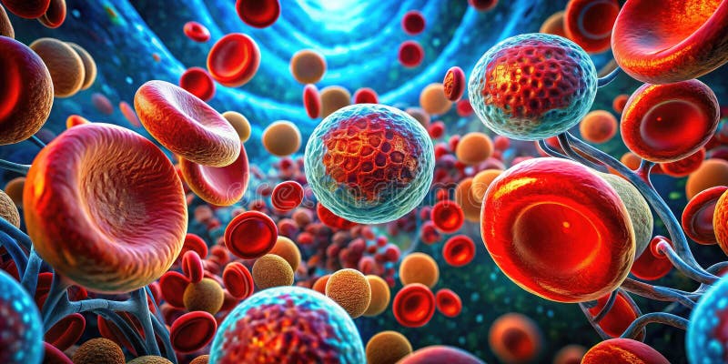
Free with trial The image shows a high-resolution, detailed view of red blood cells (erythrocytes) in circulation. The red blood cells are depicted as biconcave discs, which is their characteristic shape, allowing for efficient oxygen transport throughout the body. The image is interesting and relevant as it provides a close-up look at the essential components of blood, highlighting their structure and function within the cardiovascular system. This visualization is crucial for medical research, education, and understanding various blood-related health conditions. Microscopy visualization illustrations High-Resolution Image of Red Blood Cells in Circulation. The image shows a high-resolution, detailed view of red blood cells (erythrocytes) in circulation. The. The image shows a high-resolution, detailed view of red blood cells (erythrocytes) in circulation. The red blood cells are depicted as biconcave discs, which is their characteristic shape, allowing for efficient oxygen transport throughout the body. The image is interesting and relevant as it provides a close-up look at the essential components of blood, highlighting their structure and function within the cardiovascular system. This visualization is crucial for medical research, education, and understanding various blood-related health conditions.
Free with trial A detailed visualization showcases a virus navigating through human blood, illustrating its structure and interaction with cells. Microscopy visualization illustrations Intricate depiction of a virus interacting with human cells in a bloodstream environment. A detailed visualization showcases a virus navigating through human. A detailed visualization showcases a virus navigating through human blood, illustrating its structure and interaction with cells
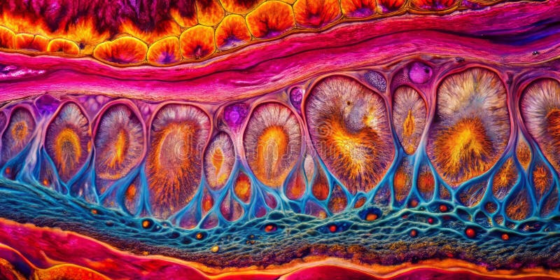
Free with trial This captivating microscopic image reveals the intricate details of acanthosis, a skin condition characterized by thickened epidermal layers. The advanced microscopy technique employed allows for a profound exploration of the cellular architecture within the epidermis. Noticeable are the elongated rete ridges, a hallmark of acanthosis. These ridges, formed by the proliferation of keratinocytes. Microscopy visualization illustrations Unveiling the Microscopic Structure of Acanthotic Epidermis: A Deep Dive into Thickened Skin Layers Under. This captivating microscopic image reveals the. This captivating microscopic image reveals the intricate details of acanthosis, a skin condition characterized by thickened epidermal layers. The advanced microscopy technique employed allows for a profound exploration of the cellular architecture within the epidermis. Noticeable are the elongated rete ridges, a hallmark of acanthosis. These ridges, formed by the proliferation of keratinocytes
Free with trial Detailed microscopic showcasing the intricate structure of a human lymph node, highlighting the cortex, medulla, and lymphatic follicles. Illustrates immune system function, cellular organization, and tissue architecture. Provides insight into lymphoid tissue. Medical and scientific visualization. Spacious empty area designed for product displays and advertisements. Perfect for banners, branding, and marketing visuals. . Generative AI. Microscopy visualization illustrations Microscopic View of Human Lymph Node Cortex Medulla and Follicles Detailed microscopic showcasing the intricate structure of. Detailed microscopic showcasing the. Detailed microscopic showcasing the intricate structure of a human lymph node, highlighting the cortex, medulla, and lymphatic follicles. Illustrates immune system function, cellular organization, and tissue architecture. Provides insight into lymphoid tissue. Medical and scientific visualization. Spacious empty area designed for product displays and advertisements. Perfect for banners, branding, and marketing visuals. . Generative AI
Free with trial 3D rendered medical illustration showing red blood cells (erythrocytes) suspended in blood plasma. The microscopic visualization depicts the circular biconcave shape of healthy red blood cells in a stylized turquoise fluid environment representing vascular system. Microscopy visualization illustrations Blood cells floating in plasma, microscopic view. 3D rendered medical illustration showing red blood cells (erythrocytes) suspended in blood plasma. The. 3D rendered medical illustration showing red blood cells (erythrocytes) suspended in blood plasma. The microscopic visualization depicts the circular biconcave shape of healthy red blood cells in a stylized turquoise fluid environment representing vascular system.
Free with trial A Colorful abstract representation of microscopic cellular structure. ai generative. Microscopy visualization illustrations Colorful abstract representation of microscopic cellular structure. ai generative. A Colorful abstract representation of microscopic cellular structure. ai. A Colorful abstract representation of microscopic cellular structure. ai generative
Free with trial Membrane proteins interact between two parallel lipid bilayers in a detailed molecular model with contrasting colors and white background, leaving ample space for text and labels for scientific presentations. Microscopy visualization illustrations Membrane proteins interacting across lipid bilayers illustrating molecular binding. Membrane proteins interact between two parallel lipid bilayers in a detailed. Membrane proteins interact between two parallel lipid bilayers in a detailed molecular model with contrasting colors and white background, leaving ample space for text and labels for scientific presentations
Free with trial This is a stunning ultra-macro photograph focusing on the interior texture of a puffed cereal piece. The crunchy brown shell is broken open exposing numerous clear, elongated crystalline structures within the core. Rendered by Ai this dramatic perspective highlights the sugary composition of the processed food item. This detailed visualization offers a unique and scientific look at everyday. Microscopy visualization illustrations Sugar crystals magnified inside sweet cereal puff. This is a stunning ultra-macro photograph focusing on the interior texture of a puffed cereal piece. The. This is a stunning ultra-macro photograph focusing on the interior texture of a puffed cereal piece. The crunchy brown shell is broken open exposing numerous clear, elongated crystalline structures within the core. Rendered by Ai this dramatic perspective highlights the sugary composition of the processed food item. This detailed visualization offers a unique and scientific look at everyday
Free with trial Detailed close-up of cancer cells spreading through tissue, illustrating the aggressive nature of tumor growth at the microscopic level - AI-generated. Microscopy visualization illustrations Microscopic View of Cancer Cells Spreading. Detailed close-up of cancer cells spreading through tissue, illustrating the aggressive nature of tumor growth at the. Detailed close-up of cancer cells spreading through tissue, illustrating the aggressive nature of tumor growth at the microscopic level - AI-generated
Free with trial Isolated malignant cancer cells under a microscope. Image of isolated malignant cancer cells under a microscope with illuminated and active effect of the cells. Microscopy visualization illustrations Isolated malignant cancer cells under a microscope. Image of isolated malignant cancer cells under a microscope with illuminated and active effect of the cells
Free with trial This detailed illustration depicts the immune system in action, showcasing cells actively fighting disease pathogens. Ideal for biomedical and healthcare applications. Microscopy visualization illustrations Immune System Illustration of Cells Fighting Disease Pathogens. This detailed illustration depicts the immune system in action, showcasing cells actively. This detailed illustration depicts the immune system in action, showcasing cells actively fighting disease pathogens. Ideal for biomedical and healthcare applications
Free with trial E. coli (Escherichia coli), in a petri dish. The enlarged image shows the rod-shaped shape and texture of the shell in an orange nutrient solution. Microscopy visualization illustrations E. coli (Escherichia coli), in a petri dish. The enlarged image shows the rod-shaped shape and texture of the shell in an orange nutrient solution
Free with trial Detailed neuron cell anatomy with labeled dendrites soma axon nucleus mitochondria schwann cells and neural biology education concept for medical learning. Microscopy visualization illustrations AI generated detailed neuron cell anatomy labeled dendrites soma axon nucleus mitochondria schwann cells neural biology education. AI generated detailed neuron. detailed neuron cell anatomy with labeled dendrites soma axon nucleus mitochondria schwann cells and neural biology education concept for medical learning
Free with trial Halobacterium salinarum Microscopic View Isolated on Pure White Background with Square and Rod Shaped Halophilic Archaea Detail High Resolution Image. Microscopy visualization illustrations Halobacterium salinarum Microscopic View Isolated on Pure White Background with Square and Rod Shaped Halophilic Archaea Detail. High Resolution Image
Free with trial Colorful abstract patterns illustrate the intricate world of T-lymphocytes, highlighting their cellular interactions and significance in the immune response. Microscopy visualization illustrations Colorful Abstract of T-lymphocytes Highlighting Dynamic Cellular Interactions. Colorful abstract patterns illustrate the intricate world of T-lymphocytes. Colorful abstract patterns illustrate the intricate world of T-lymphocytes, highlighting their cellular interactions and significance in the immune response.
Free with trial Explore intricate details of cell culture in advanced media through this vibrant macro view. Ideal for scientific articles, presentations, and educational materials. Microscopy visualization illustrations Cell Culture in Advanced Media: Growth and Development Insights. Explore intricate details of cell culture in advanced media through this vibrant macro view. Explore intricate details of cell culture in advanced media through this vibrant macro view. Ideal for scientific articles, presentations, and educational materials
Free with trial Explore the innovative field of bio sensing with quantum dots designed for detecting pathogens. This image represents advanced research and technology in diagnostics. Microscopy visualization illustrations Bio Sensing Quantum Dots for Detecting Pathogens in Future Technologies. Explore the innovative field of bio sensing with quantum dots designed for detecting. Explore the innovative field of bio sensing with quantum dots designed for detecting pathogens. This image represents advanced research and technology in diagnostics
Free with trial 3d rendering of scattered urate crystals Uric acid or kidneys excrete too little uric acid, it can build up, forming sharp, needle-like urate crystals in a joint or surrounding tissue. Microscopy visualization illustrations 3d rendering of scattered urate or uric acid crystals. 3d rendering of scattered urate crystals Uric acid or kidneys excrete too little uric acid, it can build. 3d rendering of scattered urate crystals Uric acid or kidneys excrete too little uric acid, it can build up, forming sharp, needle-like urate crystals in a joint or surrounding tissue.
Free with trial Illustration of cancer cell division. Microscopy visualization illustrations Illustration of cancer cell division. Illustration of cancer cell division
Free with trial Explore an ultra-detailed 3D rendering of microglia, showcasing their vital role in the nervous system's immune response. This captivating illustration highlights the intricate structure and dynamic activity of these essential cells. Microscopy visualization illustrations Ultra Detailed 3D Rendering of Microglia in Action. Explore an ultra-detailed 3D rendering of microglia, showcasing their vital role in the nervous system's. Explore an ultra-detailed 3D rendering of microglia, showcasing their vital role in the nervous system's immune response. This captivating illustration highlights the intricate structure and dynamic activity of these essential cells
Free with trial The close-up view captures numerous red blood cells suspended in a clear liquid, creating an interesting visual effect against a light background. Microscopy visualization illustrations The close-up view captures numerous red blood cells suspended in a clear liquid,. Creating an interesting visual effect against a light background
Free with trial A detailed close-up image of microscope lenses positioned above a mound of powdered substance, illustrating the technology behind scientific observation and Generative Aianalysis. Microscopy visualization illustrations Close-Up of a Microscope Lens Focusing on a Powdered Substance. A detailed close-up image of microscope lenses positioned above a mound of powdered substance. A detailed close-up image of microscope lenses positioned above a mound of powdered substance, illustrating the technology behind scientific observation and Generative Aianalysis
Free with trial High-resolution image of colorful pluripotent stem cells viewed under a microscope, showcasing their dynamic structures and vibrant colors, ideal for scientific presentations and educational materials. Microscopy visualization illustrations Colorful Pluripotent Stem Cells Observed Under a Microscope. High-resolution image of colorful pluripotent stem cells viewed under a microscope, showcasing their. High-resolution image of colorful pluripotent stem cells viewed under a microscope, showcasing their dynamic structures and vibrant colors, ideal for scientific presentations and educational materials
Free with trial Primary lung tuberculosis, 3D illustration featuring the Ghon complex and mediastinal lymphadenitis, alongside with close-up view of Mycobacterium tuberculosis bacteria. Microscopy visualization illustrations Primary lung tuberculosis, 3D illustration. Featuring the Ghon complex and mediastinal lymphadenitis, alongside with close-up view of Mycobacterium tuberculosis
Free with trial Highly detailed 3D representation of the West Nile Virus interacting with a human body. The vivid colors and detailed virus structure highlight the complexity of viral infections and their potential health effects. Microscopy visualization illustrations West Nile Virus within a human body emphasizing its intricate structure and impact on health. Highly detailed 3D representation of the West Nile Virus. Highly detailed 3D representation of the West Nile Virus interacting with a human body. The vivid colors and detailed virus structure highlight the complexity of viral infections and their potential health effects.
Free with trial Microscopic 3D rendering of neuroinflammation process. Detailed illustration shows neurons, glial cells, and inflammatory molecules interacting within the brain. The image uses a dark blue background for contrast and highlights the inflammatory process with bright colors. Labels indicate various key components of the neuroinflammation pathway. Microscopy visualization illustrations Neuroinflammation: 3D Microscopic Rendering of Brain\'s Inflammatory Response. Microscopic 3D rendering of neuroinflammation process. Detailed illustration shows. Microscopic 3D rendering of neuroinflammation process. Detailed illustration shows neurons, glial cells, and inflammatory molecules interacting within the brain. The image uses a dark blue background for contrast and highlights the inflammatory process with bright colors. Labels indicate various key components of the neuroinflammation pathway
Free with trial Inflammation, a crucial part of the body's defense mechanism, triggers a microscopic battleground where specialized immune cells wage war against invading pathogens. This intricate process, visible only under a powerful microscope, showcases the remarkable complexity of the human immune system. The image reveals a dynamic interplay of different immune cells, including macrophages, neutrophils,. Microscopy visualization illustrations Unveiling the Microscopic War Immune Cells Combat Pathogens During Inflammation. Inflammation, a crucial part of the body's defense mechanism, triggers a. Inflammation, a crucial part of the body's defense mechanism, triggers a microscopic battleground where specialized immune cells wage war against invading pathogens. This intricate process, visible only under a powerful microscope, showcases the remarkable complexity of the human immune system. The image reveals a dynamic interplay of different immune cells, including macrophages, neutrophils,
Free with trial Stunning 3D microscopic view of cell division, showcasing cytokinesis, cellular reproduction, and binary fission. Microscopy visualization illustrations Cell Division: Stunning 3D Microscopic View of Cytokinesis, Cellular Reproduction, and Binary Fission. Stunning 3D microscopic view of cell division, showcasing. Stunning 3D microscopic view of cell division, showcasing cytokinesis, cellular reproduction, and binary fission
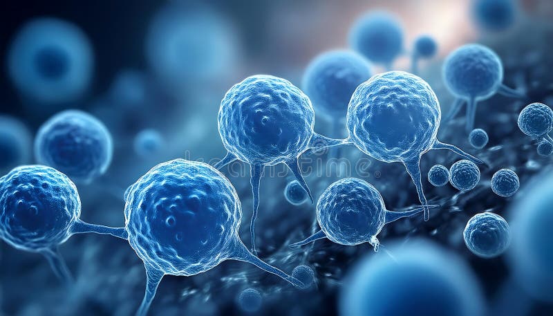
Free with trial Several blue cells are shown in a microscopic view background is blurry. Microscopy visualization illustrations Several blue cells are shown in a microscopic view background is blurry.
Free with trial This captivating tilt-shift microscopic image showcases the intricate flow of red blood cells within a vascular graft. The technique subtly blurs the background, drawing the viewer's eye to the precise movement of individual cells. This detailed view reveals the microarchitecture of the graft, highlighting the delicate interplay between the newly formed blood vessel and the circulating blood. Microscopy visualization illustrations A Captivating TiltShift Microscopic View of Red Blood Cells Flowing Through a Vascular Graft Unveiling the. This captivating tilt-shift microscopic image. This captivating tilt-shift microscopic image showcases the intricate flow of red blood cells within a vascular graft. The technique subtly blurs the background, drawing the viewer's eye to the precise movement of individual cells. This detailed view reveals the microarchitecture of the graft, highlighting the delicate interplay between the newly formed blood vessel and the circulating blood.
Free with trial This stunning, photorealistic illustration meticulously depicts the intricate cellular damage caused by oxidative stress. The image showcases the impact of free radicals on various cellular components, including the cell membrane, mitochondria, and the cell itself. Observe the subtle yet significant alterations in the cell's structure, highlighting the progressive nature of oxidative stress. Microscopy visualization illustrations Visualizing Oxidative Stress Cell Damage A Detailed Photorealistic Illustration of Cellular Aging and Disease. This stunning, photorealistic illustration. This stunning, photorealistic illustration meticulously depicts the intricate cellular damage caused by oxidative stress. The image showcases the impact of free radicals on various cellular components, including the cell membrane, mitochondria, and the cell itself. Observe the subtle yet significant alterations in the cell's structure, highlighting the progressive nature of oxidative stress.
Free with trial Ion schematic icon, outline style. Ion schematic icon. Outline ion schematic vector icon for web design isolated on white background. Microscopy visualization vectors Ion schematic icon, outline style. Ion schematic icon. Outline ion schematic vector icon for web design isolated on white background
Free with trial This captivating macro photograph delves into the intricate world of nanofiber textiles. The image showcases the mesmerizing woven fabric texture at a microscopic level, revealing a stunning array of details previously unseen. The meticulous craftsmanship and precision of the material's construction are evident in the image's sharp focus. Each nanofiber, a fundamental element of this advanced. Microscopy visualization illustrations Unveiling the Microscopic Marvels of a Nanofiber Textile A HighMagnification Look at Woven Fabric Structure and. This captivating macro photograph delves into. This captivating macro photograph delves into the intricate world of nanofiber textiles. The image showcases the mesmerizing woven fabric texture at a microscopic level, revealing a stunning array of details previously unseen. The meticulous craftsmanship and precision of the material's construction are evident in the image's sharp focus. Each nanofiber, a fundamental element of this advanced
Free with trial Microscopic view of cells. Cellular aging, oxidative stress visualized. Differences in young, old cells shown. Changes in structure size visible. Focus on aging effects. Illustration. Microscopy visualization illustrations Microscopic view of cells. Cellular aging, oxidative stress visualized. Differences in young, old cells shown. Changes in. Structure size visible. Focus on. Microscopic view of cells. Cellular aging, oxidative stress visualized. Differences in young, old cells shown. Changes in structure size visible. Focus on aging effects. Illustration.
Free with trial Intricate macro image of a cancer tumor puzzle with diverse textures and dramatic lighting effects, Generated by AI. Microscopy visualization illustrations Intricate macro image of a cancer tumor puzzle with diverse textures and dramatic lighting effects. Generated by AI
Free with trial Vibrant 3D representation of viruses displaying intricate structures and colors, ideal for scientific discussions, educational material, and health awareness campaigns related to infectious diseases. Microscopy visualization illustrations Colorful 3D Rendering of Viruses with Complex Structures Against a Dark Background for Scientific and Educational Use. Vibrant 3D representation of viruses. Vibrant 3D representation of viruses displaying intricate structures and colors, ideal for scientific discussions, educational material, and health awareness campaigns related to infectious diseases
Free with trial Illustration shows nerve cell damage caused by immune response in multiple sclerosis. Orange colored cell signifies inflammation in nerve tissue. Delicate light blue extensions. Microscopy visualization illustrations Illustration shows nerve cell damage caused by immune response in multiple sclerosis. Orange colored cell signifies inflammation. In nerve tissue. Delicate
Free with trial Magnified view of salmonella bacteria displays its complex surface features and unique morphology. This observation highlights the microscopic world of pathogens and their impact on health. Microscopy visualization illustrations Zoomed-in Revelation of Salmonella Bacteria Showcasing Its Intricate Structure and Behavior. Magnified view of salmonella bacteria displays its complex surface. Magnified view of salmonella bacteria displays its complex surface features and unique morphology. This observation highlights the microscopic world of pathogens and their impact on health.
Free with trial Microscopic examination reveals the intricate and orderly process of apoptosis occurring in bacteria and viruses, highlighting vital cellular events during infection and disease research. Microscopy visualization illustrations Exploring Orderly Apoptosis Process in Bacteria and Viruses Amidst Scientific Research. Microscopic examination reveals the intricate and orderly process of. Microscopic examination reveals the intricate and orderly process of apoptosis occurring in bacteria and viruses, highlighting vital cellular events during infection and disease research.
Free with trial 3d atom illustration, study and research, imaging and research, 3d imaging, physics. Microscopy visualization illustrations 3D Images of Silver Atoms for Study and Research, Silver Atoms, Atoms, Scientific Image, Study and Research, 3D Images. 3d atom illustration, study and research. 3d atom illustration, study and research, imaging and research, 3d imaging, physics
Free with trial A scientist engages in cancer research within a vibrant laboratory filled with colorful test tubes, beakers, and scientific symbols representing innovation and discovery. Microscopy visualization vectors A scientist engages in cancer research within a vibrant laboratory filled with colorful test tubes, beakers, and scientific. Symbols representing innovation and
Free with trial This captivating microscopic image reveals the dynamic process of blood flow at the cellular level. Observe red blood cells navigating the narrow confines of capillaries, the tiniest blood vessels in the human circulatory system. This detailed illustration showcases the intricate interplay of hemodynamics, highlighting the crucial role of microcirculation in oxygen and carbon dioxide transport. Microscopy visualization illustrations Unveiling the Intricate Dance of Red Blood Cells Within Capillaries A Microscopic Journey Through Microcirculation. This captivating microscopic image reveals. This captivating microscopic image reveals the dynamic process of blood flow at the cellular level. Observe red blood cells navigating the narrow confines of capillaries, the tiniest blood vessels in the human circulatory system. This detailed illustration showcases the intricate interplay of hemodynamics, highlighting the crucial role of microcirculation in oxygen and carbon dioxide transport
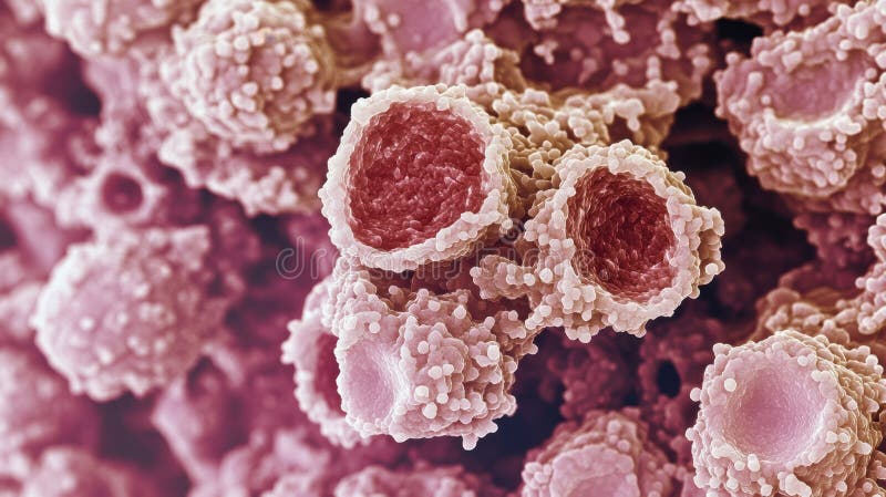
Free with trial Microscopic view of intestinal lining showing pink villi structure. This surface acts as a barrier for nutrient absorption in the digestive system. Medical science visualization of gut. Microscopy visualization illustrations Microscopic view of intestinal lining, showing pink villi structure. This surface acts as a barrier for nutrient absorption in. Microscopic view of intestinal. Microscopic view of intestinal lining showing pink villi structure. This surface acts as a barrier for nutrient absorption in the digestive system. Medical science visualization of gut.
Free with trial This captivating microscopic image showcases the detailed cellular structures of Salmonella and E. coli bacteria. The vibrant colors highlight the intricate details of these pathogenic microorganisms, revealing their unique cell walls, flagella, and other key components. This detailed view offers a glimpse into the complex world of microbiology, providing valuable insights for research into. Microscopy visualization illustrations A Microscopic Deep Dive into Salmonella and E coli Unveiling the Intricate Structures of Pathogenic Bacteria. This captivating microscopic image showcases the. This captivating microscopic image showcases the detailed cellular structures of Salmonella and E. coli bacteria. The vibrant colors highlight the intricate details of these pathogenic microorganisms, revealing their unique cell walls, flagella, and other key components. This detailed view offers a glimpse into the complex world of microbiology, providing valuable insights for research into
Free with trial Realistic image of laser targeting cells representing precision in medical technology innovation, Generated by AI. Microscopy visualization illustrations Realistic image of laser targeting cells representing precision in medical technology innovation. Generated by AI
Free with trial This captivating microscopic image delves into the fascinating world of microcirculation. Observe the intricate network of blood vessels, specifically capillaries, showcasing the remarkable structure and function of the circulatory system. Red blood cells, crucial for oxygen transport, are clearly visible traversing the narrow channels of these vessels, alongside plasma, the fluid component of. Microscopy visualization illustrations Unveiling the Intricate Microcirculation A Detailed Microscopic View of Blood Vessels Red Blood Cells and Plasma. This captivating microscopic image delves into. This captivating microscopic image delves into the fascinating world of microcirculation. Observe the intricate network of blood vessels, specifically capillaries, showcasing the remarkable structure and function of the circulatory system. Red blood cells, crucial for oxygen transport, are clearly visible traversing the narrow channels of these vessels, alongside plasma, the fluid component of
Free with trial Explore a high fidelity lattice visualization using microscopy techniques, showcasing intricate crystal structures and detailed molecular arrangements in a dynamic setting. Microscopy visualization illustrations High Fidelity Lattice Visualization through Microscopy Techniques. Explore a high fidelity lattice visualization using microscopy techniques, showcasing. Explore a high fidelity lattice visualization using microscopy techniques, showcasing intricate crystal structures and detailed molecular arrangements in a dynamic setting
Free with trial Explore a high fidelity visualization of lattice structures created through advanced microscopy techniques. The intricate details reveal unique patterns and their scientific significance. Microscopy visualization illustrations High Fidelity Visualization of Lattice Structures Through Microscopy. Explore a high fidelity visualization of lattice structures created through advanced. Explore a high fidelity visualization of lattice structures created through advanced microscopy techniques. The intricate details reveal unique patterns and their scientific significance
Free with trial Human kidney glomerulus in renal tissue, multiplex fluorescence microscopy visualization. Illustration. Microscopy visualization illustrations Human kidney glomerulus in renal tissue, multiplex fluorescence microscopy visualization. Illustration
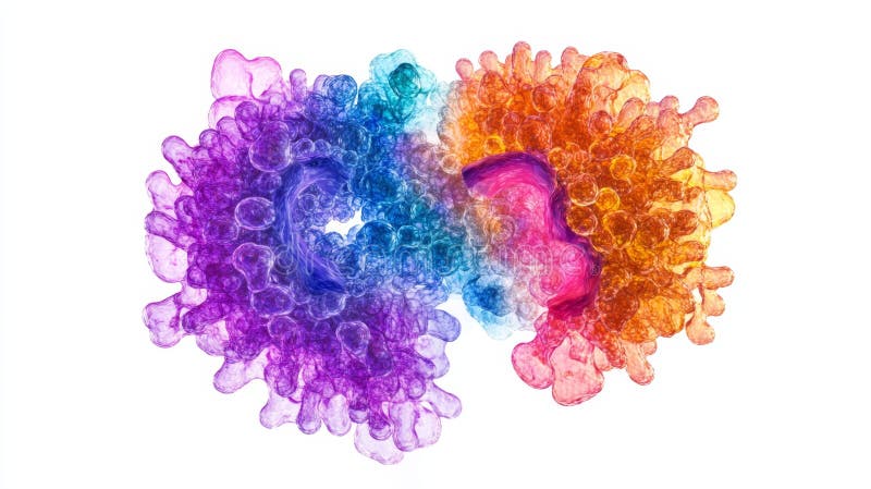
Free with trial This image showcases an intricate and colorful visualization likely representing a molecular or biological structure, possibly a protein complex or cellular network. The vivid hues and detailed branching patterns suggest it might be an electron microscopy image or a computational model used in fields like structural biology, bioinformatics, or molecular biology. Such visualizations are essential. Microscopy visualization illustrations A detailed visualization of a complex molecular or biological structure under advanced microscopy. This image showcases an intricate and colorful visualization. This image showcases an intricate and colorful visualization likely representing a molecular or biological structure, possibly a protein complex or cellular network. The vivid hues and detailed branching patterns suggest it might be an electron microscopy image or a computational model used in fields like structural biology, bioinformatics, or molecular biology. Such visualizations are essential
Free with trial Explore an artistic visualization showcasing islet cell regeneration through microscopy. This vibrant illustration depicts the dynamic structure and function of cells in a scientific context. Microscopy visualization illustrations Artistic Visualization of Islet Cell Regeneration Under Microscopy. Explore an artistic visualization showcasing islet cell regeneration through microscopy. This. Explore an artistic visualization showcasing islet cell regeneration through microscopy. This vibrant illustration depicts the dynamic structure and function of cells in a scientific context
Free with trial Explore high fidelity microstructure visualization using advanced microscopy technology. Significant detail and vibrant colors enhance microstructural analysis. Microscopy visualization illustrations High Fidelity Microstructure Visualization Using Advanced Microscopy. Explore high fidelity microstructure visualization using advanced microscopy technology. Explore high fidelity microstructure visualization using advanced microscopy technology. Significant detail and vibrant colors enhance microstructural analysis
Free with trial This detailed 3D rendering showcases the intricate cellular structure of red blood cells, or erythrocytes, as observed through advanced electron microscopy. The high-definition image reveals the complex morphology of these crucial blood components, highlighting their unique shape and internal organization. This visualization provides a profound look at the erythrocyte membrane structure,. Microscopy visualization illustrations Unveiling the 3D Microstructure of Red Blood Cells A HighDefinition Electron Microscopy Visualization. This detailed 3D rendering showcases the intricate. This detailed 3D rendering showcases the intricate cellular structure of red blood cells, or erythrocytes, as observed through advanced electron microscopy. The high-definition image reveals the complex morphology of these crucial blood components, highlighting their unique shape and internal organization. This visualization provides a profound look at the erythrocyte membrane structure,
Free with trial Detailed Cancer Cells Microscopy Visualization 3D Illustration. AI generated. Microscopy visualization illustrations Detailed Cancer Cells Microscopy Visualization 3D Illustration. AI generated
Free with trial DNA microscopy research with molecular visualization advanced laboratory analytics biotechnology innovation and precision scientific discovery for healthcare. Microscopy visualization illustrations AI generated DNA microscopy research molecular visualization advanced laboratory analytics biotechnology innovation precision. AI generated DNA microscopy. DNA microscopy research with molecular visualization advanced laboratory analytics biotechnology innovation and precision scientific discovery for healthcare
Free with trial Fluorescent microscopy of a cell's genetic components and membrane, Genetics Visualization, Scientific Imaging. Generative AI. Microscopy visualization illustrations Fluorescent microscopy of a cell's genetic components and membrane, Genetics Visualization, Scientific Imaging. Generative AI
Free with trial Fluorescent microscopy of a cell's genetic components and membrane, Genetics Visualization, Scientific Imaging. Generative AI. Microscopy visualization illustrations Fluorescent microscopy of a cell's genetic components and membrane, Genetics Visualization, Scientific Imaging. Generative AI
Free with trial This captivating 3D rendering showcases the intricate structure of stem cell DNA as observed through advanced low-light microscopy. The image provides a unique perspective on the complex interplay within stem cells, crucial for understanding their potential in cellular therapies and regenerative medicine. The low-light technique allows for enhanced visualization of subtle details within the. Microscopy visualization illustrations 3D Visualization of Stem Cell DNA Structure Under LowLight Microscopy Implications for Cellular Therapy and. This captivating 3D rendering showcases the. This captivating 3D rendering showcases the intricate structure of stem cell DNA as observed through advanced low-light microscopy. The image provides a unique perspective on the complex interplay within stem cells, crucial for understanding their potential in cellular therapies and regenerative medicine. The low-light technique allows for enhanced visualization of subtle details within the
Free with trial This image showcases a state-of-the-art microscope integrated with AI-driven data visualization, featuring multiple lenses and a digital screen displaying intricate hexagonal molecular patterns. Perfect for scientific publications, tech company websites, educational content, and research institution marketing materials. The sleek metallic design of the microscope, combined with vibrant blue lighting and futuristic screen elements, conveys precision, innovation, and the intersection of biotechnology and artificial intelligence. Ideal as a background or header for themes like laboratory research, high-tech instrumentation, and scientific discovery. This image captures the essence of modern scientific exploration, making it versatile for promoting advancements in microscopy, data analytics, and precision tools within educational and commercial contexts. Microscopy visualization illustrations Precision Microscopy Meets AI: Unveiling Scientific Frontiers. This image showcases a state-of-the-art microscope integrated with AI-driven data visualization. This image showcases a state-of-the-art microscope integrated with AI-driven data visualization, featuring multiple lenses and a digital screen displaying intricate hexagonal molecular patterns. Perfect for scientific publications, tech company websites, educational content, and research institution marketing materials. The sleek metallic design of the microscope, combined with vibrant blue lighting and futuristic screen elements, conveys precision, innovation, and the intersection of biotechnology and artificial intelligence. Ideal as a background or header for themes like laboratory research, high-tech instrumentation, and scientific discovery. This image captures the essence of modern scientific exploration, making it versatile for promoting advancements in microscopy, data analytics, and precision tools within educational and commercial contexts.
Free with trial Electron or confocal microscopy image revealing a close-up view of biological tissue structure. Cells and the extracellular matrix are highlighted by fluorescent colors: lime green structures surrounding dense clusters of nuclei or organelles colored bright magenta. This detailed visualization is essential for research in cell biology, histology, medicine, pathology, and understanding biological processes at the microscopic level. Microscopy visualization illustrations Cellular Microscopy: Fluorescent Organic Tissues. Electron or confocal microscopy image revealing a close-up view of biological tissue structure. Cells and the. Electron or confocal microscopy image revealing a close-up view of biological tissue structure. Cells and the extracellular matrix are highlighted by fluorescent colors: lime green structures surrounding dense clusters of nuclei or organelles colored bright magenta. This detailed visualization is essential for research in cell biology, histology, medicine, pathology, and understanding biological processes at the microscopic level.
Free with trial Microscopy zooming atom illustration nanotechnology visualization, magnification resolution, imaging electron microscopy zooming atom. Microscopy visualization illustrations Microscopy zooming atom. Illustration nanotechnology visualization, magnification resolution, imaging electron AI generated. microscopy zooming atom illustration nanotechnology visualization, magnification resolution, imaging electron microscopy zooming atom
Free with trial Microscopy hyper d holographic microscope projection illustration 3 imaging, visualization technology, optics resolution microscopy hyper d holographic microscope projection. Microscopy visualization illustrations Microscopy hyper d holographic microscope projection. Illustration 3 imaging, visualization technology, optics resolution AI generated. microscopy hyper d holographic microscope projection illustration 3 imaging, visualization technology, optics resolution microscopy hyper d holographic microscope projection
Free with trial This image displays a highly detailed and intricate visualization of a protein structure, likely a cryo-electron microscopy (cryo-EM) map. The dense, cloud-like appearance showcases the complex folding and intricate arrangement of the protein's subunits. The purple hue and texture suggest a three-dimensional rendering that highlights the protein's convoluted surface and potential binding sites or. Microscopy visualization illustrations A detailed visualization of a dense protein structure resembling a complex molecular cloud. This image displays a highly detailed and intricate visualization of. This image displays a highly detailed and intricate visualization of a protein structure, likely a cryo-electron microscopy (cryo-EM) map. The dense, cloud-like appearance showcases the complex folding and intricate arrangement of the protein's subunits. The purple hue and texture suggest a three-dimensional rendering that highlights the protein's convoluted surface and potential binding sites or
 Sign in