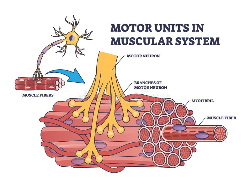
Free with trial Motor units in muscular system with fibers neuron anatomy outline diagram. Labeled educational medical scheme with myofibril and muscle fiber closeup vector illustration. Nerve functional contraction. Muscle contraction vectors Motor units in muscular system with fibers neuron anatomy outline diagram
Free with trial Woman having asthma using the asthma inhaler for being. Muscle contraction vectors Asthma
Free with trial The esophagus or oesophagus is an organ in vertebrates through which food passes, aided by peristaltic contractions, from the pharynx to the stomach. The esophagus is a fibromuscular tube, that travels behind the trachea and heart. During swallowing, the epiglottis tilts backwards to prevent food from going down the larynx and lungs. The trachea is the long tube that connects your larynx to your bronchi. Bronchi send air to your lungs. Trachea is a key part of respiratory system. The trachea is made of rings of cartilage. It is lined with cells that produce mucus. Muscle contraction illustrations Anatomy of Trachea and Esophagus. The esophagus or oesophagus is an organ in vertebrates through which food passes, aided by peristaltic contractions, from the pharynx to the stomach. The esophagus is a fibromuscular tube, that travels behind the trachea and heart. During swallowing, the epiglottis tilts backwards to prevent food from going down the larynx and lungs.The trachea is the long tube that connects your larynx to your bronchi. Bronchi send air to your lungs. Trachea is a key part of respiratory system. The trachea is made of rings of cartilage. It is lined with cells that produce mucus.
Free with trial Smooth Muscle Human Cell Vector Illustration. Muscle contraction vectors Smooth Muscle Human Cell
Free with trial Muscle icons set cartoon. Fiber tissue. Muscular body. Muscle contraction illustrations Muscle icons set cartoon . Fiber tissue
Free with trial 3D Illustration, Muscle is a soft tissue, Muscle cells contain proteins , producing a contraction that changes both the length and the shape of the cell. Muscles function to produce force and motion. Muscle contraction illustrations Omohyoideus Muscle Anatomy For Medical Concept 3D Illustration. 3D Illustration, Muscle is a soft tissue, Muscle cells contain proteins , producing a contraction that changes both the length and the shape of the cell. Muscles function to produce force and motion
Free with trial 3D Illustration, Muscle is a soft tissue, Muscle cells contain proteins , producing a contraction that changes both the length and the shape of the cell. Muscles function to produce force and motion. Muscle contraction illustrations Abductor Digiti Quinti Brevis Muscle Of Hand Anatomy For Medical Concept 3D Illustration. 3D Illustration, Muscle is a soft tissue, Muscle cells contain proteins , producing a contraction that changes both the length and the shape of the cell. Muscles function to produce force and motion
Free with trial Muscle is a soft tissue, Muscle cells contain proteins , producing a contraction that changes both the length and the shape of the cell. Muscles function to produce force and motion. Muscle contraction illustrations Flexor Carpi Radialis Muscle Anatomy For Medical Concept 3D Illustration. Muscle is a soft tissue, Muscle cells contain proteins , producing a contraction that changes both the length and the shape of the cell. Muscles function to produce force and motion
Free with trial 3D Illustration, Muscle is a soft tissue, Muscle cells contain proteins , producing a contraction that changes both the length and the shape of the cell. Muscles function to produce force and motion. Muscle contraction illustrations Palmaris Longus Muscle Anatomy For Medical Concept 3D Illustration. 3D Illustration, Muscle is a soft tissue, Muscle cells contain proteins , producing a contraction that changes both the length and the shape of the cell. Muscles function to produce force and motion
Free with trial 3D Illustration, Muscle is a soft tissue, Muscle cells contain proteins , producing a contraction that changes both the length and the shape of the cell. Muscles function to produce force and motion. Muscle contraction illustrations Biceps Brachii Muscle Anatomy For Medical Concept 3D Illustration. 3D Illustration, Muscle is a soft tissue, Muscle cells contain proteins , producing a contraction that changes both the length and the shape of the cell. Muscles function to produce force and motion
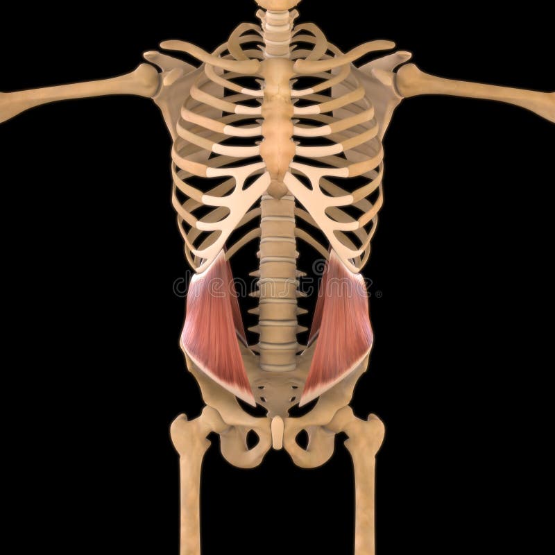
Free with trial 3D Illustration, Muscle is a soft tissue, Muscle cells contain proteins , producing a contraction that changes both the length and the shape of the cell. Muscles function to produce force and motion. Muscle contraction illustrations Pectineus Muscle Anatomy For Medical Concept 3D Illustration. 3D Illustration, Muscle is a soft tissue, Muscle cells contain proteins , producing a contraction that changes both the length and the shape of the cell. Muscles function to produce force and motion
Free with trial 3D Illustration, Muscle is a soft tissue, Muscle cells contain proteins , producing a contraction that changes both the length and the shape of the cell. Muscles function to produce force and motion. Muscle contraction illustrations Teres Major Muscle Anatomy For Medical Concept 3D Illustration. 3D Illustration, Muscle is a soft tissue, Muscle cells contain proteins , producing a contraction that changes both the length and the shape of the cell. Muscles function to produce force and motion
Free with trial 3D Illustration, Muscle is a soft tissue, Muscle cells contain proteins , producing a contraction that changes both the length and the shape of the cell. Muscles function to produce force and motion. Muscle contraction illustrations Rhomboid Major Muscle Anatomy For Medical Concept 3D Illustration. 3D Illustration, Muscle is a soft tissue, Muscle cells contain proteins , producing a contraction that changes both the length and the shape of the cell. Muscles function to produce force and motion
Free with trial Muscle is a soft tissue, Muscle cells contain proteins , producing a contraction that changes both the length and the shape of the cell. Muscles function to produce force and motion. Muscle contraction illustrations Extensor Pollicis Longus Muscle Anatomy For Medical Concept 3D Illustration. Muscle is a soft tissue, Muscle cells contain proteins , producing a contraction that changes both the length and the shape of the cell. Muscles function to produce force and motion
Free with trial 3D Illustration, Muscle is a soft tissue, Muscle cells contain proteins , producing a contraction that changes both the length and the shape of the cell. Muscles function to produce force and motion. Muscle contraction illustrations Pectineus Muscle Anatomy For Medical Concept 3D Illustration. 3D Illustration, Muscle is a soft tissue, Muscle cells contain proteins , producing a contraction that changes both the length and the shape of the cell. Muscles function to produce force and motion
Free with trial 3D Illustration, Muscle is a soft tissue, Muscle cells contain proteins , producing a contraction that changes both the length and the shape of the cell. Muscles function to produce force and motion. Muscle contraction illustrations Quadriceps Femoris Muscle Anatomy For Medical Concept 3D Illustration. 3D Illustration, Muscle is a soft tissue, Muscle cells contain proteins , producing a contraction that changes both the length and the shape of the cell. Muscles function to produce force and motion
Free with trial 3D Illustration, Muscle is a soft tissue, Muscle cells contain proteins , producing a contraction that changes both the length and the shape of the cell. Muscles function to produce force and motion. Muscle contraction illustrations Rhomboid Minor Muscle Anatomy For Medical Concept 3D Illustration. 3D Illustration, Muscle is a soft tissue, Muscle cells contain proteins , producing a contraction that changes both the length and the shape of the cell. Muscles function to produce force and motion
Free with trial Muscle is a soft tissue, Muscle cells contain proteins , producing a contraction that changes both the length and the shape of the cell. Muscles function to produce force and motion. Muscle contraction illustrations Coracobrachialis Muscle Anatomy For Medical Concept 3D Illustration. Muscle is a soft tissue, Muscle cells contain proteins , producing a contraction that changes both the length and the shape of the cell. Muscles function to produce force and motion
Free with trial 3D Illustration, Muscle is a soft tissue, Muscle cells contain proteins , producing a contraction that changes both the length and the shape of the cell. Muscles function to produce force and motion. Muscle contraction illustrations Rectus Abdominis Muscle Anatomy For Medical Concept 3D Illustration. 3D Illustration, Muscle is a soft tissue, Muscle cells contain proteins , producing a contraction that changes both the length and the shape of the cell. Muscles function to produce force and motion
Free with trial 3D Illustration, Muscle is a soft tissue, Muscle cells contain proteins , producing a contraction that changes both the length and the shape of the cell. Muscles function to produce force and motion. Muscle contraction illustrations Pectoralis Minor Muscle Anatomy For Medical Concept 3D Illustration. 3D Illustration, Muscle is a soft tissue, Muscle cells contain proteins , producing a contraction that changes both the length and the shape of the cell. Muscles function to produce force and motion
Free with trial Muscle Fibers tiny threads inside muscle tissue, creating a striped pattern. Muscle contraction illustrations Muscle Fibers tiny threads inside muscle tissue, creating a striped pattern
Free with trial 3D Illustration, Muscle is a soft tissue, Muscle cells contain proteins , producing a contraction that changes both the length and the shape of the cell. Muscles function to produce force and motion. Muscle contraction illustrations Piriformis Muscle Anatomy For Medical Concept 3D Illustration. 3D Illustration, Muscle is a soft tissue, Muscle cells contain proteins , producing a contraction that changes both the length and the shape of the cell. Muscles function to produce force and motion
Free with trial 3D Illustration, Muscle is a soft tissue, Muscle cells contain proteins , producing a contraction that changes both the length and the shape of the cell. Muscles function to produce force and motion. Muscle contraction illustrations Levator Scapulae Muscle Anatomy For Medical Concept 3D Illustration. 3D Illustration, Muscle is a soft tissue, Muscle cells contain proteins , producing a contraction that changes both the length and the shape of the cell. Muscles function to produce force and motion
Free with trial Vector design of muscle and cells symbol. Set of muscle and anatomy vector icon for stock. Muscle contraction vectors Isolated object of muscle and cells icon. Collection of muscle and anatomy stock vector illustration. Vector design of muscle and cells symbol. Set of muscle and anatomy vector icon for stock.
Free with trial A myocyte is the type of cell found in muscle tissue. Myocytes are long, tubular cells that develop from myoblasts to form muscles in a process known as myogenesis. Muscle contraction illustrations Mycoyte. A myocyte is the type of cell found in muscle tissue. Myocytes are long, tubular cells that develop from myoblasts to form muscles in a process known as myogenesis.
Free with trial Muscle is a soft tissue found in most animals. Muscle cells contain protein filaments of actin and myosin that slide past one another, producing a contraction that changes both the length and the shape of the cell. Muscles function to produce force and motion. Muscle contraction illustrations Movement of muscles. Muscle is a soft tissue found in most animals. Muscle cells contain protein filaments of actin and myosin that slide past one another, producing a contraction that changes both the length and the shape of the cell. Muscles function to produce force and motion.
Free with trial It connects the scapula and the lower arm ), and consists of three sections. The upper extremity consists of a rounded head, a narrow neck, and two short processes. Its body is cylindrical in its upper portion, and more prismatic below. The lower extremity consists of 2 epicondyles, 2 processes (trochlea & capitulum), and 3 fossae (radial fossa, coronoid fossa, and olecranon fossa). As well as its true anatomical neck, the constriction below the greater and lesser tubercles of the humerus is referred to as its surgical neck due to its tendency to commonly get fractured, thus often becoming the focus of surgeons. Muscle contraction illustrations Bicep muscle. It connects the scapula and the lower arm ), and consists of three sections. The upper extremity consists of a rounded head, a narrow neck, and two short processes . Its body is cylindrical in its upper portion, and more prismatic below. The lower extremity consists of 2 epicondyles, 2 processes (trochlea & capitulum), and 3 fossae (radial fossa, coronoid fossa, and olecranon fossa). As well as its true anatomical neck, the constriction below the greater and lesser tubercles of the humerus is referred to as its surgical neck due to its tendency to commonly get fractured, thus often becoming the focus of surgeons.
Free with trial A neuromuscular junction or myoneural junction is a chemical synapse between a motor neuron and a muscle fiber. It allows the motor neuron to transmit a signal to the muscle fiber, causing muscle contraction. Muscle contraction illustrations Synapse of the nervous system. Neuromuscular. A neuromuscular junction or myoneural junction is a chemical synapse between a motor neuron and a muscle fiber. It allows the motor neuron to transmit a signal to the muscle fiber, causing muscle contraction.
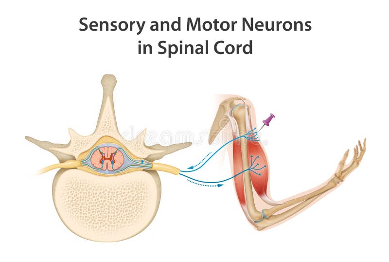
Free with trial Motor neurons of the spinal cord are part of the central nervous system and connect to muscles, glands and organs throughout the body. These neurons transmit impulses from the spinal cord to skeletal and smooth muscles, and so directly control all of our muscle movements. Muscle contraction illustrations Sensory and Motor Neurons in Spinal Cord. Motor neurons of the spinal cord are part of the central nervous system and connect to muscles, glands and organs throughout the body. These neurons transmit impulses from the spinal cord to skeletal and smooth muscles, and so directly control all of our muscle movements.
Free with trial Interior to the muscle layer is a fluid-filled chamber called a coelom that by its pressurization provides structure to the worm`s boneless body. The segments are separated from each other by septa which are perforated transverse walls, allowing the coelomic fluid to pass between segments. Muscle contraction illustrations Earthworm septum is a membrane that is flexible enough to allow for contraction of the internal muscles. Interior to the muscle layer is a fluid-filled chamber called a coelom that by its pressurization provides structure to the worm`s boneless body. The segments are separated from each other by septa which are perforated transverse walls, allowing the coelomic fluid to pass between segments
Free with trial Reflex is an involuntary, stereotyped response of an effector tissue that occurs when a receptor is stimulated. These reflexes are carried out by the sequential activation of a certain number of neurons that are interconnected. The last neuron usually innervates the effector tissue, which is usually a muscle. Muscle contraction illustrations Spinal Reflex Arc Anatomical Scheme. Reflex is an involuntary, stereotyped response of an effector tissue that occurs when a receptor is stimulated. These reflexes are carried out by the sequential activation of a certain number of neurons that are interconnected. The last neuron usually innervates the effector tissue, which is usually a muscle
Free with trial Medical illustration depicting an ectopic heartbeat. It shows three ECG readings: normal sinus rhythm, premature ventricular contraction (PVC), and premature atrial contraction (PAC). Each reading features labeled elements like the P wave. A heart diagram accompanies these graphs, highlighting anatomical structures and arteries. The illustration provides educational insight into the differences in cardiac electrical activity, focusing on irregular heartbeats. Muscle contraction vectors Ectopic heartbeat flashcard medical illustration. Medical illustration depicting an ectopic heartbeat. It shows three ECG readings: normal sinus rhythm, premature ventricular contraction (PVC), and premature atrial contraction (PAC). Each reading features labeled elements like the P wave. A heart diagram accompanies these graphs, highlighting anatomical structures and arteries. The illustration provides educational insight into the differences in cardiac electrical activity, focusing on irregular heartbeats.
Free with trial Histamine. Immune response and Allergic reaction. Histamine-releasing cells produce of Histamine then Stomach secretes gastric acid, smooth muscle is reduced and in the bronchi there are difficulties with breathing, Goblet cells secrete mucus, and Skin becomes redness and itching. Vector poster. Isometric flat illustration. Muscle contraction vectors Histamine. Immune response and Allergic reaction
Free with trial Molecule of oxytocin, a hormone released from the neurohypophysis, 3D illustration. It causes uterine contraction and milk ejection, used in gynecology and lactation treatment. Muscle contraction illustrations Molecule of oxytocin, a hormone released from the neurohypophysis
Free with trial Physical strength building concept icon. Sports energy drinks idea thin line illustration. Exercises. Training. Enhancing muscle contraction. Vector isolated outline RGB color drawing. Muscle contraction vectors Physical strength building concept icon
Free with trial Physical strength building concept icon. Sports energy drinks idea thin line illustration. Power performance. Enhancing muscle contraction. Vector isolated outline RGB color drawing. Editable stroke. Muscle contraction vectors Physical strength building concept icon
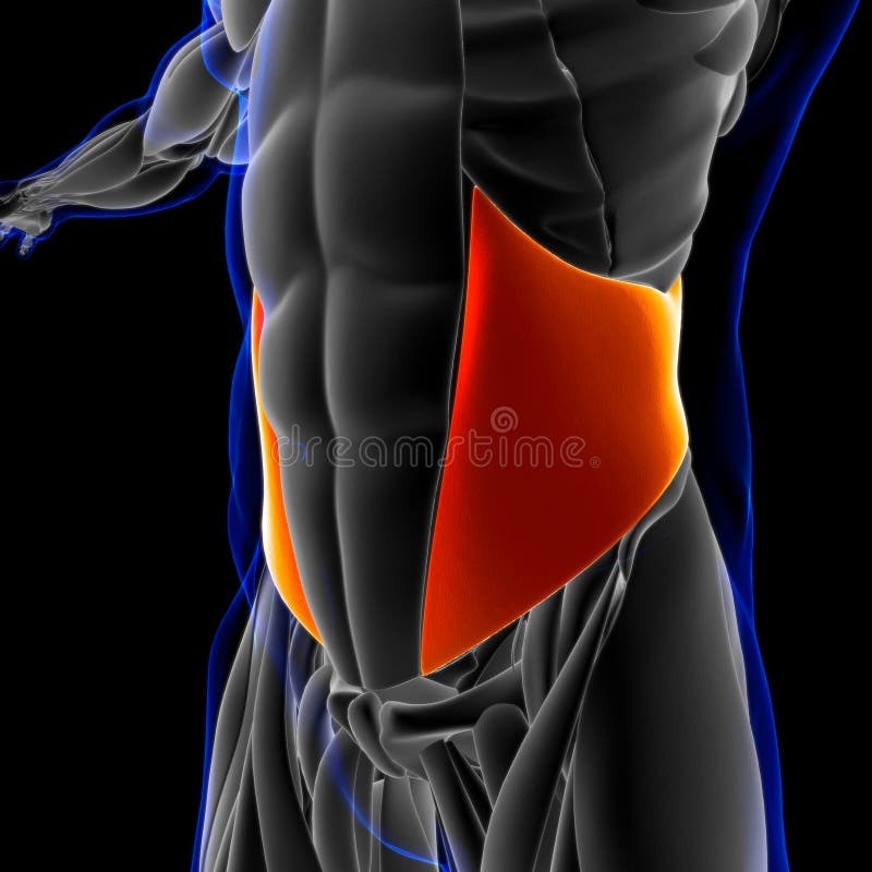
Free with trial 3D Illustration, Muscle is a soft tissue, Muscle cells contain proteins , producing a contraction that changes both the length and the shape of the cell. Muscles function to produce force and motion. Muscle contraction illustrations Internal Oblique Anatomy For Medical Concept 3D Illustration. 3D Illustration, Muscle is a soft tissue, Muscle cells contain proteins , producing a contraction that changes both the length and the shape of the cell. Muscles function to produce force and motion
Free with trial 3D Illustration, Muscle is a soft tissue, Muscle cells contain proteins , producing a contraction that changes both the length and the shape of the cell. Muscles function to produce force and motion. Muscle contraction illustrations Internal Oblique Anatomy For Medical Concept 3D Illustration. 3D Illustration, Muscle is a soft tissue, Muscle cells contain proteins , producing a contraction that changes both the length and the shape of the cell. Muscles function to produce force and motion
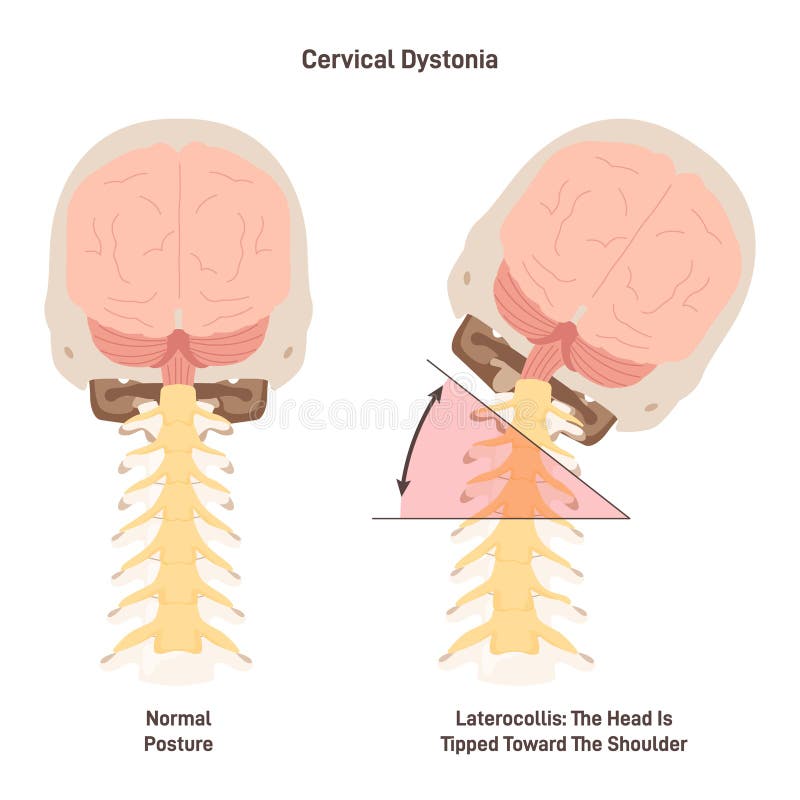
Free with trial Cervical dystonia. Spasmodic torticollis, inflamatory condition when neck muscles contract involuntarily. Muscle spasms or tremor. Flat vector illustration. Muscle contraction vectors Cervical dystonia. Spasmodic torticollis, inflamatory condition when neck
Free with trial Amyotrophic lateral sclerosis (ALS), also known as motor neurone disease (MND) or Lou Gehrig's disease in the United States, is a rare, terminal neurodegenerative disorder that results in the progressive loss of both upper and lower motor neurons that normally control voluntary muscle contraction. Muscle contraction illustrations Motor neuron damaged by Amyotrophic Lateral Sclerosis (ALS) - 3d illustration closeup view. Amyotrophic lateral sclerosis (ALS), also known as motor neurone disease (MND) or Lou Gehrig's disease in the United States, is a rare, terminal neurodegenerative disorder that results in the progressive loss of both upper and lower motor neurons that normally control voluntary muscle contraction.
Free with trial This detailed illustration reveals the complex interplay of the human cardiovascular system during the systolic and diastolic phases of the heart's rhythmic contraction and relaxation. The intricate network of blood vessels, including arteries, veins, and capillaries, is clearly depicted, showcasing the vital pathways for oxygen and nutrient transport throughout the body. Observe the precise. Muscle contraction illustrations A Detailed Visual Guide to the Human Hearts Systolic and Diastolic Cycles Unveiling the Intricate Network of Blood. This detailed illustration reveals the complex interplay of the human cardiovascular system during the systolic and diastolic phases of the heart's rhythmic contraction and relaxation. The intricate network of blood vessels, including arteries, veins, and capillaries, is clearly depicted, showcasing the vital pathways for oxygen and nutrient transport throughout the body. Observe the precise
Free with trial Blood clot formation, or hemostasis, is a rapid, multistep process that seals damaged blood vessels to stop bleeding. It involves direct vasoconstriction, the formation of a temporary platelet plug and the activation of clotting factors to form a stable fibrin mesh. Muscle contraction illustrations Blood clot formation, or hemostasis. Is a rapid, multistep process that seals damaged blood vessels to stop bleeding. It involves direct vasoconstriction, the. Blood clot formation, or hemostasis, is a rapid, multistep process that seals damaged blood vessels to stop bleeding. It involves direct vasoconstriction, the formation of a temporary platelet plug and the activation of clotting factors to form a stable fibrin mesh
Free with trial Esophageal Peristalsis Process with Food Bolus Moving Toward the Stomach. Human Digestive System Illustration Showing Esophagus Peristalsis Mechanism. Muscle contraction vectors Esophageal Peristalsis Process with Food Bolus Moving Toward the Stomach. Human Digestive System Illustration Showing Esophagus
Free with trial Motor neurons are large, multipolar lower motor neurons of the brainstem and spinal cord. Muscle contraction illustrations Motor neurons
Free with trial Fibrillin glycoprotein molecule on a white background. Muscle contraction illustrations Fibrillin glycoprotein molecule
Free with trial The body will use nutrients that provide energy when exercising, By based on factors from the blood system And primarily muscles, Waste such as perspiration and heat is released. Muscle contraction vectors Changing energy into active muscles. The body will use nutrients that provide energy when exercising, By based on factors from the blood system And primarily muscles, Waste such as perspiration and heat is released
Free with trial Gastrointestinal motility brief illustrates peristalsis and segmentation in the digestive tract, focusing on esophagus, intestines, and food bolus flow for mixing and propulsion. Outline diagram. Muscle contraction vectors Gastrointestinal motility brief illustrates peristalsis and segmentation in the digestive ... Gastrointestinal motility brief illustrates peristalsis and segmentation in the digestive tract, focusing on esophagus, intestines, and food bolus flow for mixing and propulsion. Outline diagram
Free with trial Motor neurons are large, multipolar lower motor neurons of the brainstem and spinal cord. Muscle contraction illustrations Motor neurons
Free with trial Congenital disease of the heart: Ventricular septal defect, Right ventricular hypertrophy, Pulmonic stenosis. Muscle contraction vectors Congenital disease of the heart
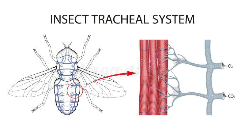
Free with trial For insects, respiration is separate from the circulatory system. Oxygen and carbon dioxide gases are exchanged through a network of tubes called tracheae. Instead of nostrils, insects breathe through openings in the thorax and abdomen called spiracles. Muscle contraction illustrations The Structure of the Tracheae of Insects. For insects, respiration is separate from the circulatory system. Oxygen and carbon dioxide gases are exchanged through a network of tubes called tracheae. Instead of nostrils, insects breathe through openings in the thorax and abdomen called spiracles
Free with trial The calcium channel is composed of a hexameric assembly or Orai subunits around a central ion pore. The channel shows selective permeability to calcium ions. Muscle contraction illustrations Pore of the calcium released-activated calcium CRAC channel. The calcium channel is composed of a hexameric assembly or Orai subunits around a central ion pore. The channel shows selective permeability to calcium ions.
Free with trial Motor neurons are large, multipolar lower motor neurons of the brainstem and spinal cord. Muscle contraction illustrations Motor neurons
Free with trial Motor neurons are large, multipolar lower motor neurons of the brainstem and spinal cord. Muscle contraction illustrations Motor neurons
Free with trial Motor neurons are large, multipolar lower motor neurons of the brainstem and spinal cord. Muscle contraction illustrations Motor neurons
Free with trial Motor neurons are large, multipolar lower motor neurons of the brainstem and spinal cord. Muscle contraction illustrations Motor neurons
Free with trial Myasthenia gravis. Girl with neuromuscular disease. patient with ptosis and Left lower facial weakness. Symptoms of Autoimmune disease. Vector poster. Muscle contraction vectors Myasthenia gravis. Girl with neuromuscular disease
Free with trial Motor neurons are large, multipolar lower motor neurons of the brainstem and spinal cord. Muscle contraction illustrations Neuron Motor. Motor neurons are large, multipolar lower motor neurons of the brainstem and spinal cord.
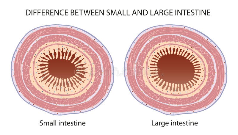
Free with trial Small intestine is narrow in width of around 3. 5 – 4. 5 cm. Large intestine has width of around 4 – 6 cm in diameter. Villi is present in small intestine. Villi is absent in large intestine. Muscle contraction illustrations Difference Between Small and Large Intestine. Small intestine is narrow in width of around 3.5 – 4.5 cm. Large intestine has width of around 4 – 6 cm in diameter. Villi is present in small intestine. Villi is absent in large intestine.
Free with trial Medical Concept: Scleroderma - Text on Black Chalkboard with Orange Stethoscope. Medical Concept: Scleroderma Handwritten on Black Chalkboard. 3D Rendering. Muscle contraction illustrations Scleroderma on Chalkboard. 3D Illustration. Medical Concept: Scleroderma - Text on Black Chalkboard with Orange Stethoscope. Medical Concept: Scleroderma Handwritten on Black Chalkboard. 3D Rendering.
Free with trial The esophagus or oesophagus is an organ in vertebrates through which food passes, aided by peristaltic contractions, from the pharynx to the stomach. The esophagus is a fibromuscular tube, that travels behind the trachea and heart. During swallowing, the epiglottis tilts backwards to prevent food from going down the larynx and lungs. The trachea is the long tube that connects your larynx to your bronchi. Bronchi send air to your lungs. Trachea is a key part of respiratory system. The trachea is made of rings of cartilage. It is lined with cells that produce mucus. Muscle contraction illustrations Trachea and Esophagus cross section. The esophagus or oesophagus is an organ in vertebrates through which food passes, aided by peristaltic contractions, from the pharynx to the stomach. The esophagus is a fibromuscular tube, that travels behind the trachea and heart. During swallowing, the epiglottis tilts backwards to prevent food from going down the larynx and lungs.The trachea is the long tube that connects your larynx to your bronchi. Bronchi send air to your lungs. Trachea is a key part of respiratory system. The trachea is made of rings of cartilage. It is lined with cells that produce mucus
Free with trial Different muscles in human body and muscular classification outline diagram. Labeled educational physiological parts scheme with anatomic skeletal, smooth and cardiac division vector illustration. Muscle contraction vectors Different muscles in human body and muscular classification outline diagram
Free with trial Reflex reaction with knee stimulus test process explanation outline diagram. Labeled educational scheme with anatomical body reaction to impulse vector illustration. Receptors or sensory neuron check. Muscle contraction vectors Reflex reaction with knee stimulus test process explanation outline diagram.
Free with trial Congenital heart disease awareness poster with sad cartoon heart on white background. Human body organs anatomy icon. Medical concept. Vector illustration. Muscle contraction vectors Congenital heart disease poster. Congenital heart disease awareness poster with sad cartoon heart on white background. Human body organs anatomy icon. Medical concept. Vector illustration.
Free with trial The heart is a muscular organ that pumps blood around the body by circulating it through the circulatory system. It is found in the middle mediastinum, wrapped in a two-layered serous sac called the pericardium. Muscle contraction illustrations Anatomy of the Human Heart. The heart is a muscular organ that pumps blood around the body by circulating it through the circulatory system. It is found in the middle mediastinum, wrapped in a two-layered serous sac called the pericardium
Free with trial The structure of the motor neuron. Infographics. Vector illustration on isolated background. Muscle contraction vectors The structure of the motor neuron. Infographics. Vector illustration on isolated background
Free with trial Degradation of motor neurons, conceptual 3D illustration. Motor neuron diseases are a group of neurodegenerative disorders including amyotrophic lateral sclerosis, progressive bulbar palsy and other. Muscle contraction illustrations Degradation of motor neurons, conceptual 3D illustration
Free with trial 3d Illustration of heart strings Tendons, inside the human heart. Muscle contraction illustrations 3d Illustration of heart strings Tendons, inside the human heart
Free with trial Motor neuron diseases, 3D illustration showing degeneration of motor neurons in anterior horns of spinal cord. Amyotrophic lateral sclerosis and other motor neuron disorders. Muscle contraction illustrations Motor neuron diseases, 3D illustration
Free with trial Demyelination of neuron, the damage of the neuron myelin sheath seen in demyelinating diseases, 3D illustration. Multiple sclerosis and other demyelinating myelinoclastic and leukodystrophic diseases. Muscle contraction illustrations Demyelination of neuron, the damage of the neuron myelin sheath seen in demyelinating diseases
Free with trial Demyelination of neuron, the damage of the neuron myelin sheath seen in demyelinating diseases, 3D illustration. Multiple sclerosis and other demyelinating myelinoclastic and leukodystrophic diseases. Muscle contraction illustrations Demyelination of a neuron, the damage of the neuron myelin sheath seen in demyelinating diseases. Demyelination of neuron, the damage of the neuron myelin sheath seen in demyelinating diseases, 3D illustration. Multiple sclerosis and other demyelinating myelinoclastic and leukodystrophic diseases
Free with trial Running man muscles knee and ankle Human Body. Muscle contraction illustrations Running man muscles
Free with trial Degradation of motor neurons, conceptual 3D illustration. Motor neuron diseases are a group of neurodegenerative disorders including amyotrophic lateral sclerosis, progressive bulbar palsy and other. Muscle contraction illustrations Degradation of motor neurons, conceptual 3D illustration
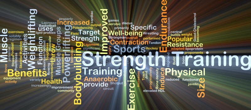
Free with trial Background concept wordcloud illustration of strength training glowing light. Muscle contraction illustrations Strength training background concept glowing. Background concept wordcloud illustration of strength training glowing light
Free with trial Degradation of motor neurons, conceptual 3D illustration. Motor neuron diseases are a group of neurodegenerative disorders including amyotrophic lateral sclerosis, progressive bulbar palsy and other. Muscle contraction illustrations Degradation of motor neurons, conceptual 3D illustration
Free with trial Demyelination of neuron, the damage of the neuron myelin sheath seen in demyelinating diseases, 3D illustration. Multiple sclerosis and other demyelinating myelinoclastic and leukodystrophic diseases. Muscle contraction illustrations Demyelination of neuron, the damage of the neuron myelin sheath seen in demyelinating diseases
Free with trial Men's skeleton, race knee inflammation pain. Muscle contraction illustrations Men's skeleton, knee inflammation. Men's skeleton, race knee inflammation pain
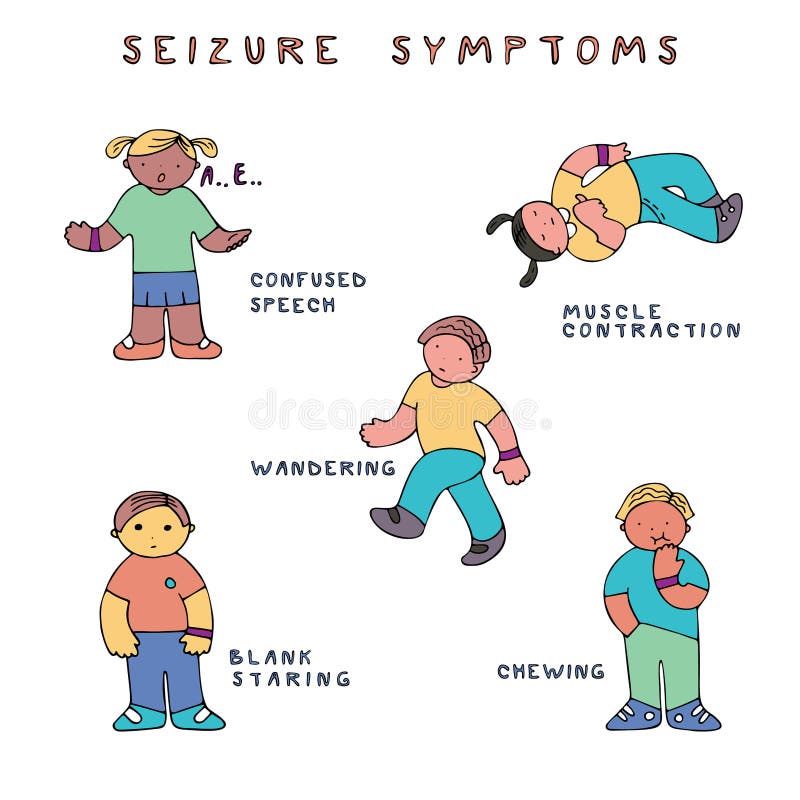
Free with trial Set of kids with seizure symptoms. Fine for medical infobrochures for kids and teenagers, public sites about epilepsy and medical checks, banners for sites about epilepsy. Muscle contraction vectors Epilepsy seizure symptoms. Set of kids with seizure symptoms. Fine for medical infobrochures for kids and teenagers, public sites about epilepsy and medical checks, banners for sites about epilepsy.
Free with trial Motor neuron diseases, 3D illustration showing degeneration of motor neurons in anterior horns of spinal cord. Amyotrophic lateral sclerosis and other motor neuron disorders. Muscle contraction illustrations Motor neuron diseases, 3D illustration
Free with trial Close-up of a muscular black man, back view of a bodybuilder athlete, isolated on a black background with copy space. Generative Ai. Muscle contraction illustrations Muscular Man Back View of a Bodybuilder Athlete on a Black Background - Generative Ai. Close-up of a muscular black man, back view of a bodybuilder athlete, isolated on a black background with copy space. Generative Ai
Free with trial Running man muscles ankle Human Body. Muscle contraction illustrations Running man muscles ankle