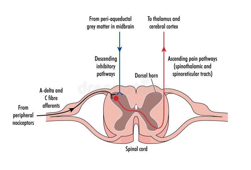
Free with trial Cross section of spinal cord showing ascending and descending nerve tracts, plus incoming A-delta and C-fibre afferent nerves from peripheral nociceptors. Nociceptors vectors Cross section of spinal cord
Free with trial Noxious and pain receptors in skin and the nerve pathways to the brain, via the spinal cord and thalamus. Created in Adobe Illustrator. Contains transparencies. EPS 10. Nociceptors vectors Nerve response to pain and touch. Noxious and pain receptors in skin and the nerve pathways to the brain, via the spinal cord and thalamus. Created in Adobe Illustrator. Contains transparencies. EPS 10.
Free with trial Cells act as external stimuli in different environments. change to act as internal stimuli. When the senses are sent signal information to the nervous system. Which will eventually reach the brain or spinal cord. Nociceptors vectors Sensory neuron. Cells act as external stimuli in different environments. Cells act as external stimuli in different environments. change to act as internal stimuli. When the senses are sent signal information to the nervous system. Which will eventually reach the brain or spinal cord
Free with trial Detailed illustration of the somatosensory pathway, depicting the different types of sensory neurons and their connections to the brain. The diagram shows the pathways for both noxious stimuli (pain) and touch, highlighting the roles of nociceptors and mechanoreceptors. The diagram also illustrates the connections to the thalamus and cortex, as well as to the limbic system. Nociceptors illustrations Diagram illustrating the somatosensory pathway. Detailed illustration of the somatosensory pathway, depicting the different types of sensory neurons and their connections to the brain. The diagram shows the pathways for both noxious stimuli (pain) and touch, highlighting the roles of nociceptors and mechanoreceptors. The diagram also illustrates the connections to the thalamus and cortex, as well as to the limbic system
Free with trial Detailed anatomical illustration of the somatosensory pathway, depicting the transmission of nerve signals from a hand to the brain, including nociceptors, primary afferents, and different fiber types. Nociceptors illustrations Somatosensory Pathway Diagram Illustrating Nerve Signal Transmission. Detailed anatomical illustration of the somatosensory pathway, depicting the transmission. Detailed anatomical illustration of the somatosensory pathway, depicting the transmission of nerve signals from a hand to the brain, including nociceptors, primary afferents, and different fiber types
Free with trial Delve into the intricate network of sensory cells within the human skin. This microscopic image showcases the delicate nerve endings and receptors responsible for our sense of touch, pressure, temperature, and pain. The bokeh effect beautifully isolates these cellular structures, highlighting their complex morphology and the intricate connections that translate external stimuli into sensory. Nociceptors illustrations Unveiling the Microscopic World of Skin Sensory Cells A Deep Dive into Nerve Endings Receptors and Sensory Perception. Delve into the intricate network of sensory cells within the human skin. This microscopic image showcases the delicate nerve endings and receptors responsible for our sense of touch, pressure, temperature, and pain. The bokeh effect beautifully isolates these cellular structures, highlighting their complex morphology and the intricate connections that translate external stimuli into sensory
Free with trial Nerve ending transmitting pain signals,intricate structures and electrical impulses. Nociceptors illustrations Nerve ending transmitting pain signals,intricate structures and electrical impulses
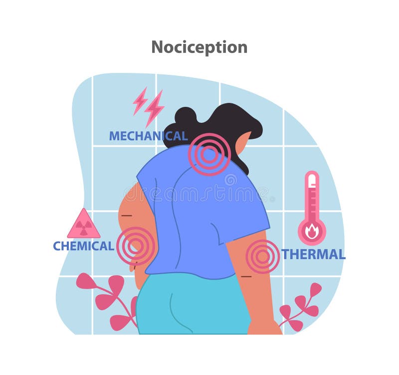
Free with trial Nociception illustration. Visualization of the body's response to mechanical, chemical, and thermal stimuli. Pain perception and sensory signals. Flat vector illustration. Nociceptors vectors Nociception illustration. Visualization of the
Free with trial This illustration depicts the various receptors found in human skin, including Meissner's corpuscles, Ruffini endings, Merkel corpuscles, and Pacinian corpuscles. These receptors play a crucial role in touch, pressure, temperature, and other sensations. Nociceptors illustrations Human Skin Receptors. This illustration depicts the various receptors found in human skin, including Meissner's corpuscles, Ruffini endings, Merkel corpuscles, and Pacinian corpuscles. These receptors play a crucial role in touch, pressure, temperature, and other sensations.
Free with trial Nociception illustration. Visualization of the body's response to mechanical, chemical, and thermal stimuli. Pain perception and sensory signals. Flat vector illustration. Nociceptors vectors Nociception illustration. Visualization of the body's response to mechanical. Nociception illustration. Visualization of the body's response to mechanical, chemical, and thermal stimuli. Pain perception and sensory signals. Flat vector illustration.
Free with trial Explore the intricate network of sensory receptors beneath the surface of human skin in stunning detail. This captivating long exposure photograph showcases the astonishing texture and delicate architecture of the skin at a microscopic level. The extended exposure time allows us to visualize the subtle variations in skin topography, highlighting the complex interplay of epidermal layers, dermal. Nociceptors illustrations Unveiling the Microscopic Wonders of Human Skin Sensory Receptors Revealed Through Long Exposure Photography. Explore the intricate network of sensory receptors beneath the surface of human skin in stunning detail. This captivating long exposure photograph showcases the astonishing texture and delicate architecture of the skin at a microscopic level. The extended exposure time allows us to visualize the subtle variations in skin topography, highlighting the complex interplay of epidermal layers, dermal
Free with trial 3D image of Piperine skeletal formula - molecular chemical structure of alkaloid Bioperine isolated on white background. Nociceptors illustrations 3D image of Piperine skeletal formula