Free with trial Two types of skin melanoma on the neck and back of a man. Nodular surface vectors Two types of melanoma. Two types of skin melanoma on the neck and back of a man
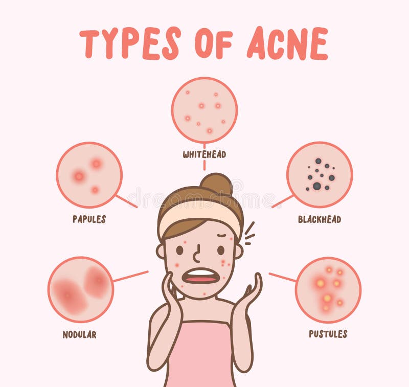
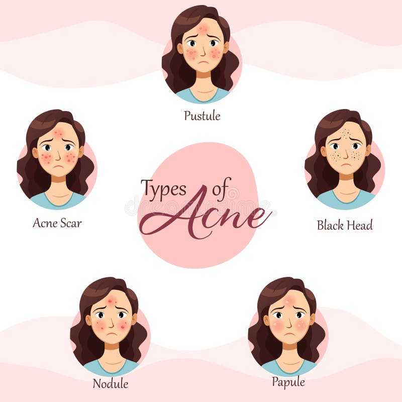
Free with trial Types of acne with woman cartoon illustration vector on pink background. Beauty concept. Nodular surface vectors Types of acne with woman cartoon illustration vector on pink background. Beauty concept.
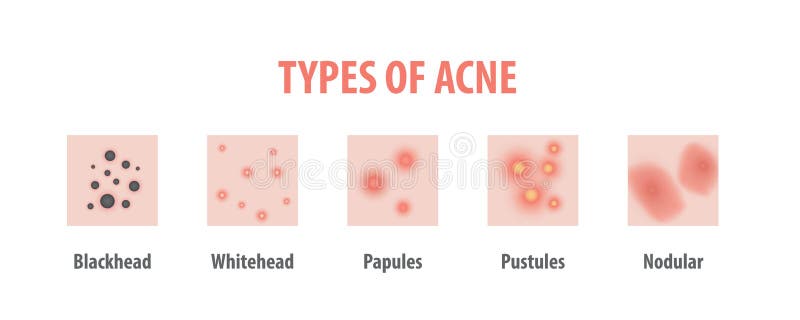
Free with trial Types of acne diagram illustration vector on white background, Beauty concept. Nodular surface vectors Types of acne diagram illustration vector on white background, B
Free with trial Types of acne diagram illustration vector on white background, Beauty concept. Nodular surface vectors Types of acne diagram illustration vector on white background, B
Free with trial Types of acne infographics. Vector illustration types of acne, pimples, skin pores, blackhead, whitehead, scar comedones. Nodular surface vectors Types of acne infographics. Vector illustration types of acne, pimples, skin pores, blackhead, whitehead, scar
Free with trial Types of acne vector on white background. Formation of noninflammatory acne, whitehead, blackhead, inflammatory acne, papule, pustule, nodule and cyst. Skin care and beauty concept illustration. Nodular surface illustrations Types of acne vector on white background. Formation of noninflammatory and inflammatory acne. Types of acne vector on white background. Formation of noninflammatory acne, whitehead, blackhead, inflammatory acne, papule, pustule, nodule and cyst. Skin care and beauty concept illustration.
Free with trial This captivating image showcases the intricate and rugged exterior of a stony capsule shell. The surface exhibits a diverse array of textures, providing a close-up view of its unique characteristics. From coarse, granular, and pebbled areas to nodular lumps and pitted depressions, the shell's exterior is a testament to its complex formation and possible history. The irregular, uneven topography. Nodular surface illustrations Examining the Rugged Exterior of a Stony Capsule Shell A Detailed Look at Texture Surface Features and Potential. This captivating image showcases the intricate and rugged exterior of a stony capsule shell. The surface exhibits a diverse array of textures, providing a close-up view of its unique characteristics. From coarse, granular, and pebbled areas to nodular lumps and pitted depressions, the shell's exterior is a testament to its complex formation and possible history. The irregular, uneven topography
Free with trial Detailed botanical illustration of a cactus with a bumpy, nodular surface and muted green tones on a beige background. Nodular surface illustrations Botanical Study of Bumpy Cactus Body. Detailed botanical illustration of a cactus with a bumpy, nodular surface and muted green tones on a beige background.
Free with trial An unusual cactus variety with a compact, egg-like silhouette and richly detailed surface structure, drawn in soft natural hues. Nodular surface illustrations Textured Cactus Form with Lumpy Surface. An unusual cactus variety with a compact, egg-like silhouette and richly detailed surface structure, drawn in soft natural hues.
Free with trial Tie-dye pattern of indigo color on white silk, nodular batik. Shibori dyeing, Generative AI. Nodular surface illustrations Tie-dye pattern of indigo color on white silk, nodular batik.
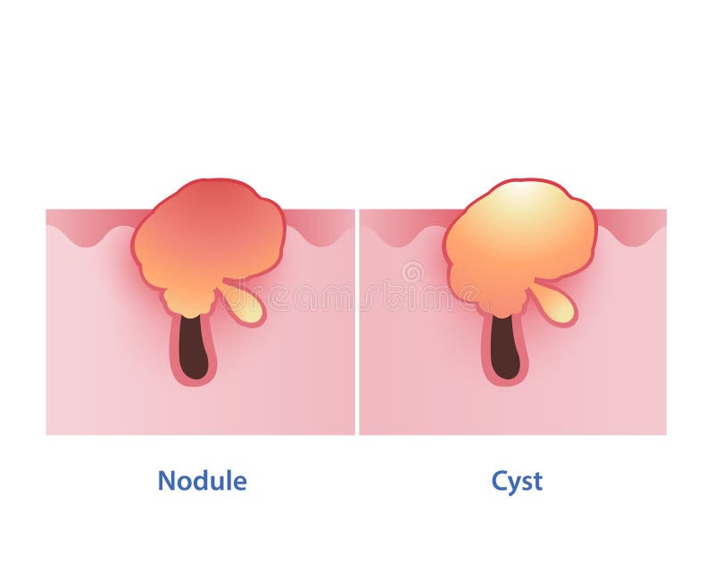
Free with trial The difference between nodule and cyst vector on white background. Nodular is firm, painful, red bump on the surface skin. Cystic acne consist of deep, pus filled. The both are inflammatory severe. Nodular surface illustrations The difference between nodule and cystic acne vector isolated on white background. The difference between nodule and cyst vector on white background. Nodular is firm, painful, red bump on the surface skin. Cystic acne consist of deep, pus filled. The both are inflammatory severe.
Free with trial Tie-dye pattern of indigo color on white silk, nodular batik. Shibori dyeing, Generative AI. Nodular surface illustrations Tie-dye pattern indigo and white silk. nodular batik fabrics. Shibori dyeing. tie-dye pattern of indigo color on white silk, nodular batik. Shibori dyeing, Generative AI
Free with trial Patterns and banners backgrounds: Seamless tie-dye pattern of indigo color on white silk. Hand painting fabrics - nodular batik. Shibori dyeing. Nodular surface vectors Seamless tie-dye pattern of indigo color on white silk. Hand painting fabrics - nodular batik. Shibori dyeing
Free with trial Patterns and banners backgrounds: Seamless tie-dye pattern of pink and red color on white silk. Hand painting fabrics - nodular batik. Shibori dyeing. Nodular surface vectors Seamless tie-dye pattern of pink and red color on white silk. Hand painting fabrics - nodular batik. Shibori dyeing
Free with trial AI-generated intricate, fractal sculpture resembling an abstract cosmic structure with a swirling, hollow center. The sculpture features complex, organic shapes and textures, primarily in shades of pink and red, suggesting dynamic movement and energy. The surface is adorned with bulbous, nodular patterns, enhancing its three-dimensional appearance. Warm light emanates from within, creating a glowing effect that highlights the detailed contours. This art piece merges natural forms with mathematical precision, evoking the expansive and intricate nature of cosmic phenomena. Nodular surface illustrations Fractal sculpture of a supernova. AI-generated intricate, fractal sculpture resembling an abstract cosmic structure with a swirling, hollow center. The sculpture features complex, organic shapes and textures, primarily in shades of pink and red, suggesting dynamic movement and energy. The surface is adorned with bulbous, nodular patterns, enhancing its three-dimensional appearance. Warm light emanates from within, creating a glowing effect that highlights the detailed contours. This art piece merges natural forms with mathematical precision, evoking the expansive and intricate nature of cosmic phenomena.
Free with trial Microscopic view of a malignant melanoma, showcasing its irregular shape and nodular texture. The image displays the lesion's reddish-pink hue against a solid blue background. High-resolution detail reveals the melanoma's cellular structure and uneven surface. The lighting is even, providing a clear and detailed view of the cancerous cells. Nodular surface illustrations Microscopic View of Malignant Melanoma: Reddish-Pink Nodular Lesion on Blue Background. Microscopic view of a malignant melanoma, showcasing its irregular shape and nodular texture. The image displays the lesion's reddish-pink hue against a solid blue background. High-resolution detail reveals the melanoma's cellular structure and uneven surface. The lighting is even, providing a clear and detailed view of the cancerous cells
Free with trial A macro perspective reveals the intricate and chaotic beauty of severe metal decay. Spherical rust tubercles burst forth from a corroded surface, creating a dynamic landscape of rich, earthy tones. This image captures the powerful process of oxidation, highlighting the abstract patterns found in industrial aging and decomposition. Nodular surface illustrations Intense Rust Tubercles - An Abstract Corrosion Background. A macro perspective reveals the intricate and chaotic beauty of severe metal decay. Spherical rust tubercles burst forth from a corroded surface, creating a dynamic landscape of rich, earthy tones. This image captures the powerful process of oxidation, highlighting the abstract patterns found in industrial aging and decomposition
Free with trial A naturalistic depiction of a cactus featuring clustered bulges across its form, resembling a fusion of botanical and mineral textures. Nodular surface illustrations Egg-Shaped Cactus with Nodular Texture. A naturalistic depiction of a cactus featuring clustered bulges across its form, resembling a fusion of botanical and mineral textures.
Free with trial Artistic study of an exotic cactus species, highlighting the uneven surface, complex texture, and organic symmetry. Nodular surface illustrations Natural Illustration of Rare Cactus Structure. Artistic study of an exotic cactus species, highlighting the uneven surface, complex texture, and organic symmetry.
Free with trial This striking image reveals a high-magnification view of two distinct, densely textured materials or biological tissues meeting along a diagonal frontier. The structures are vividly rendered in contrasting false colors, deep magenta and neon green, highlighting their intricate, sponge-like surface morphology. Rendered by Ai, this visualization emphasizes the complexity and roughness of the. Nodular surface illustrations Detailed electron micrograph of a microscopic interface. This striking image reveals a high-magnification view of two distinct, densely textured materials or biological tissues meeting along a diagonal frontier. The structures are vividly rendered in contrasting false colors, deep magenta and neon green, highlighting their intricate, sponge-like surface morphology. Rendered by Ai, this visualization emphasizes the complexity and roughness of the
Free with trial Acne types include whiteheads, blackheads, papules, pustules, nodules, and cysts. Each varies in severity, from mild surface blemishes to deep, painful lumps. Nodular surface vectors Decoding Acne A Comprehensive Guide to Different Types and Effective Treatments. Acne types include whiteheads, blackheads, papules, pustules, nodules, and cysts. Each varies in severity, from mild surface blemishes to deep, painful lumps
Free with trial This striking macro photograph captures a heavily textured metallic geological object shaped like a closed human fist resting on a sleek black pedestal. Rendered by Ai this fascinating specimen exhibits deep fissures and a rough, nodular surface, suggesting immense pressure and ancient origin. The dark background and powerful overhead illumination highlight the object's imposing form and rugged. Nodular surface illustrations A dramatic spotlight illuminates a cracked iron fist sculpture. This striking macro photograph captures a heavily textured metallic geological object shaped like a closed human fist resting on a sleek black pedestal. Rendered by Ai this fascinating specimen exhibits deep fissures and a rough, nodular surface, suggesting immense pressure and ancient origin. The dark background and powerful overhead illumination highlight the object's imposing form and rugged
Free with trial This striking image reveals a high-magnification view of two distinct, densely textured materials or biological tissues meeting along a diagonal frontier. The structures are vividly rendered in contrasting false colors, deep magenta and neon green, highlighting their intricate, sponge-like surface morphology. Rendered by Ai, this visualization emphasizes the complexity and roughness of the. Nodular surface illustrations Detailed electron micrograph of a microscopic interface. This striking image reveals a high-magnification view of two distinct, densely textured materials or biological tissues meeting along a diagonal frontier. The structures are vividly rendered in contrasting false colors, deep magenta and neon green, highlighting their intricate, sponge-like surface morphology. Rendered by Ai, this visualization emphasizes the complexity and roughness of the
Free with trial Diagram of inflammatory acne types vector illustration isolated on white background. Papule, pustule, nodule, nodular, nodulocystic cystic acne and cyst. Skin care and beauty concept. Nodular surface illustrations Diagram of inflammatory acne types vector illustration isolated on white background.
Free with trial This captivating image reveals a profound narrative etched into weathered wooden planks. Thick, rough layers of paint, in a vibrant array of colors, cling to the wood's surface, showcasing a rich history of application and wear. The peeling paint reveals the underlying wood grain, a subtle texture that speaks volumes about the passage of time. Each layer of paint tells a story, with brushstrokes. Nodular surface illustrations A Tapestry of Time Intriguing Weathered Wooden Planks Showcasing a History of Color and Texture Through Layers of. This captivating image reveals a profound narrative etched into weathered wooden planks. Thick, rough layers of paint, in a vibrant array of colors, cling to the wood's surface, showcasing a rich history of application and wear. The peeling paint reveals the underlying wood grain, a subtle texture that speaks volumes about the passage of time. Each layer of paint tells a story, with brushstrokes
Free with trial Plantar fibroma. Plantar fascial fibromatosis. Ledderhoses disease. Morbus Ledderhose. Plantar fibromatosis. Detailed Vector poster. Nodular surface vectors Plantar fibroma. Plantar fascial fibromatosis
Free with trial Plantar fascial fibromatosis. Plantar fibroma. Ledderhoses disease. Morbus Ledderhose. Skeleton of foot. Medial aspect of bones and plantar fascia with non-malignant thickening of connective tissue. Detailed Vector poster. Nodular surface vectors Plantar fascial fibromatosis. Plantar fibroma
Free with trial Thyroid gland 3d model, two angles. 3D rendering isolated on white background. Nodular surface illustrations Thyroid gland 3d model, two angles. 3D rendering
Free with trial This vintage artwork showcases a rare cactus form with a dense pattern of lumps and a smooth, matte texture. Nodular surface illustrations Vintage Drawing of Knobby Green Succulent. This vintage artwork showcases a rare cactus form with a dense pattern of lumps and a smooth, matte texture.
Free with trial Comparison of a healthy liver and a cirrhotic liver. On the left, the healthy liver appears smooth and uniform with a glossy surface. Blood vessels and bile ducts are visible in blue and yellow. On the right, the cirrhotic liver is irregular and nodular, indicating damage and scarring. A circular inset shows a closer view of the liver tissue, highlighting fibrous scar tissue and regenerating nodules. The contrast illustrates the effects of liver disease on organ texture and structure. Nodular surface illustrations Healthy vs. Cirrhotic Liver. Comparison of a healthy liver and a cirrhotic liver. On the left, the healthy liver appears smooth and uniform with a glossy surface. Blood vessels and bile ducts are visible in blue and yellow. On the right, the cirrhotic liver is irregular and nodular, indicating damage and scarring. A circular inset shows a closer view of the liver tissue, highlighting fibrous scar tissue and regenerating nodules. The contrast illustrates the effects of liver disease on organ texture and structure.
Free with trial Sea urchin skeleton displayed against a black background, highlighting its intricate texture. The shell is round with a central hole, featuring a symmetrical pattern of raised, nodular structures. The surface coloration is a pale, off-white, emphasizing the detailed geometric ridges and depressions. The stark contrast with the dark backdrop accentuates the skeleton's delicate and complex design, showcasing its natural beauty and structural intricacy. Nodular surface illustrations Exotic sea urchin skeleton with intricate texture against a stark black background. Sea urchin skeleton displayed against a black background, highlighting its intricate texture. The shell is round with a central hole, featuring a symmetrical pattern of raised, nodular structures. The surface coloration is a pale, off-white, emphasizing the detailed geometric ridges and depressions. The stark contrast with the dark backdrop accentuates the skeleton's delicate and complex design, showcasing its natural beauty and structural intricacy.
Free with trial Close-up view of bauxite highlighting rounded pisolites, porous surface texture, and iron oxide staining on an aluminum ore rock. Nodular surface illustrations Natural bauxite rock showing rounded nodules and iron rich staining. Close-up view of bauxite highlighting rounded pisolites, porous surface texture, and iron oxide staining on an aluminum ore rock.
Free with trial Mineralogy documentation photo of bauxite ore illustrating pisolitic fabric and natural surface relief for textbooks and catalogs. Nodular surface illustrations Studio closeup of bauxite illustrating lateritic ore formation textures. Mineralogy documentation photo of bauxite ore illustrating pisolitic fabric and natural surface relief for textbooks and catalogs.
Free with trial Macro view documents fracture network geometry and brown host rock remnants, supporting mineralogy reference layouts and teaching materials. Nodular surface illustrations Copper aluminum phosphate gemstone rough with mottled surface and veined pattern. Macro view documents fracture network geometry and brown host rock remnants, supporting mineralogy reference layouts and teaching materials.
Free with trial This image displays a three-dimensional anatomical model of a human liver featuring multiple nodular growths on its surface. The liver is shown with attached blood vessels, including the hepatic artery and portal vein, which appear to be connected to the liver's lower section. The nodules suggest potential pathological changes such as cysts or tumors, providing a clear visual representation for. Nodular surface illustrations Detailed anatomical illustration of a human liver with visible nodules and blood vessels. This image displays a three-dimensional anatomical model of a human liver featuring multiple nodular growths on its surface. The liver is shown with attached blood vessels, including the hepatic artery and portal vein, which appear to be connected to the liver's lower section. The nodules suggest potential pathological changes such as cysts or tumors, providing a clear visual representation for
Free with trial This image displays a three-dimensional anatomical model of a human liver featuring multiple nodular growths on its surface. The liver is shown with attached blood vessels, including the hepatic artery and portal vein, which appear to be connected to the liver's lower section. The nodules suggest potential pathological changes such as cysts or tumors, providing a clear visual representation for. Nodular surface illustrations Detailed anatomical illustration of a human liver with visible nodules and blood vessels. This image displays a three-dimensional anatomical model of a human liver featuring multiple nodular growths on its surface. The liver is shown with attached blood vessels, including the hepatic artery and portal vein, which appear to be connected to the liver's lower section. The nodules suggest potential pathological changes such as cysts or tumors, providing a clear visual representation for
Free with trial A beige limestone with smooth, embedded gray chert nodules. Isolated on transparent background. Nodular surface vectors A beige limestone with smooth, embedded gray chert nodules
Free with trial Types of acne 3D icons - pimples, blackhead, whitehead. Problems in skin pores. Nodular surface vectors Types of acne pimples, blackhead, whitehead. Types of acne 3D icons - pimples, blackhead, whitehead. Problems in skin pores
Free with trial Highly detailed and scientifically accurate illustration of a human liver affected by cirrhosis, showcasing the characteristic nodular lesions and fibrotic scarring, with a subtle gradient of blues and purples to convey a sense of disease and toxicity, set against a muted, creamy background to represent the liver's natural texture, with precise anatomical features, including the gallbladder and hepatic veins, with subtle hints of redness and inflammation around the damaged areas, and a faint network of blood vessels visible beneath the surface. Nodular surface illustrations Realistic illustration of cirrhosis of human liverRealistic illustration of cirrhosis of human liver AI generated. Highly detailed and scientifically accurate illustration of a human liver affected by cirrhosis, showcasing the characteristic nodular lesions and fibrotic scarring, with a subtle gradient of blues and purples to convey a sense of disease and toxicity, set against a muted, creamy background to represent the liver's natural texture, with precise anatomical features, including the gallbladder and hepatic veins, with subtle hints of redness and inflammation around the damaged areas, and a faint network of blood vessels visible beneath the surface.
Free with trial This captivating close-up image reveals the remarkable detail of the human tongue's surface. The high contrast accentuates the diverse textures, from velvety smoothness to rough, bumpy areas. Tiny papillary structures, ridges, and subtle wrinkles are clearly visible, showcasing the intricate anatomy of the oral cavity. The image highlights the nuanced interplay of different surface. Nodular surface illustrations Unveiling the Intricate Anatomy of the Human Tongue A Closeup Exploration of Textures and Structures. This captivating close-up image reveals the remarkable detail of the human tongue's surface. The high contrast accentuates the diverse textures, from velvety smoothness to rough, bumpy areas. Tiny papillary structures, ridges, and subtle wrinkles are clearly visible, showcasing the intricate anatomy of the oral cavity. The image highlights the nuanced interplay of different surface
Free with trial Nodule, type of inflammatory acne vector on white background. Normal skin and nodular acne is firm, painful lump to form under your skin and red bump is inflamed to appear on the skin surface. Nodular surface illustrations Normal skin and Nodule, type of inflammatory acne vector on white background. Nodule, type of inflammatory acne vector on white background. Normal skin and nodular acne is firm, painful lump to form under your skin and red bump is inflamed to appear on the skin surface.
Free with trial Types of Acne, Pimples Blackheads, Whitehead, Papules, Cystic and Nodular eps file. Nodular surface vectors Types of Acne, Pimples Blackheads, Whitehead, Papules, Cystic and Nodular
Free with trial Different types of acne. Acne skin types - blackheads, whiteheads, papules, pustules, cystic and nodular. Skincare problems and inflammation. Inflamed skin with scars. Nodular surface vectors Different types of acne. Acne skin types. Blackheads, whiteheads, papules, pustules, cystic and nodular. Skincare. Different types of acne. Acne skin types - blackheads, whiteheads, papules, pustules, cystic and nodular. Skincare problems and inflammation. Inflamed skin with scars
Free with trial Acne types set. Skin pimples, blackheads and face comedones. Skincare problems and inflammation. Acne skin types - blackheads, whiteheads, papules, pustules, cystic and nodular. Nodular surface vectors Acne types set. Skin pimples, blackheads and face comedones. Acne skin types - blackheads, whiteheads, papules, pustules. Acne types set. Skin pimples, blackheads and face comedones. Skincare problems and inflammation. Acne skin types - blackheads, whiteheads, papules, pustules, cystic and nodular
Free with trial Types of acne illustration. Pimples, skin pores, blackhead, whitehead, scar, comedone. Skincare problems and inflammation. Acne skin types - blackheads, whiteheads, papules pustules cystic nodular. Nodular surface vectors Types of acne illustration. Pimples, skin pores, blackhead, whitehead, scar, comedone. Skincare problems and
Free with trial Stages of development of acne. Inflamed skin with scars, acne and pimples. Skincare problems and inflammation. Blackheads, whiteheads, papules, pustules, cystic and nodular. Nodular surface vectors Stages of development of acne. Inflamed skin with scars, acne and pimples. Blackheads, whiteheads, papules, pustules. Stages of development of acne. Inflamed skin with scars, acne and pimples. Skincare problems and inflammation. Blackheads, whiteheads, papules, pustules, cystic and nodular
Free with trial Grade of acne severity. Medical diagram different types acne. Acne skin types - blackheads, whiteheads, papules, pustules, cystic and nodular. Nodular surface vectors Grade of acne severity. Medical diagram different types acne. Blackheads, whiteheads, papules, pustules, cystic and. Grade of acne severity. Medical diagram different types acne. Acne skin types - blackheads, whiteheads, papules, pustules, cystic and nodular.
Free with trial Types of acne illustration vector on white background, Beauty concept. Nodular surface vectors Types of acne illustration vector on white background, Beauty co
Free with trial Aged type & woman cartoon action half body banner illustration vector on pink background. Beauty concept. Nodular surface vectors Aged type & woman cartoon action half body banner illustration vector on pink background. Beauty concept
Free with trial Acne treatments diagram illustration vector on white background, Beauty concept. Nodular surface vectors Acne treatments diagram illustration vector on white background, Beauty concept.
Free with trial Batik background, textile batik structure. Blue color. Nodular surface illustrations Batik background, textile batik structure. Blue color.
Free with trial Acne treatments diagram illustration vector on white background, Beauty concept. Nodular surface vectors Acne treatments diagram illustration vector on white background, Beauty concept.
Free with trial Textile background and polygon structure, color background. Nodular surface illustrations Textile background and polygon structure, color background.
Free with trial Batik background, textile batik structure. Blue color. Nodular surface illustrations Batik background, textile batik structure. Blue color.
Free with trial Textile background - abstract blue and violet pattern on colored silk batik. Nodular surface illustrations Abstract blue and violet pattern on silk batik. Textile background - abstract blue and violet pattern on colored silk batik
Free with trial Handpainted red line in abstract green background in cold Tie-dye batik technique on silk fabric. Nodular surface illustrations Red line in abstract green background in batik. Handpainted red line in abstract green background in cold Tie-dye batik technique on silk fabric
Free with trial Cystic acne, the most severe type of inflammatory acne vector on white background. Normal skin and cyst develop pus filled pimple deep under the skin, often painful, large and to cause scarring. Nodular surface illustrations Cystic acne, the most severe type of inflammatory acne vector on white background.
Free with trial Batik background, textile batik structure. Blue color. Nodular surface illustrations Batik background, textile batik structure. Blue color.
Free with trial Textile background - abstract blue and violet colored stitched silk batik. Nodular surface illustrations Abstract blue and violet colored stitched batik. Textile background - abstract blue and violet colored stitched silk batik
Free with trial Textile background - abstract yellow colored silk batik. Nodular surface illustrations Abstract yellow colored silk batik
Free with trial Abstract pattern on silk nodosa batik. Nodular surface illustrations Abstract pattern on silk batik. Abstract pattern on silk nodosa batik
Free with trial Textile background - abstract blue colored silk batik. Nodular surface illustrations Abstract blue colored silk batik
Free with trial Dye effect. Tribal boho pattern. Batik brush. Summer rainbow Ikat zig zag. Retro Aquarelle hand drawn. Tie dye effect background. Shibori texture. Geo tiles. Ethnic boho rug ikat. Nodular surface illustrations Dye effect. Tribal boho pattern. Batik brush
Free with trial Textile background - abstract blue and yellow pattern on colored silk batik. Nodular surface illustrations Abstract blue and yellow pattern on silk batik. Textile background - abstract blue and yellow pattern on colored silk batik
Free with trial Illustrations Types of acne occur on a Woman Face. Nodular surface vectors Types-of-acne-Woman. Illustrations Types of acne occur on a Woman Face
Free with trial Illustrations Types of acne occur on a man Face. Nodular surface vectors Types-of-acne-man. Illustrations Types of acne occur on a man Face