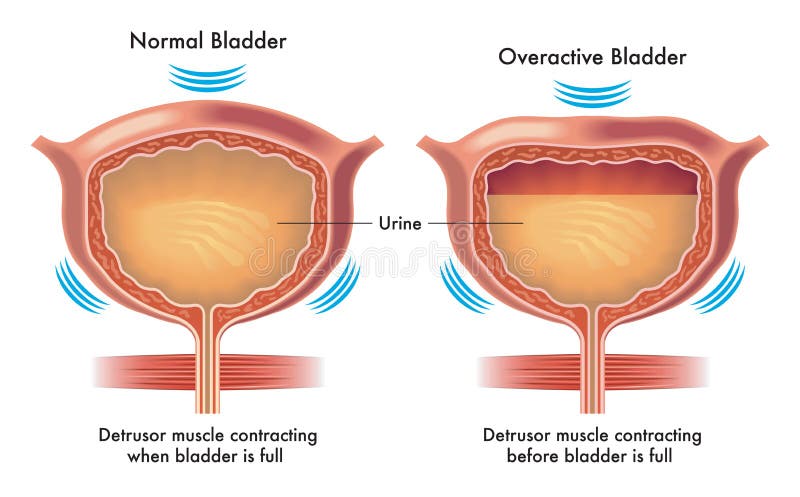
Free with trial A medical illustration with a normal urinary bladder compared to an overactive bladder. Normal structures vectors Normal and overactive bladder. A medical illustration with a normal urinary bladder compared to an overactive bladder. A medical illustration with a normal urinary bladder compared to an overactive bladder.
Free with trial Medical diagram illustrating cell transformation from healthy to cancerous. Three circular illustrations include: "Healthy" cell with uniform purple nucleus, smooth cytoplasm "Dysplastic" cell showing irregular shape, dense orange and purple regions "Malignant" cell with distorted, scattered structures, and darker nucleus, representing cancer. Each stage visually highlights cellular changes, aiding in understanding cancer development. Normal structures illustrations Normal Cell to Cancer Cell Development Medical Diagram. Medical diagram illustrating cell transformation from healthy to cancerous. Three circular illustrations. Medical diagram illustrating cell transformation from healthy to cancerous. Three circular illustrations include: "Healthy" cell with uniform purple nucleus, smooth cytoplasm "Dysplastic" cell showing irregular shape, dense orange and purple regions "Malignant" cell with distorted, scattered structures, and darker nucleus, representing cancer. Each stage visually highlights cellular changes, aiding in understanding cancer development.
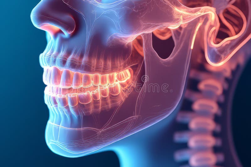
Free with trial This detailed 3D anatomical model showcases the intricate structure of the human jaw and teeth, highlighting their precise alignment and relationships. The model provides a clear visualization of the maxillofacial bones, the dental arches, and the complex interplay of tooth roots, enamel, and dentin. Ideal for dental education, it assists in understanding normal anatomical structures and. Normal structures illustrations Comprehensive 3D Jaw and Teeth Model Visualizing Structure Alignment and Dental Health for Education and Diagnosis. This detailed 3D anatomical model showcases. This detailed 3D anatomical model showcases the intricate structure of the human jaw and teeth, highlighting their precise alignment and relationships. The model provides a clear visualization of the maxillofacial bones, the dental arches, and the complex interplay of tooth roots, enamel, and dentin. Ideal for dental education, it assists in understanding normal anatomical structures and
Free with trial Flat foot anatomy comparison diagram showing fallen arch and normal foot alignment with labeled structures for medical education orthopedic healthcare reference. Normal structures illustrations AI generated flat foot anatomy comparison diagram showing fallen arch normal foot alignment labeled structures for medical. AI generated flat foot anatomy. flat foot anatomy comparison diagram showing fallen arch and normal foot alignment with labeled structures for medical education orthopedic healthcare reference
Free with trial Flat foot anatomy comparison diagram showing fallen arch and normal foot alignment with labeled structures for medical education orthopedic healthcare reference. Normal structures illustrations AI generated flat foot anatomy comparison diagram showing fallen arch normal foot alignment labeled structures for medical. AI generated flat foot anatomy. flat foot anatomy comparison diagram showing fallen arch and normal foot alignment with labeled structures for medical education orthopedic healthcare reference
Free with trial Comparison diagram of a healthy epithelial cell and a malignant cancer cell. The healthy cell is smooth, with distinct organelles, including mitochondria and a prominent nucleus. The cancer cell is irregularly shaped, with protrusions and altered organelle distribution. Both cells are depicted with transparent membranes, emphasizing internal structures. The diagram highlights anatomical differences, such as the uniform shape of the healthy cell versus the distorted contour of the cancer cell. Normal structures illustrations Normal Cell Versus Cancer Cell Anatomy Comparison Diagram. Comparison diagram of a healthy epithelial cell and a malignant cancer cell. The healthy cell is. Comparison diagram of a healthy epithelial cell and a malignant cancer cell. The healthy cell is smooth, with distinct organelles, including mitochondria and a prominent nucleus. The cancer cell is irregularly shaped, with protrusions and altered organelle distribution. Both cells are depicted with transparent membranes, emphasizing internal structures. The diagram highlights anatomical differences, such as the uniform shape of the healthy cell versus the distorted contour of the cancer cell.
Free with trial The process of mutagenesis, showing a single normal cell transforming into a cluster of tumor cells through a mutagenic process. The normal cell is depicted as a single red and orange circular structure on the left, while the tumor cells are shown as a dense cluster of similar red and orange circular structures on the right. The background is a plain orange color, and the process is indicated by a. Normal structures vectors Mutagenesis process showing normal cell to tumor cell transformation. The process of mutagenesis, showing a single normal cell transforming into a cluster of. The process of mutagenesis, showing a single normal cell transforming into a cluster of tumor cells through a mutagenic process. The normal cell is depicted as a single red and orange circular structure on the left, while the tumor cells are shown as a dense cluster of similar red and orange circular structures on the right. The background is a plain orange color, and the process is indicated by a
Free with trial This detailed X-ray image showcases a normal elbow joint, providing a crucial visual reference for professionals in traumatology, orthopedics, and sports medicine. The image clearly depicts the anatomy of the humerus, radius, and ulna, highlighting the crucial articular surfaces and soft tissue structures surrounding the elbow. Proper alignment of these bones is essential for optimal elbow. Normal structures illustrations Normal Elbow XRay A Comprehensive Overview for Traumatology Orthopedics and Sports Medicine Professionals. This detailed X-ray image showcases a normal elbow. This detailed X-ray image showcases a normal elbow joint, providing a crucial visual reference for professionals in traumatology, orthopedics, and sports medicine. The image clearly depicts the anatomy of the humerus, radius, and ulna, highlighting the crucial articular surfaces and soft tissue structures surrounding the elbow. Proper alignment of these bones is essential for optimal elbow
Free with trial Multi slice CT scan of the chest showing normal study, normal appearance of the lungs, parenchyma, pulmonary vasculature, mediastinal structures, no adenopathy, no pleural effusion, no abnormality. Normal structures illustrations Multi slice CT scan of the chest showing normal study, normal appearance of the lungs, parenchyma, pulmonary vasculature,. Mediastinal structures, no adenopathy. Multi slice CT scan of the chest showing normal study, normal appearance of the lungs, parenchyma, pulmonary vasculature, mediastinal structures, no adenopathy, no pleural effusion, no abnormality
Free with trial Detailed microscopic view of cellular structures, with one cell distinctly colored orange, suggesting a focus or anomaly. Normal structures illustrations Close up of microscopic cells with one highlighted in orange. Detailed microscopic view of cellular structures, with one cell distinctly colored orange. Detailed microscopic view of cellular structures, with one cell distinctly colored orange, suggesting a focus or anomaly
Free with trial The image displays a histological section stained primarily in shades of blue and pink, showing multiple glandular structures with distinct circular and oval shapes. The glands are surrounded by a fibrous stroma, and the nuclei of the cells appear darkly stained in blue. The tissue architecture suggests a glandular or epithelial origin, potentially indicative of a normal or pathological state such. Normal structures illustrations Histological section showing glandular structures in tissue under magnification. The image displays a histological section stained primarily in shades of blue. The image displays a histological section stained primarily in shades of blue and pink, showing multiple glandular structures with distinct circular and oval shapes. The glands are surrounded by a fibrous stroma, and the nuclei of the cells appear darkly stained in blue. The tissue architecture suggests a glandular or epithelial origin, potentially indicative of a normal or pathological state such
Free with trial A prominent geological phenomenon unfolds within this urban expanse—an extensive crack slicing through the city skyline. This fracture traverses highways, roads, and concrete structures, representing a complex network of faults and rifts. It encompasses various types of ruptures, from seismic and tectonic to surface and land cracks, each contributing to the city's geological discontinuity. Geologically, it encompasses numerous fault types, including normal, reverse, thrust, strike-slip, transform, extensional, compressional, dip-slip, oblique-slip, and horst and graben structures. This profound tilt-shift effect with bokeh and grain texture accentuates the apocalyptic atmosphere, highlighting the devastation caused by the earthquake-induced cracking. The cracked cityscape serves as a stark reminder of the Earth's geological dynamism and the potential consequences of tectonic forces. Normal structures illustrations A large cracked hole pit in the center of a post apocalyptic city. geologic fault, normal fault, reverse fault. A prominent geological phenomenon unfolds within. A prominent geological phenomenon unfolds within this urban expanse—an extensive crack slicing through the city skyline. This fracture traverses highways, roads, and concrete structures, representing a complex network of faults and rifts. It encompasses various types of ruptures, from seismic and tectonic to surface and land cracks, each contributing to the city's geological discontinuity.Geologically, it encompasses numerous fault types, including normal, reverse, thrust, strike-slip, transform, extensional, compressional, dip-slip, oblique-slip, and horst and graben structures. This profound tilt-shift effect with bokeh and grain texture accentuates the apocalyptic atmosphere, highlighting the devastation caused by the earthquake-induced cracking. The cracked cityscape serves as a stark reminder of the Earth's geological dynamism and the potential consequences of tectonic forces.
Free with trial A set of 3D renderings comparing the structures of healthy and damaged skin, in both normal and dry states. The. A set of 3D renderings comparing the structures. Normal structures illustrations A set of 3D renderings comparing the structures of healthy and damaged skin, in both normal and dry states. The. A set of 3D renderings comparing the structures
Free with trial Illustration comparing a healthy epithelial cell and a malignant cancer cell. The healthy cell appears oval with a smooth membrane, organized organelles, and clearly defined structures like mitochondria and a nucleus. The malignant cancer cell is irregular with a distorted membrane, displaying an abnormal, chaotic arrangement of organelles and multiple nuclei. The textures reflect the complexity and disorganization typical of cancerous cells, highlighting differences in structure and function. Normal structures illustrations Normal Cell Compared With Malignant Cancer Cell Medical Illustration. Illustration comparing a healthy epithelial cell and a malignant cancer cell. The healthy. Illustration comparing a healthy epithelial cell and a malignant cancer cell. The healthy cell appears oval with a smooth membrane, organized organelles, and clearly defined structures like mitochondria and a nucleus. The malignant cancer cell is irregular with a distorted membrane, displaying an abnormal, chaotic arrangement of organelles and multiple nuclei. The textures reflect the complexity and disorganization typical of cancerous cells, highlighting differences in structure and function.
Free with trial Crystals acting as psychic amplifiers, their lattice structures vibrating and pulsating with energy in a spectrum outside of normal sight or sound. Generative AI. Normal structures illustrations Crystals acting as psychic amplifiers, their lattice structures vibrating and pulsating with energy in a spectrum outside of. Normal sight or sound. Generative
Free with trial Crystals acting as psychic amplifiers, their lattice structures vibrating and pulsating with energy in a spectrum outside of normal sight or sound. Generative AI. Normal structures illustrations Crystals acting as psychic amplifiers, their lattice structures vibrating and pulsating with energy in a spectrum outside of. Normal sight or sound. Generative
Free with trial Crystals acting as psychic amplifiers, their lattice structures vibrating and pulsating with energy in a spectrum outside of normal sight or sound. Generative AI. Normal structures illustrations Crystals acting as psychic amplifiers, their lattice structures vibrating and pulsating with energy in a spectrum outside of. Normal sight or sound. Generative
Free with trial This hyper realistic medical illustration depicts human lungs affected by tuberculosis, highlighting fibrosis and scar tissue formation. Hardened areas of lung tissue reduce elasticity, impairing normal respiratory function. Detailed macro anatomical rendering shows bronchi, alveoli, and surrounding lung structures with scientific accuracy, emphasizing areas of fibrotic tissue and structural deformation. Cinematic medical lighting enhances contrast between healthy and diseased regions, while the dark background emphasizes pathological changes. This illustration is ideal for healthcare education, pulmonary disease awareness, medical research, and scientific publications explaining the progression of tuberculosis-related lung fibrosis and its impact on respiratory mechanics. Normal structures illustrations Hyper Realistic Lungs Showing Tuberculosis-Induced Fibrosis and Scar Tissue Formation with Reduced Elasticity. This hyper realistic medical illustration depicts. This hyper realistic medical illustration depicts human lungs affected by tuberculosis, highlighting fibrosis and scar tissue formation. Hardened areas of lung tissue reduce elasticity, impairing normal respiratory function. Detailed macro anatomical rendering shows bronchi, alveoli, and surrounding lung structures with scientific accuracy, emphasizing areas of fibrotic tissue and structural deformation. Cinematic medical lighting enhances contrast between healthy and diseased regions, while the dark background emphasizes pathological changes. This illustration is ideal for healthcare education, pulmonary disease awareness, medical research, and scientific publications explaining the progression of tuberculosis-related lung fibrosis and its impact on respiratory mechanics.
Free with trial Organic Nerve Network Normal Map Texture. Intricate normal map of an organic nerve network or biological vein structures for detailed 3D surface shaders. Normal structures illustrations Organic Nerve Network Normal Map Texture. Intricate normal map of an organic nerve network or biological vein structures for detailed 3D surface shaders
Free with trial A high detail purple normal map with biological organic cell structures and fluid patterns for realistic game materials. Normal structures illustrations Biological Cell Structure Organic Normal Map. A high detail purple normal map with biological organic cell structures and fluid patterns for realistic game. A high detail purple normal map with biological organic cell structures and fluid patterns for realistic game materials
Free with trial This hyper realistic medical illustration depicts bronchial airways severely inflamed due to tuberculosis infection, resulting in narrowed passages that restrict normal airflow. The image highlights swollen airway walls, irritated respiratory lining, and reduced airway diameter, demonstrating how pulmonary tuberculosis can impair breathing. Detailed anatomical rendering reveals bronchial structures and surrounding tissues with high scientific accuracy. Cinematic medical lighting enhances the contrast between healthy and inflamed areas, while the dark background emphasizes the severity of airway obstruction. The visualization effectively represents respiratory distress caused by infection and inflammation. This illustration is ideal for healthcare education, respiratory disease awareness, medical research, and scientific publications explaining how tuberculosis affects bronchial airways and breathing function. Normal structures illustrations Hyper Realistic Medical Illustration of Inflamed Bronchial Airways with Narrowed Passages Caused by Tuberculosis Infection. This hyper realistic medical. This hyper realistic medical illustration depicts bronchial airways severely inflamed due to tuberculosis infection, resulting in narrowed passages that restrict normal airflow. The image highlights swollen airway walls, irritated respiratory lining, and reduced airway diameter, demonstrating how pulmonary tuberculosis can impair breathing. Detailed anatomical rendering reveals bronchial structures and surrounding tissues with high scientific accuracy. Cinematic medical lighting enhances the contrast between healthy and inflamed areas, while the dark background emphasizes the severity of airway obstruction. The visualization effectively represents respiratory distress caused by infection and inflammation. This illustration is ideal for healthcare education, respiratory disease awareness, medical research, and scientific publications explaining how tuberculosis affects bronchial airways and breathing function.
Free with trial Normal brain anatomy, MRI scan. Axial T2-weighted MRI of a 40-year-old patient showing normal brain anatomy with labelled main structures. Normal structures illustrations Normal brain anatomy, MRI scan. Axial T2-weighted MRI of a 40-year-old patient showing normal brain anatomy with labelled main structures
Free with trial A side-by-side diagram a normal eye cross-section on the left and an eye with keratoconus on the right. The normal eye a smooth, rounded cornea. The keratoconus eye a bulging, cone-shaped cornea labeled "Keratoconus" and "Corner". Both diagrams feature simplified internal structures of the eye in shades of pink, yellow, and white, with blue outer layers. Normal structures illustrations Normal eye versus keratoconus eye cross section diagram. A side-by-side diagram a normal eye cross-section on the left and an eye with keratoconus on the right. A side-by-side diagram a normal eye cross-section on the left and an eye with keratoconus on the right. The normal eye a smooth, rounded cornea. The keratoconus eye a bulging, cone-shaped cornea labeled "Keratoconus" and "Corner". Both diagrams feature simplified internal structures of the eye in shades of pink, yellow, and white, with blue outer layers
Free with trial 3D models of free radical, antioxidant, and normal molecules on blue pedestals, showcasing their molecular structures,. Normal structures illustrations Free Radical Antioxidant and Normal Molecules. 3D models of free radical, antioxidant, and normal molecules on blue pedestals, showcasing their molecular. 3D models of free radical, antioxidant, and normal molecules on blue pedestals, showcasing their molecular structures,.
Free with trial This ultra realistic medical illustration depicts lungs affected by pleural effusion resulting from tuberculosis infection. The image highlights fluid accumulation around the lung tissue within the pleural cavity, compressing the lungs and restricting normal breathing function. Inflamed surrounding tissues and reduced lung expansion demonstrate the severity of the condition. Detailed anatomical rendering shows the relationship between lung structures and the pleural space with high scientific accuracy. Cinematic medical lighting enhances contrast between fluid-filled areas and healthy tissue, while the dark clinical background emphasizes the pathological changes. This illustration is ideal for healthcare education, respiratory disease awareness, medical research, and scientific publications explaining pleural effusion as a complication of tuberculosis infection. Normal structures illustrations Ultra Realistic Medical Illustration of Lungs with Pleural Effusion Caused by Tuberculosis Infection and Fluid Accumulation. This ultra realistic medical. This ultra realistic medical illustration depicts lungs affected by pleural effusion resulting from tuberculosis infection. The image highlights fluid accumulation around the lung tissue within the pleural cavity, compressing the lungs and restricting normal breathing function. Inflamed surrounding tissues and reduced lung expansion demonstrate the severity of the condition. Detailed anatomical rendering shows the relationship between lung structures and the pleural space with high scientific accuracy. Cinematic medical lighting enhances contrast between fluid-filled areas and healthy tissue, while the dark clinical background emphasizes the pathological changes. This illustration is ideal for healthcare education, respiratory disease awareness, medical research, and scientific publications explaining pleural effusion as a complication of tuberculosis infection.
Free with trial The image showcases two aerial views of the same tropical island, one in normal color and the other in an artistic, pixelated style. The island features dense green vegetation, including palm trees, with urban structures and buildings concentrated along the coastline. The shoreline is lined with clear blue waters, and the surrounding area includes smaller landmasses or islets. The pixelated. Normal structures illustrations Aerial view of a tropical island with dense greenery and urban development. The image showcases two aerial views of the same tropical island, one in normal color. The image showcases two aerial views of the same tropical island, one in normal color and the other in an artistic, pixelated style. The island features dense green vegetation, including palm trees, with urban structures and buildings concentrated along the coastline. The shoreline is lined with clear blue waters, and the surrounding area includes smaller landmasses or islets. The pixelated
Free with trial This image is a radiographic X-ray of the human thoracic cavity, displaying the lungs, heart shadow, ribs, clavicles, and part of the spine. The lung fields appear mostly clear, indicating normal aeration, while the bony structures such as the ribs, spine, and clavicles are distinctly visible. This type of imaging is commonly used to diagnose conditions related to the chest, including lung. Normal structures illustrations X-ray image showing the human thoracic cavity with clear lung fields and ribcage structure. This image is a radiographic X-ray of the human thoracic cavity. This image is a radiographic X-ray of the human thoracic cavity, displaying the lungs, heart shadow, ribs, clavicles, and part of the spine. The lung fields appear mostly clear, indicating normal aeration, while the bony structures such as the ribs, spine, and clavicles are distinctly visible. This type of imaging is commonly used to diagnose conditions related to the chest, including lung
Free with trial This clear and detailed post-operative X-ray image showcases a successful subtalar arthrodesis procedure. The image demonstrates a stable fusion of the subtalar joint, also known as the talocalcaneal joint, with two strategically placed screws firmly anchoring the bone fragments. The ankle joint itself displays no abnormalities, with normal anatomical structures and bone density evident. The. Normal structures illustrations PostOperative Subtalar Fusion XRay Showing Stable Fusion and Normal Bone Healing Detailed Analysis of Ankle Joint and. This clear and detailed post-operative X. This clear and detailed post-operative X-ray image showcases a successful subtalar arthrodesis procedure. The image demonstrates a stable fusion of the subtalar joint, also known as the talocalcaneal joint, with two strategically placed screws firmly anchoring the bone fragments. The ankle joint itself displays no abnormalities, with normal anatomical structures and bone density evident. The
Free with trial The image showcases various shapes of red blood cells as seen under a microscope. The cells exhibit different forms including normal disc-shaped cells, elongated cells, and some abnormal shapes which may indicate certain health conditions. The vibrant red color highlights the cells against a stark white background, providing a clear and detailed view of their unique structures. Normal structures illustrations Diverse shapes of red blood cells under microscopic view isolated on white background. The image showcases various shapes of red blood cells as seen under a. The image showcases various shapes of red blood cells as seen under a microscope. The cells exhibit different forms including normal disc-shaped cells, elongated cells, and some abnormal shapes which may indicate certain health conditions. The vibrant red color highlights the cells against a stark white background, providing a clear and detailed view of their unique structures
Free with trial Diagram comparing a normal heart and one with cardiomegaly. The normal heart shows standard anatomical features like the right atrium (RA), left ventricle (LV), and superior vena cava. The heart with cardiomegaly illustrates enlarged ventricles, likely indicating increased heart size. Both diagrams label structures such as the aorta, pulmonary artery (PA), and valves. The normal heart is on the left, with cardiomegaly on the right. The condition involves significant structural changes, emphasizing the size difference in the ventricles. Normal structures vectors Cardiomegaly (Enlarged Heart). Diagram comparing a normal heart and one with cardiomegaly. The normal heart shows standard anatomical features like the right. Diagram comparing a normal heart and one with cardiomegaly. The normal heart shows standard anatomical features like the right atrium (RA), left ventricle (LV), and superior vena cava. The heart with cardiomegaly illustrates enlarged ventricles, likely indicating increased heart size. Both diagrams label structures such as the aorta, pulmonary artery (PA), and valves. The normal heart is on the left, with cardiomegaly on the right. The condition involves significant structural changes, emphasizing the size difference in the ventricles.
Free with trial Diagram contrasting a normal heart with one experiencing aortic valve regurgitation. The normal heart illustration shows a properly functioning aortic valve between the left ventricle and aorta, preventing backflow. The regurgitation example highlights a faulty aortic valve allowing blood to leak backward into the left ventricle, disrupting efficient circulation. The heart structures are color-coded, with red indicating oxygenated sections like the aorta and left ventricle, and blue for deoxygenated areas like the right atrium and ventricle. Normal structures vectors Aortic Valve Regurgitation Heart. Diagram contrasting a normal heart with one experiencing aortic valve regurgitation. The normal heart illustration shows a. Diagram contrasting a normal heart with one experiencing aortic valve regurgitation. The normal heart illustration shows a properly functioning aortic valve between the left ventricle and aorta, preventing backflow. The regurgitation example highlights a faulty aortic valve allowing blood to leak backward into the left ventricle, disrupting efficient circulation. The heart structures are color-coded, with red indicating oxygenated sections like the aorta and left ventricle, and blue for deoxygenated areas like the right atrium and ventricle.
Free with trial This science vector illustration presents three distinct forms of nucleic acid bases: normal base, nucleotide, and nucleoside, set against a pristine white background. These molecular entities play vital roles in DNA and RNA structure, functioning as the building blocks of genetic material. With precise molecular structures and clean design, this vector graphic serves as an invaluable educational tool for students, educators, and researchers in the fields of biochemistry, genetics, and molecular biology. Ideal for presentations, educational materials, and scientific publications, this illustration offers insights into the molecular intricacies of nucleic acid composition and structure. Normal structures vectors Illustration of Three Different Forms of Nucleic Acid Bases Normal Base, Nucleotide, Nucleoside on White Background. This science vector illustration presents. This science vector illustration presents three distinct forms of nucleic acid bases: normal base, nucleotide, and nucleoside, set against a pristine white background. These molecular entities play vital roles in DNA and RNA structure, functioning as the building blocks of genetic material. With precise molecular structures and clean design, this vector graphic serves as an invaluable educational tool for students, educators, and researchers in the fields of biochemistry, genetics, and molecular biology. Ideal for presentations, educational materials, and scientific publications, this illustration offers insights into the molecular intricacies of nucleic acid composition and structure.
Free with trial This ultra-detailed AI-generated medical illustration presents human kidneys with microscopic crystal-like toxin particles damaging renal tissue. The hyper realistic 3D render highlights the delicate structure of the kidneys, including the renal cortex, medulla, and intricate filtration pathways. Tiny crystalline toxin particles are shown interacting with and penetrating renal tissue, symbolizing toxic stress and potential damage to the kidney's filtration system. The particles appear sharp and reflective, representing harmful substances that can disrupt normal kidney function and injure delicate cellular structures. Dramatic medical lighting enhances the depth and texture of the anatomical structures while emphasizing the presence of the damaging crystal particles. A dark medical laboratory-style background creates a focused scientific atmosphere and increases contrast for clearer visualization of the pathology. Created in 5000x5000px resolution at 300dpi, this illustration is suitable for medical education, research presentations, healthcare awareness campaigns, and scientific publications related to renal toxicity and kidney health. Normal structures illustrations Ultra Detailed AI-Generated Illustration of Human Kidneys with Microscopic Crystal Toxin Particles Damaging Renal Tissue in. This ultra-detailed AI-generated. This ultra-detailed AI-generated medical illustration presents human kidneys with microscopic crystal-like toxin particles damaging renal tissue. The hyper realistic 3D render highlights the delicate structure of the kidneys, including the renal cortex, medulla, and intricate filtration pathways. Tiny crystalline toxin particles are shown interacting with and penetrating renal tissue, symbolizing toxic stress and potential damage to the kidney's filtration system. The particles appear sharp and reflective, representing harmful substances that can disrupt normal kidney function and injure delicate cellular structures. Dramatic medical lighting enhances the depth and texture of the anatomical structures while emphasizing the presence of the damaging crystal particles. A dark medical laboratory-style background creates a focused scientific atmosphere and increases contrast for clearer visualization of the pathology. Created in 5000x5000px resolution at 300dpi, this illustration is suitable for medical education, research presentations, healthcare awareness campaigns, and scientific publications related to renal toxicity and kidney health.
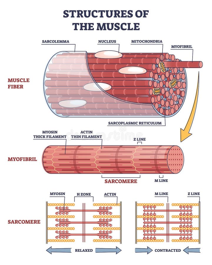
Free with trial Structures of muscle with fiber, myofibril and sarcomere contraction outline diagram. Labeled educational isolated parts closeup description from anatomical and physiology sides vector illustration. Normal structures vectors Structures of muscle with fiber, myofibril and sarcomere outline diagram. Structures of muscle with fiber, myofibril and sarcomere contraction outline diagram. Structures of muscle with fiber, myofibril and sarcomere contraction outline diagram. Labeled educational isolated parts closeup description from anatomical and physiology sides vector illustration.
Free with trial This high-magnification photomicrograph showcases the intricate histology of human gastric glands, expertly stained with Hematoxylin and Eosin (H&E). The image reveals numerous circular and oval glandular structures, each lined by epithelial cells displaying distinct purple nuclei and pink cytoplasm, surrounding a central lumen. The surrounding connective tissue is also visible in varying shades of pink. This detailed view is ideal for medical education, scientific research, and understanding the normal morphology of gastric tissue within the digestive system. Normal structures illustrations Human Gastric Glands Histology, H&E Stain Micrograph. This high-magnification photomicrograph showcases the intricate histology of human gastric glands, expertly. This high-magnification photomicrograph showcases the intricate histology of human gastric glands, expertly stained with Hematoxylin and Eosin (H&E). The image reveals numerous circular and oval glandular structures, each lined by epithelial cells displaying distinct purple nuclei and pink cytoplasm, surrounding a central lumen. The surrounding connective tissue is also visible in varying shades of pink. This detailed view is ideal for medical education, scientific research, and understanding the normal morphology of gastric tissue within the digestive system.
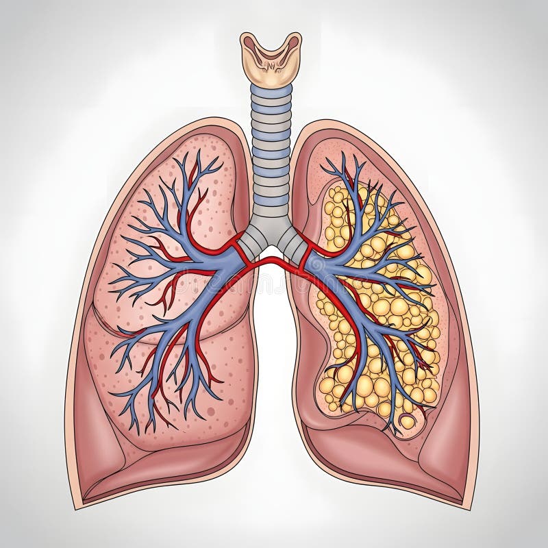
Free with trial Illustration of human lungs and respiratory system showing detailed internal structures. The trachea extends downward, branching into the bronchi within both lungs. The left lung appears normal with visible bronchi and blood vessels. The right lung displays an abnormal condition, highlighted by yellow clusters suggesting potential disease or congestion. The image includes the trachea, bronchi, and alveoli, providing insight into respiratory health and potential lung conditions. Normal structures illustrations Human lungs and respiratory system illustration highlighting internal structure. Illustration of human lungs and respiratory system showing detailed internal. Illustration of human lungs and respiratory system showing detailed internal structures. The trachea extends downward, branching into the bronchi within both lungs. The left lung appears normal with visible bronchi and blood vessels. The right lung displays an abnormal condition, highlighted by yellow clusters suggesting potential disease or congestion. The image includes the trachea, bronchi, and alveoli, providing insight into respiratory health and potential lung conditions.
Free with trial This ultra realistic medical illustration depicts human kidneys affected by tuberculosis infection, showing damaged renal tissue and visible inflammation within the organ. The image highlights how tuberculosis can spread to the kidneys, causing structural damage and disrupting normal filtration function. Inflamed regions and infected tissue areas are clearly visible, demonstrating the progression of renal tuberculosis. Macro anatomical detail reveals kidney structures including nephrons and blood vessels with high scientific accuracy. Cinematic medical lighting enhances contrast between healthy and damaged tissue, while the dark background emphasizes the pathological changes. This illustration is ideal for healthcare education, nephrology studies, infectious disease awareness, medical research, and scientific publications explaining how tuberculosis affects kidney function and tissue health. Normal structures illustrations Ultra Realistic Medical Illustration of Human Kidneys with Tuberculosis Infection Damaging Renal Tissue and Causing Inflammation. This ultra realistic medical. This ultra realistic medical illustration depicts human kidneys affected by tuberculosis infection, showing damaged renal tissue and visible inflammation within the organ. The image highlights how tuberculosis can spread to the kidneys, causing structural damage and disrupting normal filtration function. Inflamed regions and infected tissue areas are clearly visible, demonstrating the progression of renal tuberculosis. Macro anatomical detail reveals kidney structures including nephrons and blood vessels with high scientific accuracy. Cinematic medical lighting enhances contrast between healthy and damaged tissue, while the dark background emphasizes the pathological changes. This illustration is ideal for healthcare education, nephrology studies, infectious disease awareness, medical research, and scientific publications explaining how tuberculosis affects kidney function and tissue health.
Free with trial Microscopic view of biological tissue showing pink and purple cellular structures with intricate patterns and textures. Normal structures illustrations Microscopic view of biological tissue with pink and purple cellular structures biology. Microscopic view of biological tissue showing pink and purple cellular. Microscopic view of biological tissue showing pink and purple cellular structures with intricate patterns and textures
Free with trial This detailed visual comparison highlights the microscopic differences between healthy skin and skin affected by psoriasis. It illustrates the cellular structures and processes involved in both conditions, offering a clear understanding of psoriasis's impact on skin health and appearance. Normal structures illustrations Comparing normal skin anatomy and psoriasis skin development detailed microscopic medical visual comparison. This detailed visual comparison highlights the. This detailed visual comparison highlights the microscopic differences between healthy skin and skin affected by psoriasis. It illustrates the cellular structures and processes involved in both conditions, offering a clear understanding of psoriasis's impact on skin health and appearance
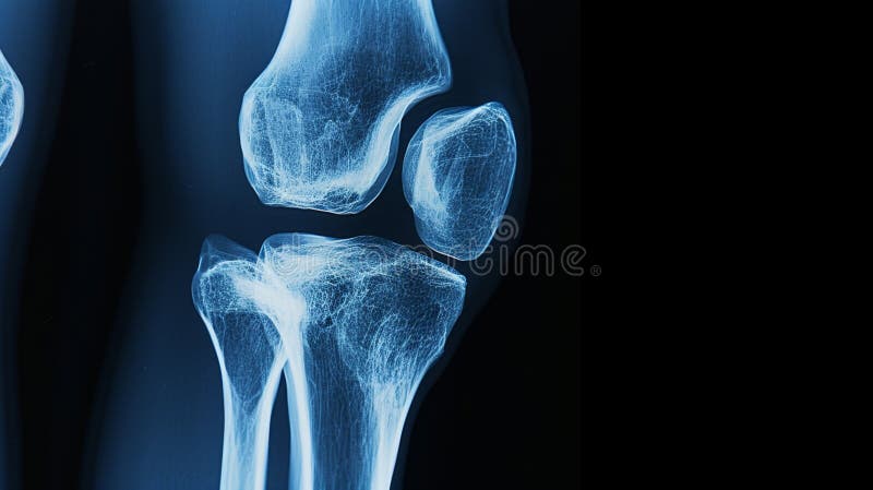
Free with trial X-ray image of the knee anatomy and joint structures. AI generated. Normal structures illustrations X-ray image of the knee anatomy and joint structures. AI generated
Free with trial Educational medical vector illustration comparing a healthy kidney with a kidney infection (pyelonephritis). The diagram labels major kidney structures, including the cortex, renal artery, renal vein, hilum, and ureter, while highlighting inflammation associated with infection. Ideal for healthcare education, nephrology, anatomy, biology, pathology, and clinical reference materials. Normal structures vectors Healthy Kidney vs Kidney Infection Medical Anatomy Comparison Vector Illustration. Educational medical vector illustration comparing a healthy kidney with a. Educational medical vector illustration comparing a healthy kidney with a kidney infection (pyelonephritis). The diagram labels major kidney structures, including the cortex, renal artery, renal vein, hilum, and ureter, while highlighting inflammation associated with infection. Ideal for healthcare education, nephrology, anatomy, biology, pathology, and clinical reference materials.
Free with trial A prominent geological phenomenon unfolds within this urban expanse—an extensive crack slicing through the city skyline. This fracture traverses highways, roads, and concrete structures, representing a complex network of faults and rifts. It encompasses various types of ruptures, from seismic and tectonic to surface and land cracks, each contributing to the city's geological discontinuity. Geologically, it encompasses numerous fault types, including normal, reverse, thrust, strike-slip, transform, extensional, compressional, dip-slip, oblique-slip, and horst and graben structures. This profound tilt-shift effect with bokeh and grain texture accentuates the apocalyptic atmosphere, highlighting the devastation caused by the earthquake-induced cracking. The cracked cityscape serves as a stark reminder of the Earth's geological dynamism and the potential consequences of tectonic forces. Normal structures illustrations A vast rumbling city viewed from a cracked concrete wall. A prominent geological phenomenon unfolds within this urban expanse—an extensive crack slicing. A prominent geological phenomenon unfolds within this urban expanse—an extensive crack slicing through the city skyline. This fracture traverses highways, roads, and concrete structures, representing a complex network of faults and rifts. It encompasses various types of ruptures, from seismic and tectonic to surface and land cracks, each contributing to the city's geological discontinuity.Geologically, it encompasses numerous fault types, including normal, reverse, thrust, strike-slip, transform, extensional, compressional, dip-slip, oblique-slip, and horst and graben structures. This profound tilt-shift effect with bokeh and grain texture accentuates the apocalyptic atmosphere, highlighting the devastation caused by the earthquake-induced cracking. The cracked cityscape serves as a stark reminder of the Earth's geological dynamism and the potential consequences of tectonic forces.
Free with trial A prominent geological phenomenon unfolds within this urban expanse—an extensive crack slicing through the city skyline. This fracture traverses highways, roads, and concrete structures, representing a complex network of faults and rifts. It encompasses various types of ruptures, from seismic and tectonic to surface and land cracks, each contributing to the city's geological discontinuity. Geologically, it encompasses numerous fault types, including normal, reverse, thrust, strike-slip, transform, extensional, compressional, dip-slip, oblique-slip, and horst and graben structures. This profound tilt-shift effect with bokeh and grain texture accentuates the apocalyptic atmosphere, highlighting the devastation caused by the earthquake-induced cracking. The cracked cityscape serves as a stark reminder of the Earth's geological dynamism and the potential consequences of tectonic forces. Normal structures illustrations Highway cracked and destroyed by a earthquake seismic event. A prominent geological phenomenon unfolds within this urban expanse—an extensive crack slicing. A prominent geological phenomenon unfolds within this urban expanse—an extensive crack slicing through the city skyline. This fracture traverses highways, roads, and concrete structures, representing a complex network of faults and rifts. It encompasses various types of ruptures, from seismic and tectonic to surface and land cracks, each contributing to the city's geological discontinuity.Geologically, it encompasses numerous fault types, including normal, reverse, thrust, strike-slip, transform, extensional, compressional, dip-slip, oblique-slip, and horst and graben structures. This profound tilt-shift effect with bokeh and grain texture accentuates the apocalyptic atmosphere, highlighting the devastation caused by the earthquake-induced cracking. The cracked cityscape serves as a stark reminder of the Earth's geological dynamism and the potential consequences of tectonic forces.
Free with trial A prominent geological phenomenon unfolds within this urban expanse—an extensive crack slicing through the city skyline. This fracture traverses highways, roads, and concrete structures, representing a complex network of faults and rifts. It encompasses various types of ruptures, from seismic and tectonic to surface and land cracks, each contributing to the city's geological discontinuity. Geologically, it encompasses numerous fault types, including normal, reverse, thrust, strike-slip, transform, extensional, compressional, dip-slip, oblique-slip, and horst and graben structures. This profound tilt-shift effect with bokeh and grain texture accentuates the apocalyptic atmosphere, highlighting the devastation caused by the earthquake-induced cracking. The cracked cityscape serves as a stark reminder of the Earth's geological dynamism and the potential consequences of tectonic forces. Normal structures illustrations A large crack cutting through a vast city skyline. A prominent geological phenomenon unfolds within this urban expanse—an extensive crack slicing through the. A prominent geological phenomenon unfolds within this urban expanse—an extensive crack slicing through the city skyline. This fracture traverses highways, roads, and concrete structures, representing a complex network of faults and rifts. It encompasses various types of ruptures, from seismic and tectonic to surface and land cracks, each contributing to the city's geological discontinuity.Geologically, it encompasses numerous fault types, including normal, reverse, thrust, strike-slip, transform, extensional, compressional, dip-slip, oblique-slip, and horst and graben structures. This profound tilt-shift effect with bokeh and grain texture accentuates the apocalyptic atmosphere, highlighting the devastation caused by the earthquake-induced cracking. The cracked cityscape serves as a stark reminder of the Earth's geological dynamism and the potential consequences of tectonic forces.
Free with trial Street with completely destroyed buildings on one side and intact homes on the other. Normal structures illustrations Street with completely destroyed buildings on one side and intact homes. On the other. AI generated. Street with completely destroyed buildings on one side and intact homes on the other.
Free with trial A prominent geological phenomenon unfolds within this urban expanse—an extensive crack slicing through the city skyline. This fracture traverses highways, roads, and concrete structures, representing a complex network of faults and rifts. It encompasses various types of ruptures, from seismic and tectonic to surface and land cracks, each contributing to the city's geological discontinuity. Geologically, it encompasses numerous fault types, including normal, reverse, thrust, strike-slip, transform, extensional, compressional, dip-slip, oblique-slip, and horst and graben structures. This profound tilt-shift effect with bokeh and grain texture accentuates the apocalyptic atmosphere, highlighting the devastation caused by the earthquake-induced cracking. The cracked cityscape serves as a stark reminder of the Earth's geological dynamism and the potential consequences of tectonic forces. Normal structures illustrations Cracked old concrete highway on the mountains. chasms, cleavages, rimas. A prominent geological phenomenon unfolds within this urban expanse—an extensive. A prominent geological phenomenon unfolds within this urban expanse—an extensive crack slicing through the city skyline. This fracture traverses highways, roads, and concrete structures, representing a complex network of faults and rifts. It encompasses various types of ruptures, from seismic and tectonic to surface and land cracks, each contributing to the city's geological discontinuity.Geologically, it encompasses numerous fault types, including normal, reverse, thrust, strike-slip, transform, extensional, compressional, dip-slip, oblique-slip, and horst and graben structures. This profound tilt-shift effect with bokeh and grain texture accentuates the apocalyptic atmosphere, highlighting the devastation caused by the earthquake-induced cracking. The cracked cityscape serves as a stark reminder of the Earth's geological dynamism and the potential consequences of tectonic forces.
Free with trial Here we are offering you a neurons and synapse-like structures depicting brain that can be used in project-related marketing. Normal structures illustrations Neurons and synapse-like structures depicting brain. Here we are offering you a neurons and synapse-like structures depicting brain that can be used in project. Here we are offering you a neurons and synapse-like structures depicting brain that can be used in project-related marketing.
Free with trial A prominent geological phenomenon unfolds within this urban expanse—an extensive crack slicing through the city skyline. This fracture traverses highways, roads, and concrete structures, representing a complex network of faults and rifts. It encompasses various types of ruptures, from seismic and tectonic to surface and land cracks, each contributing to the city's geological discontinuity. Geologically, it encompasses numerous fault types, including normal, reverse, thrust, strike-slip, transform, extensional, compressional, dip-slip, oblique-slip, and horst and graben structures. This profound tilt-shift effect with bokeh and grain texture accentuates the apocalyptic atmosphere, highlighting the devastation caused by the earthquake-induced cracking. The cracked cityscape serves as a stark reminder of the Earth's geological dynamism and the potential consequences of tectonic forces. Normal structures illustrations Giant crack in the center of a city caused by a earthquake. tilt shift effect. A prominent geological phenomenon unfolds within this urban expanse—an extensive. A prominent geological phenomenon unfolds within this urban expanse—an extensive crack slicing through the city skyline. This fracture traverses highways, roads, and concrete structures, representing a complex network of faults and rifts. It encompasses various types of ruptures, from seismic and tectonic to surface and land cracks, each contributing to the city's geological discontinuity.Geologically, it encompasses numerous fault types, including normal, reverse, thrust, strike-slip, transform, extensional, compressional, dip-slip, oblique-slip, and horst and graben structures. This profound tilt-shift effect with bokeh and grain texture accentuates the apocalyptic atmosphere, highlighting the devastation caused by the earthquake-induced cracking. The cracked cityscape serves as a stark reminder of the Earth's geological dynamism and the potential consequences of tectonic forces.
Free with trial A detailed cross-section of the human eye. The internal structures including the optic nerve, iris, pupil, and vessels. Labels indicate aqueous humor, Schlemm's canal, normal Nersur, aqueous humor flow, and increased intraocular pressure. The color palette blues, reds, pinks, and yellows against a white background. Normal structures illustrations Human eye anatomy cross section with labels optic nerve. A detailed cross-section of the human eye. The internal structures including the optic nerve, iris. A detailed cross-section of the human eye. The internal structures including the optic nerve, iris, pupil, and vessels. Labels indicate aqueous humor, Schlemm's canal, normal Nersur, aqueous humor flow, and increased intraocular pressure. The color palette blues, reds, pinks, and yellows against a white background
Free with trial A prominent geological phenomenon unfolds within this urban expanse—an extensive crack slicing through the city skyline. This fracture traverses highways, roads, and concrete structures, representing a complex network of faults and rifts. It encompasses various types of ruptures, from seismic and tectonic to surface and land cracks, each contributing to the city's geological discontinuity. Geologically, it encompasses numerous fault types, including normal, reverse, thrust, strike-slip, transform, extensional, compressional, dip-slip, oblique-slip, and horst and graben structures. This profound tilt-shift effect with bokeh and grain texture accentuates the apocalyptic atmosphere, highlighting the devastation caused by the earthquake-induced cracking. The cracked cityscape serves as a stark reminder of the Earth's geological dynamism and the potential consequences of tectonic forces. Normal structures illustrations A big crack on a concrete highway. close-up view. dusk. earthquake destruction concept. A prominent geological phenomenon unfolds within this urban expanse—an. A prominent geological phenomenon unfolds within this urban expanse—an extensive crack slicing through the city skyline. This fracture traverses highways, roads, and concrete structures, representing a complex network of faults and rifts. It encompasses various types of ruptures, from seismic and tectonic to surface and land cracks, each contributing to the city's geological discontinuity.Geologically, it encompasses numerous fault types, including normal, reverse, thrust, strike-slip, transform, extensional, compressional, dip-slip, oblique-slip, and horst and graben structures. This profound tilt-shift effect with bokeh and grain texture accentuates the apocalyptic atmosphere, highlighting the devastation caused by the earthquake-induced cracking. The cracked cityscape serves as a stark reminder of the Earth's geological dynamism and the potential consequences of tectonic forces.
Free with trial A prominent geological phenomenon unfolds within this urban expanse—an extensive crack slicing through the city skyline. This fracture traverses highways, roads, and concrete structures, representing a complex network of faults and rifts. It encompasses various types of ruptures, from seismic and tectonic to surface and land cracks, each contributing to the city's geological discontinuity. Geologically, it encompasses numerous fault types, including normal, reverse, thrust, strike-slip, transform, extensional, compressional, dip-slip, oblique-slip, and horst and graben structures. This profound tilt-shift effect with bokeh and grain texture accentuates the apocalyptic atmosphere, highlighting the devastation caused by the earthquake-induced cracking. The cracked cityscape serves as a stark reminder of the Earth's geological dynamism and the potential consequences of tectonic forces. Normal structures illustrations A deep long crack in the center of a city skyline highway. blue sunny sky. A prominent geological phenomenon unfolds within this urban expanse—an extensive. A prominent geological phenomenon unfolds within this urban expanse—an extensive crack slicing through the city skyline. This fracture traverses highways, roads, and concrete structures, representing a complex network of faults and rifts. It encompasses various types of ruptures, from seismic and tectonic to surface and land cracks, each contributing to the city's geological discontinuity.Geologically, it encompasses numerous fault types, including normal, reverse, thrust, strike-slip, transform, extensional, compressional, dip-slip, oblique-slip, and horst and graben structures. This profound tilt-shift effect with bokeh and grain texture accentuates the apocalyptic atmosphere, highlighting the devastation caused by the earthquake-induced cracking. The cracked cityscape serves as a stark reminder of the Earth's geological dynamism and the potential consequences of tectonic forces.
Free with trial A prominent geological phenomenon unfolds within this urban expanse—an extensive crack slicing through the city skyline. This fracture traverses highways, roads, and concrete structures, representing a complex network of faults and rifts. It encompasses various types of ruptures, from seismic and tectonic to surface and land cracks, each contributing to the city's geological discontinuity. Geologically, it encompasses numerous fault types, including normal, reverse, thrust, strike-slip, transform, extensional, compressional, dip-slip, oblique-slip, and horst and graben structures. This profound tilt-shift effect with bokeh and grain texture accentuates the apocalyptic atmosphere, highlighting the devastation caused by the earthquake-induced cracking. The cracked cityscape serves as a stark reminder of the Earth's geological dynamism and the potential consequences of tectonic forces. Normal structures illustrations A deep long crack in the center of a city skyline highway. blue sunny sky. A prominent geological phenomenon unfolds within this urban expanse—an extensive. A prominent geological phenomenon unfolds within this urban expanse—an extensive crack slicing through the city skyline. This fracture traverses highways, roads, and concrete structures, representing a complex network of faults and rifts. It encompasses various types of ruptures, from seismic and tectonic to surface and land cracks, each contributing to the city's geological discontinuity.Geologically, it encompasses numerous fault types, including normal, reverse, thrust, strike-slip, transform, extensional, compressional, dip-slip, oblique-slip, and horst and graben structures. This profound tilt-shift effect with bokeh and grain texture accentuates the apocalyptic atmosphere, highlighting the devastation caused by the earthquake-induced cracking. The cracked cityscape serves as a stark reminder of the Earth's geological dynamism and the potential consequences of tectonic forces.
Free with trial A series of MRI brain scans, predominantly in shades of blue and orange, are displayed in a grid pattern. The scans show varying levels of detail in brain structures. Normal structures illustrations Multiple Axial Brain MRI Scans. A series of MRI brain scans, predominantly in shades of blue and orange, are displayed in a grid pattern. The scans show varying. A series of MRI brain scans, predominantly in shades of blue and orange, are displayed in a grid pattern. The scans show varying levels of detail in brain structures
Free with trial A prominent geological phenomenon unfolds within this urban expanse—an extensive crack slicing through the city skyline. This fracture traverses highways, roads, and concrete structures, representing a complex network of faults and rifts. It encompasses various types of ruptures, from seismic and tectonic to surface and land cracks, each contributing to the city's geological discontinuity. Geologically, it encompasses numerous fault types, including normal, reverse, thrust, strike-slip, transform, extensional, compressional, dip-slip, oblique-slip, and horst and graben structures. This profound tilt-shift effect with bokeh and grain texture accentuates the apocalyptic atmosphere, highlighting the devastation caused by the earthquake-induced cracking. The cracked cityscape serves as a stark reminder of the Earth's geological dynamism and the potential consequences of tectonic forces. Normal structures illustrations Cracked highway leading to a large city skyline in the distance. fissures, ruptures, crevices. A prominent geological phenomenon unfolds within this urban. A prominent geological phenomenon unfolds within this urban expanse—an extensive crack slicing through the city skyline. This fracture traverses highways, roads, and concrete structures, representing a complex network of faults and rifts. It encompasses various types of ruptures, from seismic and tectonic to surface and land cracks, each contributing to the city's geological discontinuity.Geologically, it encompasses numerous fault types, including normal, reverse, thrust, strike-slip, transform, extensional, compressional, dip-slip, oblique-slip, and horst and graben structures. This profound tilt-shift effect with bokeh and grain texture accentuates the apocalyptic atmosphere, highlighting the devastation caused by the earthquake-induced cracking. The cracked cityscape serves as a stark reminder of the Earth's geological dynamism and the potential consequences of tectonic forces.
Free with trial A prominent geological phenomenon unfolds within this urban expanse—an extensive crack slicing through the city skyline. This fracture traverses highways, roads, and concrete structures, representing a complex network of faults and rifts. It encompasses various types of ruptures, from seismic and tectonic to surface and land cracks, each contributing to the city's geological discontinuity. Geologically, it encompasses numerous fault types, including normal, reverse, thrust, strike-slip, transform, extensional, compressional, dip-slip, oblique-slip, and horst and graben structures. This profound tilt-shift effect with bokeh and grain texture accentuates the apocalyptic atmosphere, highlighting the devastation caused by the earthquake-induced cracking. The cracked cityscape serves as a stark reminder of the Earth's geological dynamism and the potential consequences of tectonic forces. Normal structures illustrations Earthquake destruction on a city and highway. sunset. Shear zones, tectonic fractures, earthquake fissures. A prominent geological phenomenon unfolds within. A prominent geological phenomenon unfolds within this urban expanse—an extensive crack slicing through the city skyline. This fracture traverses highways, roads, and concrete structures, representing a complex network of faults and rifts. It encompasses various types of ruptures, from seismic and tectonic to surface and land cracks, each contributing to the city's geological discontinuity.Geologically, it encompasses numerous fault types, including normal, reverse, thrust, strike-slip, transform, extensional, compressional, dip-slip, oblique-slip, and horst and graben structures. This profound tilt-shift effect with bokeh and grain texture accentuates the apocalyptic atmosphere, highlighting the devastation caused by the earthquake-induced cracking. The cracked cityscape serves as a stark reminder of the Earth's geological dynamism and the potential consequences of tectonic forces.
Free with trial A prominent geological phenomenon unfolds within this urban expanse—an extensive crack slicing through the city skyline. This fracture traverses highways, roads, and concrete structures, representing a complex network of faults and rifts. It encompasses various types of ruptures, from seismic and tectonic to surface and land cracks, each contributing to the city's geological discontinuity. Geologically, it encompasses numerous fault types, including normal, reverse, thrust, strike-slip, transform, extensional, compressional, dip-slip, oblique-slip, and horst and graben structures. This profound tilt-shift effect with bokeh and grain texture accentuates the apocalyptic atmosphere, highlighting the devastation caused by the earthquake-induced cracking. The cracked cityscape serves as a stark reminder of the Earth's geological dynamism and the potential consequences of tectonic forces. Normal structures illustrations A vast city skyline. cracked earthquake highway disaster. compressional fault, dip-slip fault, oblique-slip fault. A prominent geological phenomenon unfolds. A prominent geological phenomenon unfolds within this urban expanse—an extensive crack slicing through the city skyline. This fracture traverses highways, roads, and concrete structures, representing a complex network of faults and rifts. It encompasses various types of ruptures, from seismic and tectonic to surface and land cracks, each contributing to the city's geological discontinuity.Geologically, it encompasses numerous fault types, including normal, reverse, thrust, strike-slip, transform, extensional, compressional, dip-slip, oblique-slip, and horst and graben structures. This profound tilt-shift effect with bokeh and grain texture accentuates the apocalyptic atmosphere, highlighting the devastation caused by the earthquake-induced cracking. The cracked cityscape serves as a stark reminder of the Earth's geological dynamism and the potential consequences of tectonic forces.
Free with trial This image features a high fidelity cross section comparison of organ tissues, highlighting vibrant colors and structures for diagnostic education and analysis. Normal structures illustrations High Fidelity Organ Cross Section Comparison for Diagnostic Purposes. This image features a high fidelity cross section comparison of organ tissues, highlighting. This image features a high fidelity cross section comparison of organ tissues, highlighting vibrant colors and structures for diagnostic education and analysis
Free with trial This microscopic image reveals the intricate architecture of bone tissue compromised by osteoporosis. The condition, characterized by decreased bone density and increased bone fragility, is clearly visible at this magnified scale. Notice the porous, less dense areas within the bone matrix, starkly contrasting with the healthy, robust structures found in normal bone. Osteoporosis weakens bones,. Normal structures illustrations Unveiling the Delicate Structure of OsteoporosisAffected Bone A Microscopic Exploration of Bone Loss and Fragility. This microscopic image reveals the intricate. This microscopic image reveals the intricate architecture of bone tissue compromised by osteoporosis. The condition, characterized by decreased bone density and increased bone fragility, is clearly visible at this magnified scale. Notice the porous, less dense areas within the bone matrix, starkly contrasting with the healthy, robust structures found in normal bone. Osteoporosis weakens bones,
Free with trial Abstract backgrounds of neurons working inside brain, neuron link Neurons and synapse like structures depicting brain chemistry. On human. Normal structures illustrations Abstract backgrounds of neurons working inside brain, neuron link Neurons and synapse like structures depicting brain chemistry. On human
Free with trial Abstract backgrounds of neurons working inside brain, neuron link Neurons and synapse like structures depicting brain chemistry. On human. Normal structures illustrations Abstract backgrounds of neurons working inside brain, neuron link Neurons and synapse like structures depicting brain chemistry. On human
Free with trial This image shows a microscopic view of numerous red blood cells, also known as erythrocytes, dispersed across a slide. The cells appear to be stained pink, which is typical in hematological examinations to highlight cellular structures and aid in the identification of abnormalities or normal morphology. The distribution and shape of these cells can provide valuable information for medical. Normal structures illustrations Microscopic view of red blood cells stained and distributed on a slide. This image shows a microscopic view of numerous red blood cells, also known as. This image shows a microscopic view of numerous red blood cells, also known as erythrocytes, dispersed across a slide. The cells appear to be stained pink, which is typical in hematological examinations to highlight cellular structures and aid in the identification of abnormalities or normal morphology. The distribution and shape of these cells can provide valuable information for medical
Free with trial Abstract backgrounds of neurons working inside brain, neuron link Neurons and synapse like structures depicting brain chemistry. On human. Normal structures illustrations Abstract backgrounds of neurons working inside brain, neuron link Neurons and synapse like structures depicting brain chemistry. On human
Free with trial Abstract backgrounds of neurons working inside brain, neuron link Neurons and synapse like structures depicting brain chemistry. On human. Normal structures illustrations Abstract backgrounds of neurons working inside brain, neuron link Neurons and synapse like structures depicting brain chemistry. On human
Free with trial Abstract backgrounds of neurons working inside brain, neuron link Neurons and synapse like structures depicting brain chemistry. On human. Normal structures illustrations Abstract backgrounds of neurons working inside brain, neuron link Neurons and synapse like structures depicting brain chemistry. On human
Free with trial Microscopic images compare a healthy epithelial cell and a malignant cancer cell. The healthy cell is round with well-defined structures, showing typical organized cellular components. The malignant cancer cell appears irregular and distorted, with protruding extensions and altered morphology. This comparison highlights changes in cell structure associated with cancer, illustrating the loss of normal cellular architecture in malignant transformation. The differences are key to understanding cancer pathology regarding cell shape and organization. Normal structures illustrations Cancer Cell Morphology Compared With Healthy Cell. Microscopic images compare a healthy epithelial cell and a malignant cancer cell. The healthy cell is round. Microscopic images compare a healthy epithelial cell and a malignant cancer cell. The healthy cell is round with well-defined structures, showing typical organized cellular components. The malignant cancer cell appears irregular and distorted, with protruding extensions and altered morphology. This comparison highlights changes in cell structure associated with cancer, illustrating the loss of normal cellular architecture in malignant transformation. The differences are key to understanding cancer pathology regarding cell shape and organization.
Free with trial Abstract backgrounds of neurons working inside brain, neuron link Neurons and synapse like structures depicting brain chemistry. On human. Normal structures illustrations Abstract backgrounds of neurons working inside brain, neuron link Neurons and synapse like structures depicting brain chemistry. On human
Free with trial Abstract backgrounds of neurons working inside brain, neuron link Neurons and synapse like structures depicting brain chemistry. On human. Normal structures illustrations Abstract backgrounds of neurons working inside brain, neuron link Neurons and synapse like structures depicting brain chemistry. On human
Free with trial Abstract backgrounds of neurons working inside brain, neuron link Neurons and synapse like structures depicting brain chemistry. On human. Normal structures illustrations Abstract backgrounds of neurons working inside brain, neuron link Neurons and synapse like structures depicting brain chemistry. On human
Free with trial Comparison illustration highlights differences between a healthy epithelial cell and a malignant cancer cell. The healthy cell is depicted with a regular shape and organized structures, colored in light blue with visible organelles. In contrast, the cancer cell is shown with an irregular shape and disorganized nucleus, in reddish-brown hues. Text labels emphasize the differences: "Regular shape, organized structures" for the healthy cell and "Irregular shape, disorganized nucleus" for the cancer cell. Normal structures illustrations Healthy Epithelial Cell Compared With Cancer Cell. Comparison illustration highlights differences between a healthy epithelial cell and a malignant cancer cell. Comparison illustration highlights differences between a healthy epithelial cell and a malignant cancer cell. The healthy cell is depicted with a regular shape and organized structures, colored in light blue with visible organelles. In contrast, the cancer cell is shown with an irregular shape and disorganized nucleus, in reddish-brown hues. Text labels emphasize the differences: "Regular shape, organized structures" for the healthy cell and "Irregular shape, disorganized nucleus" for the cancer cell.
Free with trial Categories of IBS stages according to bristol stool chart outline diagram. Labeled educational scheme with normal and abnormal excrement structures vector illustration. Digestive health conditions. Normal structures vectors Categories of IBS stages according to bristol stool chart outline diagram. Labeled educational scheme with normal and abnormal excrement structures vector
Free with trial This image is created by Artificial Intelligence. A detailed illustration of human feet, one showing the normal sole and the other with exposed muscular and skeletal structures, set against a plain white background, providing a clear educational visual aid for anatomy studies. Normal structures illustrations Human feet anatomy illustration, detailed muscular and skeletal structure. This image is created by Artificial Intelligence. A detailed illustration of human. This image is created by Artificial Intelligence. A detailed illustration of human feet, one showing the normal sole and the other with exposed muscular and skeletal structures, set against a plain white background, providing a clear educational visual aid for anatomy studies
Free with trial A medical 3D illustration showcasing a healthy, normal retina as observed during ophthalmoscopy, displaying clear retinal structures and vasculature. Normal structures illustrations Healthy retina, 3D illustration. A medical 3D illustration showcasing a healthy, normal retina as observed during ophthalmoscopy, displaying clear retinal. A medical 3D illustration showcasing a healthy, normal retina as observed during ophthalmoscopy, displaying clear retinal structures and vasculature.
Free with trial Comparison of a healthy epithelial cell and a malignant cancer cell. The healthy cell, on the left, is a smooth, round shape with a light blue-green color, and clearly organized internal structures such as the nucleus and organelles like mitochondria and endoplasmic reticulum. The cancerous cell, on the right, appears irregular with a rougher, uneven surface and reddish color, indicating abnormal growth. Its internal structures, including the nucleus, are distorted, reflecting the disease's invasive nature. Normal structures illustrations Healthy Stem Cell Compared With Malignant Cancer Cell. Comparison of a healthy epithelial cell and a malignant cancer cell. The healthy cell, on the left, is a. Comparison of a healthy epithelial cell and a malignant cancer cell. The healthy cell, on the left, is a smooth, round shape with a light blue-green color, and clearly organized internal structures such as the nucleus and organelles like mitochondria and endoplasmic reticulum. The cancerous cell, on the right, appears irregular with a rougher, uneven surface and reddish color, indicating abnormal growth. Its internal structures, including the nucleus, are distorted, reflecting the disease's invasive nature.
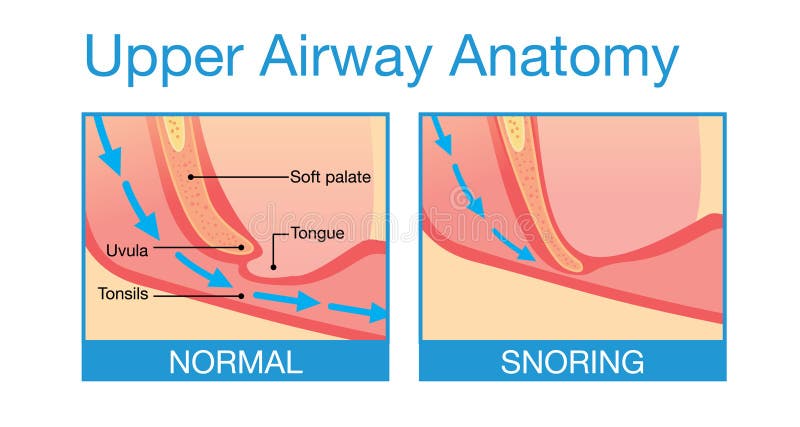
Free with trial Upper airway human anatomy in normal sleeping and have snoring. Normal structures vectors Upper airway human anatomy in sleeping. Upper airway human anatomy in normal sleeping and have snoring. Upper airway human anatomy in normal sleeping and have snoring.
Free with trial Medium-chain fatty acids, MCFAs, chemical structures. Contained in medium-chain triglycerides, who produce more ketone bodies than normal dietary fats, hence they are used in a ketogenic diet. Vector. Normal structures vectors Medium-chain fatty acids, MCFAs, chemical structures. Contained in medium-chain triglycerides, who produce more ketone bodies than normal dietary fats, hence
Free with trial 3D representation of antioxidant molecule interacting with free radical, showcasing the contrast between normal, free. Normal structures illustrations Antioxidant Molecule Neutralizing Free Radical. 3D representation of antioxidant molecule interacting with free radical, showcasing the contrast between normal. 3D representation of antioxidant molecule interacting with free radical, showcasing the contrast between normal, free.
Free with trial This AI-generated medical illustration shows a detailed anatomical view of the human liver highlighting areas affected by oxygen deprivation. Pale regions of liver tissue represent hypoxic or ischemic areas where blood flow and oxygen supply have been reduced. These areas contrast clearly with the surrounding healthy liver tissue, which appears richer in color and structurally intact. The visualization emphasizes the difference between well-perfused hepatic tissue and oxygen-starved regions that may lead to cellular damage or organ dysfunction. Subtle lighting and shading enhance the texture and three-dimensional appearance of the liver surface, revealing vascular structures and tissue patterns. The realistic anatomical style makes the image suitable for medical education, pathology studies, and healthcare awareness materials. This illustration helps viewers understand how impaired circulation or oxygen deficiency can affect liver health and highlights the importance of proper blood flow for maintaining normal liver function and cellular activity. Normal structures illustrations AI-Generated Medical Illustration of Human Liver. This AI-generated medical illustration shows a detailed anatomical view of the human liver highlighting areas. This AI-generated medical illustration shows a detailed anatomical view of the human liver highlighting areas affected by oxygen deprivation. Pale regions of liver tissue represent hypoxic or ischemic areas where blood flow and oxygen supply have been reduced. These areas contrast clearly with the surrounding healthy liver tissue, which appears richer in color and structurally intact. The visualization emphasizes the difference between well-perfused hepatic tissue and oxygen-starved regions that may lead to cellular damage or organ dysfunction. Subtle lighting and shading enhance the texture and three-dimensional appearance of the liver surface, revealing vascular structures and tissue patterns. The realistic anatomical style makes the image suitable for medical education, pathology studies, and healthcare awareness materials. This illustration helps viewers understand how impaired circulation or oxygen deficiency can affect liver health and highlights the importance of proper blood flow for maintaining normal liver function and cellular activity.
Free with trial Street bustling with orange taxis and motorbikes, lined with multi-story buildings featuring wooden balconies. Shops with colorful awnings display various goods. Vibrant greenery and trees are interspersed among the structures. Snow-capped mountains rise in the background under a bright blue sky. The scene depicts a lively urban environment, in a himalayan town in uttrakhand India, characterized by its mix of traditional and modern elements. generative AI. Normal structures illustrations Average traffic condition in a Himalayas mountain town in uttrakhand, India. Street bustling with orange taxis and motorbikes, lined with multi-story buildings. Street bustling with orange taxis and motorbikes, lined with multi-story buildings featuring wooden balconies. Shops with colorful awnings display various goods. Vibrant greenery and trees are interspersed among the structures. Snow-capped mountains rise in the background under a bright blue sky. The scene depicts a lively urban environment, in a himalayan town in uttrakhand India, characterized by its mix of traditional and modern elements. generative AI.
Free with trial This ultra detailed medical illustration depicts human lungs affected by tuberculosis, highlighting destruction of alveolar structures, collapsed air sacs, and inflamed surrounding tissue. The visualization demonstrates how TB infection compromises gas exchange and normal respiratory function. Macro medical realism emphasizes detailed lung anatomy, including bronchi, alveoli, and pulmonary tissue, with clear depiction of damaged and healthy regions. Cinematic lighting enhances contrast and depth, while the dark background focuses attention on structural lung damage. This illustration, rendered at 5000x5000px, is ideal for healthcare education, pulmonary disease awareness, medical research, and scientific publications explaining tuberculosis-induced alveolar destruction. Normal structures illustrations Ultra Detailed Lungs Showing Tuberculosis Destroying Alveoli Structures with Collapsed Air Sacs and Inflamed Tissue. This ultra detailed medical illustration. This ultra detailed medical illustration depicts human lungs affected by tuberculosis, highlighting destruction of alveolar structures, collapsed air sacs, and inflamed surrounding tissue. The visualization demonstrates how TB infection compromises gas exchange and normal respiratory function. Macro medical realism emphasizes detailed lung anatomy, including bronchi, alveoli, and pulmonary tissue, with clear depiction of damaged and healthy regions. Cinematic lighting enhances contrast and depth, while the dark background focuses attention on structural lung damage. This illustration, rendered at 5000x5000px, is ideal for healthcare education, pulmonary disease awareness, medical research, and scientific publications explaining tuberculosis-induced alveolar destruction.
 Sign in