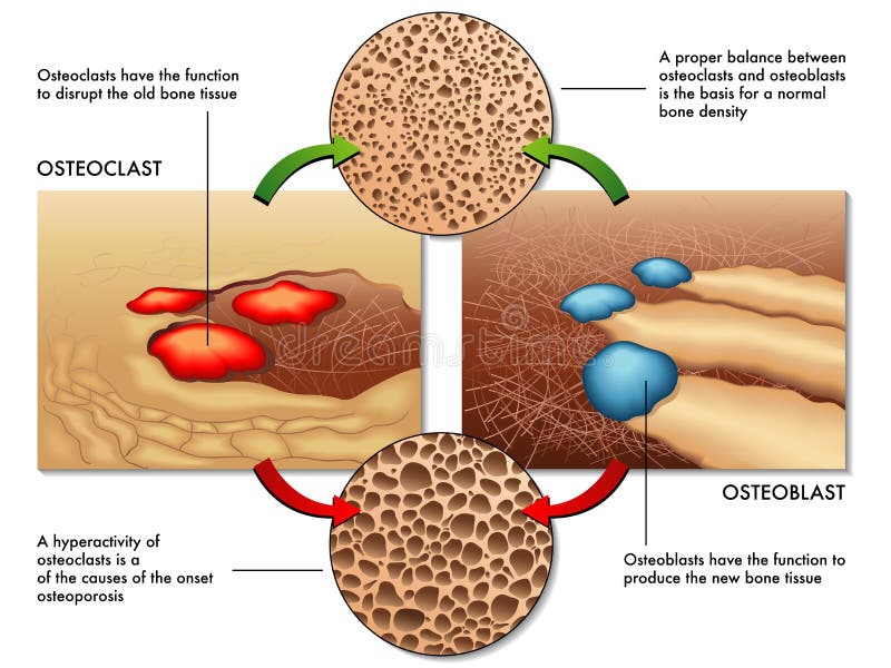
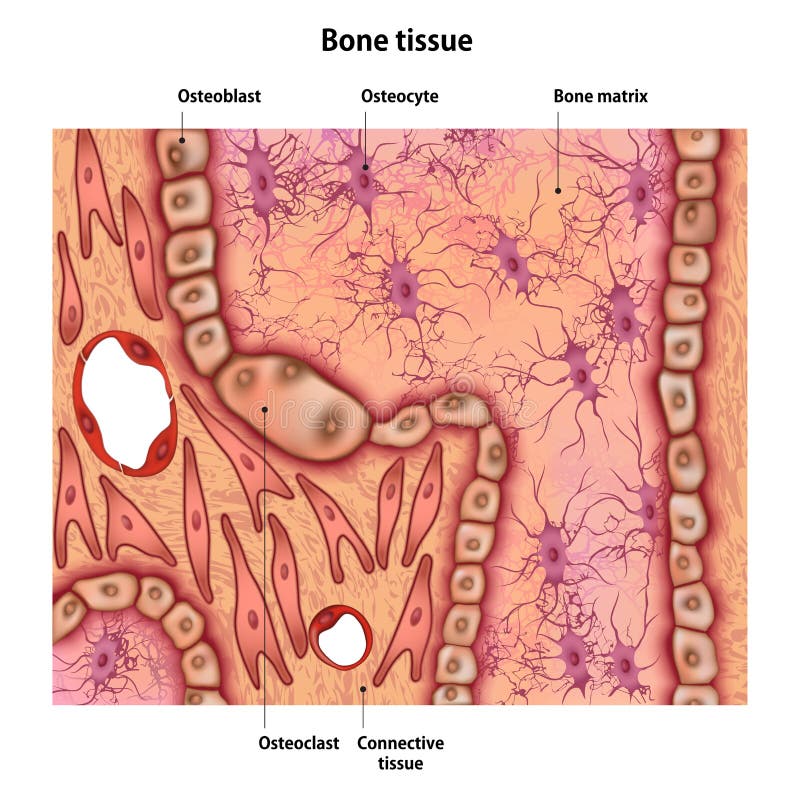
Free with trial Medical illustration of the function of osteoblasts and osteoclasts in the regeneration of bone mass. Osteoblast cell vectors Osteoblast & osteoclast. Medical illustration of the function of osteoblasts and osteoclasts in the regeneration of bone mass
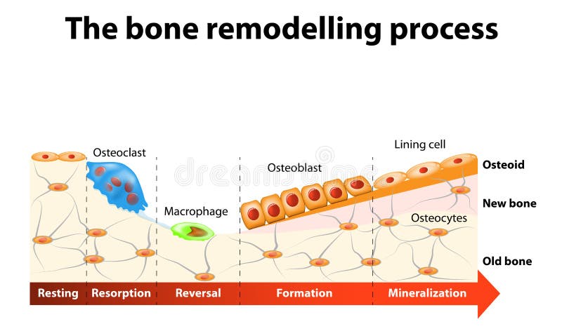
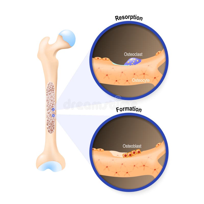
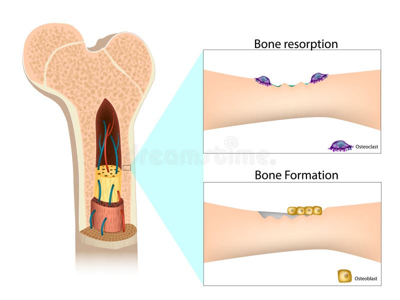
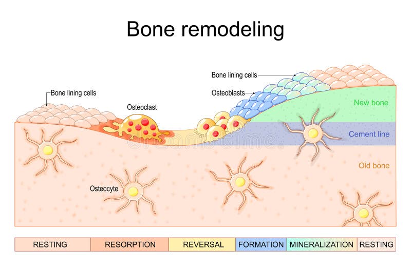
Free with trial The bone remodeling process involves the following steps: resorption, reversal, formation, mineralization and resting. In a healthy body, osteoclasts and osteoblasts work together to maintain the balance between bone loss and bone formation. Osteoblast cell vectors Bone remodelling process. The bone remodeling process involves the following steps: resorption, reversal, formation, mineralization and resting. In a healthy body, osteoclasts and osteoblasts work together to maintain the balance between bone loss and bone formation.
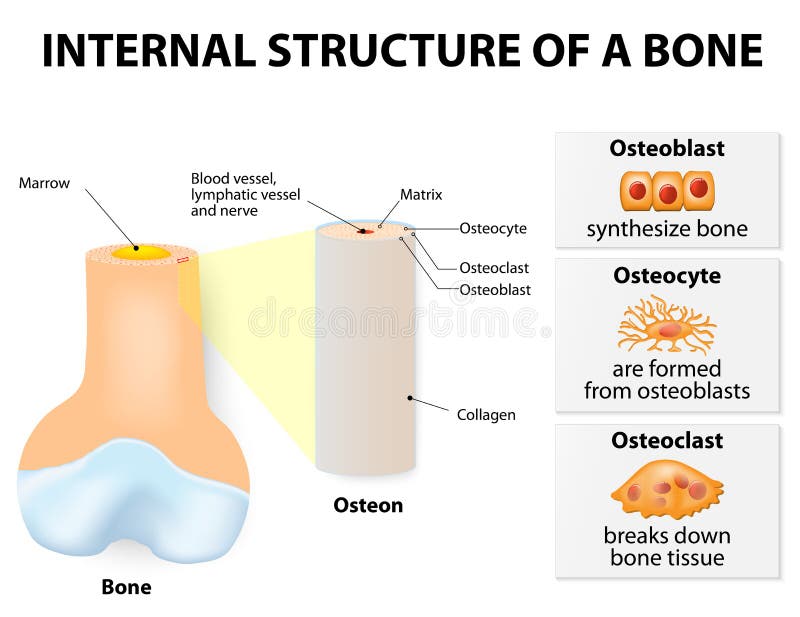
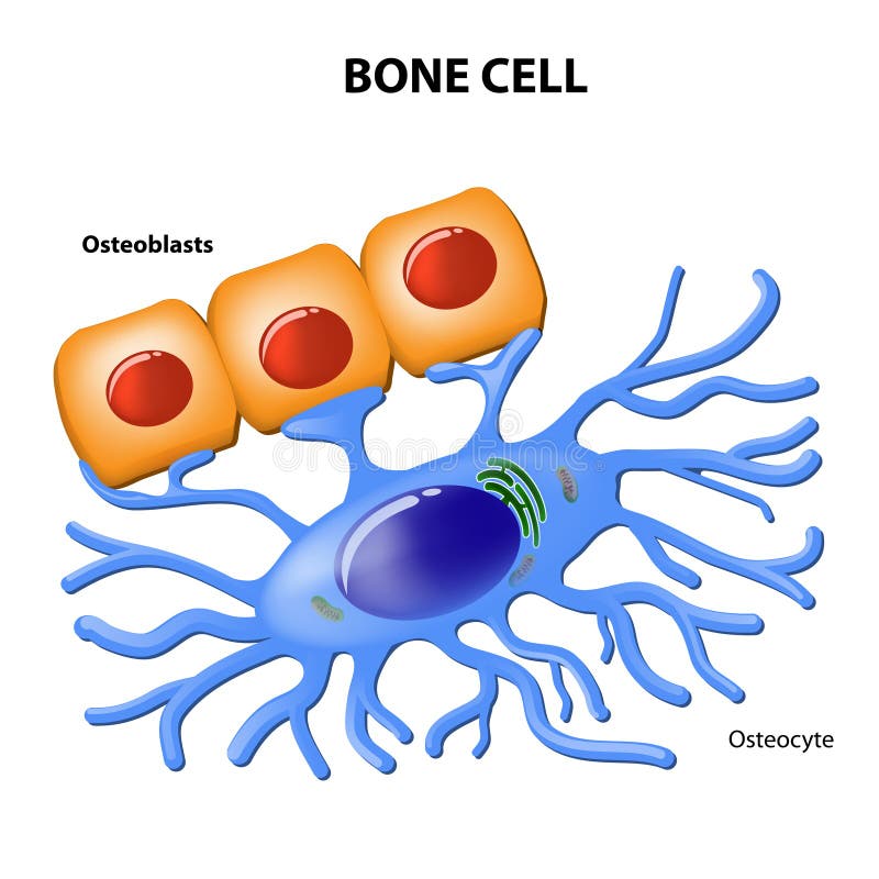
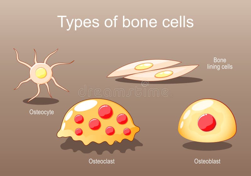
Free with trial Internal structure of a bone. 3 types of cells are found within bone tissue: Osteoblasts, Osteocytes, and Osteoclasts. Osteoblast cell vectors Internal structure of a bone
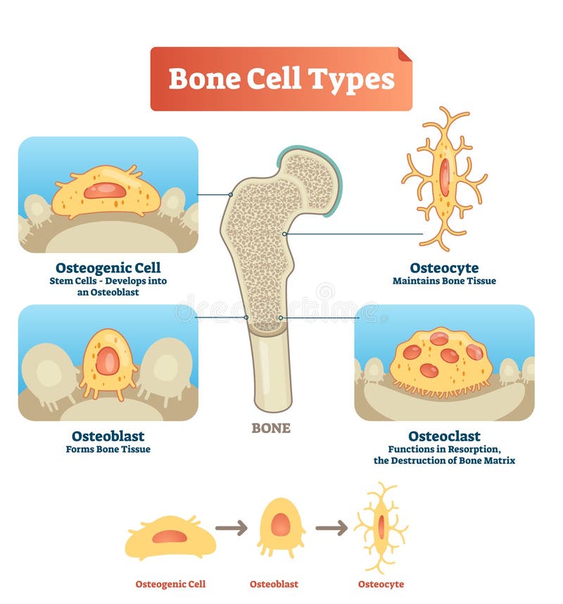
Free with trial Vector illustration of human bone cell types. Scheme of osteogenic cell, osteoblast and osteocyte. Medical diagram visualization of stem cells, bone tissue, resorption and destruction of bone matrix. Osteoblast cell vectors Vector illustration bone cell types diagram. Scheme of osteogenic cell, osteoblast, osteocyte. Medical visualization of stem cells. Vector illustration of human bone cell types. Scheme of osteogenic cell, osteoblast and osteocyte. Medical diagram visualization of stem cells, bone tissue, resorption and destruction of bone matrix.
Free with trial Osteoblast and osteoclast. The bone remodeling process. In a healthy body, osteoclasts and osteoblasts work together to maintain the balance between bone loss and bone formation. Osteoblast cell vectors Osteoblast and osteoclast
Free with trial Bone remodeling process resorption, reversal, formation, and mineralization. Osteoblast, osteoclast, and osteocyte. Vector illustration of human bone cell types. Medical diagram. Osteoblast cell vectors Bone remodeling process. Osteoblast, osteoclast, and osteocyte. Bone remodeling process resorption, reversal, formation, and mineralization. Osteoblast, osteoclast, and osteocyte. Vector illustration of human bone cell types. Medical diagram
Free with trial Osteocyte. structure of bone cell. Vector diagram. Osteoblast cell vectors Osteocyte. structure of bone cell
Free with trial Bone marrow, stem cells of human bone marrow stem cells. These cells are known as multipotential stem cells because they form the precursors to every type of blood cell. Osteoblast cell illustrations Bone marrow, stem cells
Free with trial Mesenchymal stem cells are multipotent stromal cells. differentiate into osteoblast bone, chondrocyte cartilage, myocytes muscle. Osteoblast cell vectors Mesenchymal stem cells are multipotent stromal cells
Free with trial Bone Biology. Role of RANK, RANKL, and OPG. bone remodeling. Bone is broken down by osteoclasts, and rebuilt by osteoblasts. Receptor activator of RANKL is the mediator of bone resorption. Osteoprotegerin OPG. Paracrine and endocrine actions of bone. functions of secretory proteins from osteoblasts, osteocytes, and osteoclasts. Osteoblast cell vectors Bone Biology. Role of RANK, RANKL, and OPG. bone remodeling
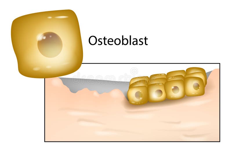
Free with trial Structure of a long bone and cells of bone tissue(useful for education in schools and clinics ) - vector illustration. Osteoblast cell vectors Structure of a long bone
Free with trial Bone remodeling. Close-up of a Osteoclast resorbs bone, and Osteoblasts synthesized bone tissue. Vector illustration. Osteoblast cell vectors Bone remodeling. Osteoclast resorbs bone, and Osteoblasts synthesized bone tissue. Bone remodeling. Close-up of a Osteoclast resorbs bone, and Osteoblasts synthesized bone tissue. Vector illustration
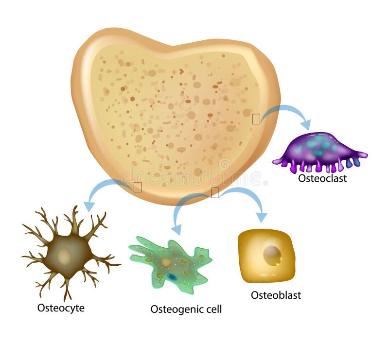
Free with trial Bone tissue has four types of cells. Osteogenic cell, Osteocyte, Osteoclast, Osteoblast. Bone Structure. Osteoblast cell vectors Bone tissue has four types of cells. Osteogenic cell, Osteocyte, Osteoclast, Osteoblast.
Free with trial Bone anatomy. Close-up of Osteoclast for Breaks down Bone tissue, Osteoblast that Synthesize Bone and Osteocyte. Cross section of a human femur. Vector poster. Osteoblast cell vectors Bone anatomy. Close-up of Osteoclast, Osteoblast, and Osteocyte. Bone anatomy. Close-up of Osteoclast for Breaks down Bone tissue, Osteoblast that Synthesize Bone and Osteocyte. Cross section of a human femur. Vector poster
Free with trial Osteoblast Osteoclast. Process Bone Formation and Resorption. Osteoblast cell vectors Process Bone Formation and Resorption. Osteoblast Osteoclast. Osteoblast Osteoclast. Process Bone Formation and Resorption.
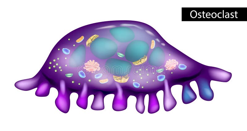
Free with trial Osteoclast is a type of bone cell that breaks down bone tissue. Structure of osteoclasts. Osteoblast cell vectors Structure of osteoclasts. Osteoclast is a type of bone cell that breaks down bone tissue. Osteoclast is a type of bone cell that breaks down bone tissue. Structure of osteoclasts
Free with trial Differentiation and activation process of bone cells, osteoclast and osteoblast development pathways diagram hand drawn schematic vector illustration. Medical science educational illustration. Osteoblast cell vectors Bone Cell Differentiation Activation Diagram. differentiation and activation process of bone cells, osteoclast and osteoblast development pathways diagram hand drawn schematic vector illustration. Medical science educational illustration
Free with trial Rheumatoid arthritis RA. Close-up of bone cells Osteocyte, Osteoblast, Osteoclast, and Fibroblast-like synoviocyte that secrete RANKL and Sclerostin. autoimmune disorder that affects joints. comparison and difference between Healthy joint and knee with Rheumatoid arthritis. Vector illustration. Osteoblast cell vectors Rheumatoid arthritis
Free with trial Resistin is a hormone from adipose tissue, regulator of inflammation, autoimmune processes, obesity and insulin resistance. Atherosclerosis and its related complications. Target cells for resistin: macrophage, PBMC, osteoclast, osteoblast, plasma and endothelial cells, adipocyte fat cells. Osteoblast cell vectors Resistin is a hormone from adipose tissue, regulator of inflammation, autoimmune processes, obesity and insulin resistance
Free with trial Bone marrow, stem cells of human bone marrow stem cells. These cells are known as multipotential stem cells because they form the precursors to every type of blood cell. Osteoblast cell illustrations Bone marrow, stem cells
Free with trial Bone marrow, stem cells of human bone marrow stem cells. These cells are known as multipotential stem cells because they form the precursors to every type of blood cell. Osteoblast cell illustrations Bone marrow, stem cells
Free with trial Bone marrow, stem cells of human bone marrow stem cells. These cells are known as multipotential stem cells because they form the precursors to every type of blood cell. Osteoblast cell illustrations Bone marrow, stem cells
Free with trial Bone marrow, stem cells of human bone marrow stem cells. These cells are known as multipotential stem cells because they form the precursors to every type of blood cell. Osteoblast cell illustrations Bone marrow, stem cells
Free with trial Bone marrow, stem cells of human bone marrow stem cells. These cells are known as multipotential stem cells because they form the precursors to every type of blood cell. Osteoblast cell illustrations Bone marrow, stem cells
Free with trial Bone marrow, stem cells of human bone marrow stem cells. These cells are known as multipotential stem cells because they form the precursors to every type of blood cell. Osteoblast cell illustrations Bone marrow, stem cells
Free with trial Bone marrow, stem cells of human bone marrow stem cells. These cells are known as multipotential stem cells because they form the precursors to every type of blood cell. Osteoblast cell illustrations Bone marrow, stem cells
Free with trial Bone marrow, stem cells of human bone marrow stem cells. These cells are known as multipotential stem cells because they form the precursors to every type of blood cell. Osteoblast cell illustrations Bone marrow, stem cells
Free with trial Bone marrow, stem cells of human bone marrow stem cells. These cells are known as multipotential stem cells because they form the precursors to every type of blood cell. Osteoblast cell illustrations Bone marrow, stem cells
Free with trial Gain a deeper understanding of bone cell anatomy through this captivating image. Witness the various types of bone cells and their roles in maintaining skeletal health. illustration created by Generative AI. Osteoblast cell illustrations An Insight into the Anatomy of Bone Cells - Generative AI. Gain a deeper understanding of bone cell anatomy through this captivating image. Witness the various types of bone cells and their roles in maintaining skeletal health. illustration created by Generative AI
Free with trial Dynamic Regeneration, Fracture Healing, Fibrocartilaginous Callus, Osteoblastogenesis, Formation and Bone Healing, Ossification Process, Scar Tissue, Chondrocyte Proliferation, Osteoblast, 3d render. Osteoblast cell illustrations Dynamic Regeneration, Fracture Healing, Fibrocartilaginous Callus, Osteoblastogenesis, Formation and Bone Healing, Ossification
Free with trial Bone remodeling. Cross section of joint. Close-up of Osteoblasts that Synthesize bone tissue, and Osteoclast removes bone tissue. Vector illustration. Medical diagram. Osteoblast cell vectors Bone remodeling. Cross section of joint. Close-up of Osteoblasts that Synthesize bone tissue, and Osteoclast removes bone tissue
Free with trial Bone tissue with the name and description of all sites. Osteoblast cell illustrations Bone tissue with the name
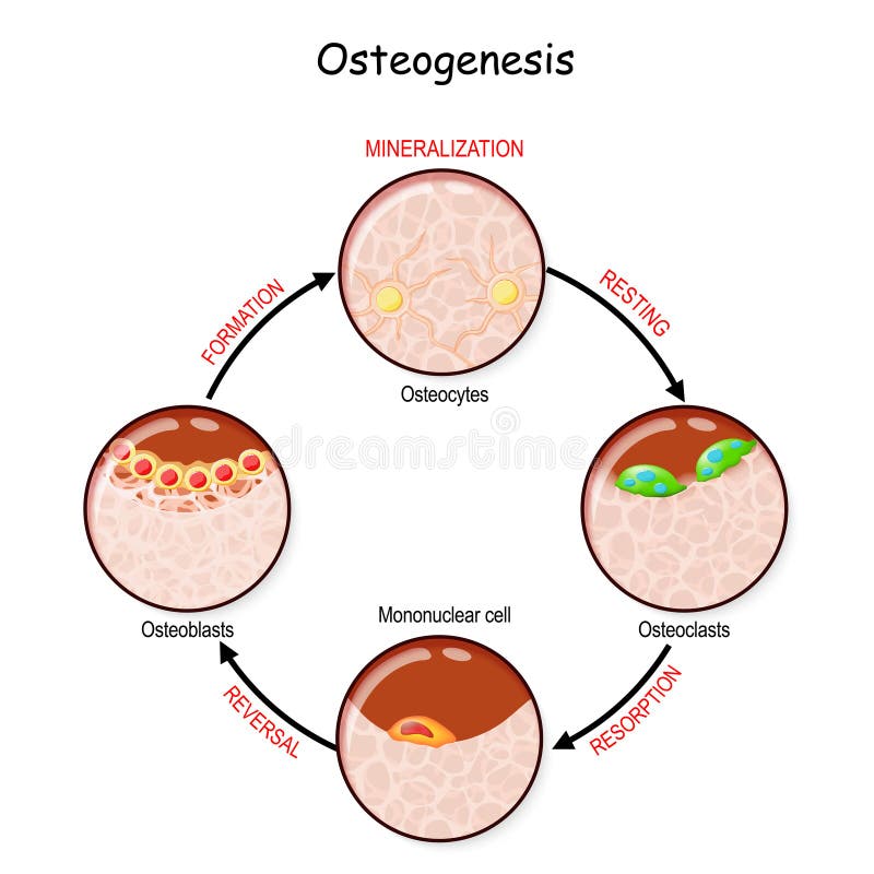
Free with trial Bone remodeling resting, resorption, reversal, mineralization, formation. Describe a process of Ossification. osteogenesis. Bone is broken down by osteoclasts, and rebuilt by osteoblasts. Osteoblast cell vectors Bone remodeling. Describe a process of Ossification. Bone remodeling resting, resorption, reversal, mineralization, formation. Describe a process of Ossification. osteogenesis. Bone is broken down by osteoclasts, and rebuilt by osteoblasts
Free with trial Bone Biology. Role of RANK, RANKL, and OPG. bone remodeling. Bone is broken down by osteoclasts, and rebuilt by osteoblasts. Receptor activator of RANKL is the mediator of bone resorption. Osteoprotegerin OPG. Paracrine and endocrine actions of bone. functions of secretory proteins from osteoblasts, osteocytes, and osteoclasts. Osteoblast cell vectors Bone Biology. Role of RANK, RANKL, and OPG. bone remodeling
Free with trial 3d rendered medically accurate illustration of a osteoclast. Osteoblast cell illustrations A osteoclast
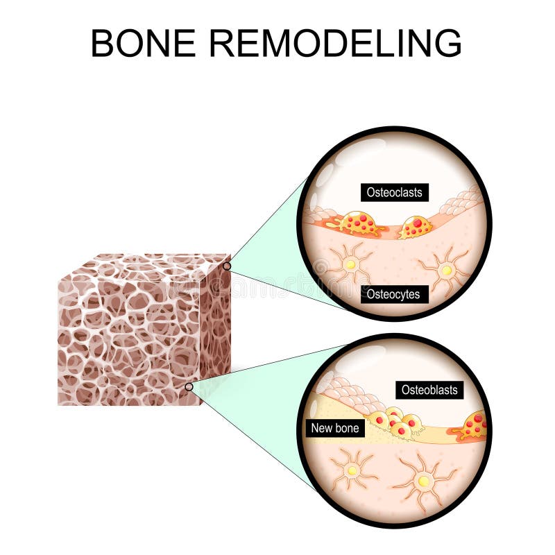
Free with trial Bone remodeling process. Close-up of Osteoblasts, Osteoclast, Osteocytes and bone lining cells. Bone resorption and matrix formation. Vector illustration. Osteoblast cell vectors Bone remodeling process. Close-up of Osteoblasts, Osteoclast, Osteocytes
Free with trial Differentiation of Primary Human Osteoblasts cells. Osteoblasts and their applications in bone tissue. Osteoblast cell vectors Differentiation of Primary Human Osteoblasts cells.
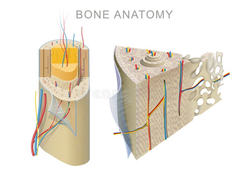
Free with trial The outer shell of the long bone is made of cortical bone also known as compact bone. This is covered by a membrane of connective tissue called the periosteum. Beneath the cortical bone layer is a layer of spongy cancellous bone. Inside this is the medullary cavity which has an inner core of bone marrow, it contains nutrients and help in formation of cells, made up of yellow marrow in the adult and red marrow in the child. Osteoblast cell illustrations Anatomy of a Long Bone. The outer shell of the long bone is made of cortical bone also known as compact bone. This is covered by a membrane of connective tissue called the periosteum. Beneath the cortical bone layer is a layer of spongy cancellous bone. Inside this is the medullary cavity which has an inner core of bone marrow, it contains nutrients and help in formation of cells, made up of yellow marrow in the adult and red marrow in the child.
Free with trial Mesenchymal stem cells or MSCs are stromal cells. Cells bone Marrow. Osteoblast cell vectors Mesenchymal stem cells or MSCs are stromal cells. Cells bone Marrow
Free with trial Bone remodeling from resorption to new bone formation. Close-up of Trabecular bone matrix, Osteoblasts, Osteoclasts and Osteocytes. Vector illustration. Osteoblast cell vectors Bone remodeling from resorption to new bone formation
Free with trial Bone marrow set anatomical poster. Human bone structure and clinic logo. Doctors appointment, consultation and medical exam flat vector illustration. Human skeleton x ray scan medical banner. Osteoblast cell vectors Bone marrow set
Free with trial 3d illustration of Osteoblaste, medically accurate, Bone modeling and remodeling, Bone tissue, Calcium absorbing cells from bone and cartilage, 3d render. Osteoblast cell illustrations Osteoblaste, medically accurate, Bone modeling and remodeling
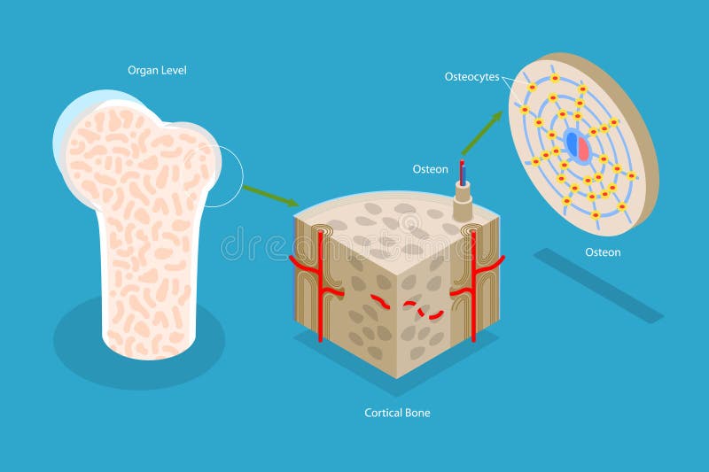
Free with trial 3D Isometric Flat Vector Illustration of Osteon, Diagram of Haversian System with Compact Bone Structure. Osteoblast cell vectors 3D Isometric Flat Vector Illustration of Osteon
Free with trial 3d illustration of Osteoblaste and Osteoclaste, medically accurate, Bone modeling and remodeling, Bone tissue, Calcium absorbing cells from bone and cartilage, 3d render. Osteoblast cell illustrations 3d illustration of Osteoblaste and Osteoclaste, medically accurate, Bone modeling and remodeling
Free with trial 3d illustration of Osteoclaste, medically accurate, Bone modeling and remodeling, Bone tissue, Calcium absorbing cells from bone and cartilage, 3d render. Osteoblast cell illustrations Osteoclaste, medically accurate, Bone modeling and remodeling, Bone tissue
Free with trial 3d illustration of Osteoblaste and Osteoclaste, medically accurate, Bone modeling and remodeling, Bone tissue, Calcium absorbing cells from bone and cartilage, 3d render. Osteoblast cell illustrations 3d illustration of Osteoblaste and Osteoclaste, medically accurate, Bone modeling and remodeling
Free with trial 3d illustration of Osteoblaste and Osteoclaste, medically accurate, Bone modeling and remodeling, Bone tissue, Calcium absorbing cells from bone and cartilage, 3d render. Osteoblast cell illustrations Osteoblaste and Osteoclaste, medically accurate, Bone modeling and remodeling, Bone tissue
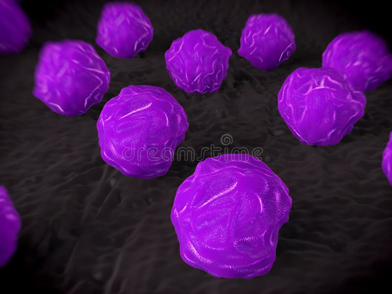
Free with trial 3d rendering of Osteoclasts, are the cells that degrade bone to initiate normal bone remodeling. Osteoblast cell illustrations 3d rendering of isolated of Osteoclasts cells. 3d rendering of Osteoclasts, are the cells that degrade bone to initiate normal bone remodeling
Free with trial Bone cell structure anatomy diagram infographic. Osteoblast and osteocyte. Labeled osteocyte, nucleus, matrix, canaliculi. Bone tissue structure, mineralizing, connective, skeletal system. Osteoblast cell vectors Bone cell structure anatomy diagram infographic. Osteoblast and osteocyte. Labeled osteocyte, nucleus, matrix, canaliculi. Bone
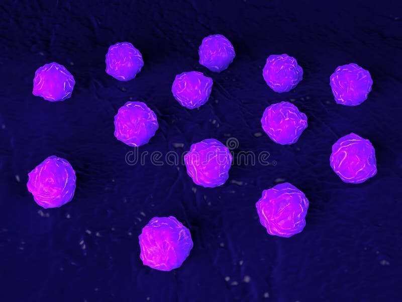
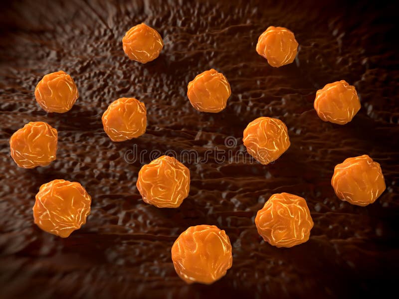
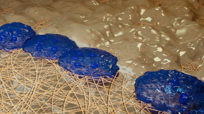
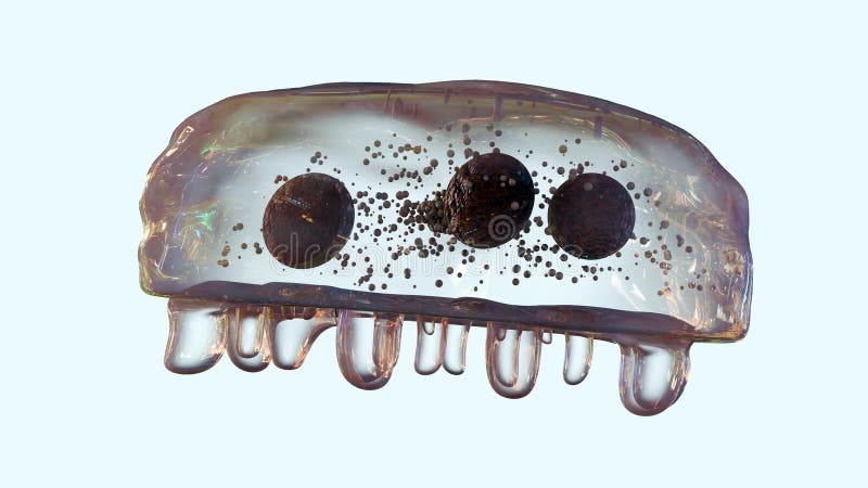
Free with trial Illustration of osteoblast cells. Detailed microscopic image shows, cell structure. Scientific background medical concept. Biology anatomy science research. Medical illustration. Osteoblast cell illustrations Illustration of osteoblast cells. Detailed microscopic image shows cell structure. Scientific background medical concept. Biology. Illustration of osteoblast cells. Detailed microscopic image shows, cell structure. Scientific background medical concept. Biology anatomy science research. Medical illustration.
Free with trial Illustration showing mesenchymal stem cells undergoing osteoblast differentiation in response to mechanical stimulation in a bone fracture healing process. New bone matrix is formed, promoting regeneration. Osteoblast cell illustrations Mesenchymal Stem Cell Differentiation for Bone Regeneration Osteoblast Formation. Illustration showing mesenchymal stem cells undergoing osteoblast differentiation in response to mechanical stimulation in a bone fracture healing process. New bone matrix is formed, promoting regeneration.
Free with trial Detailed 3D medical animation showing bone marrow anatomy, illustrating hematopoiesis and stem cell development. Depicts erythrocytes, leukocytes, thrombocytes, and osteoblast cell formation inside. Osteoblast cell illustrations Bone Marrow Anatomy 3D Medical Illustration Animation Depicting Hematopoiesis Stem Cell Development Erythrocytes Leukocytes. Detailed 3D medical animation showing bone marrow anatomy, illustrating hematopoiesis and stem cell development. Depicts erythrocytes, leukocytes, thrombocytes, and osteoblast cell formation inside
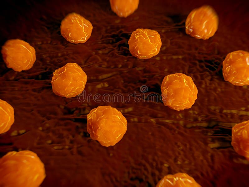
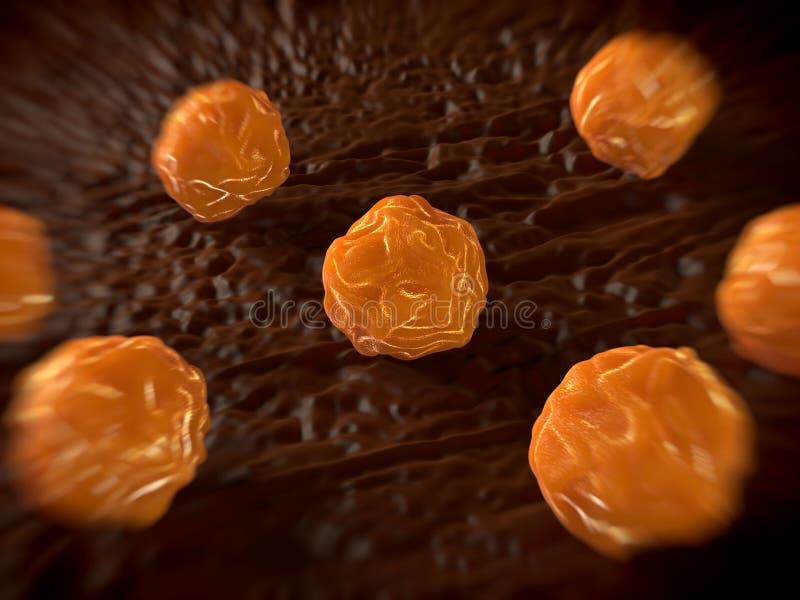
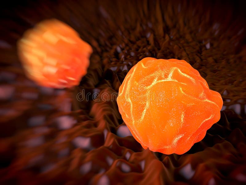
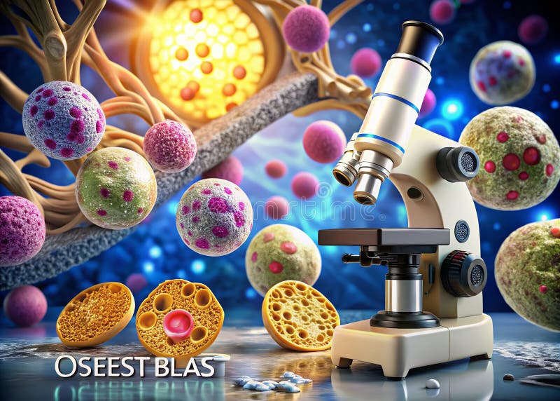
Free with trial This captivating microscopic image offers a detailed view of osteoblast cells, the key players in bone health. Osteoblasts are bone-forming cells actively involved in the intricate processes of bone formation and remodeling. Their activity is crucial for maintaining the structural integrity and strength of our skeletal system. The image showcases the complex cellular structures of osteoblasts,. Osteoblast cell illustrations Understanding Osteoblast Function A Deep Dive into Bone Cell Activity Crucial for Healthy Bone Formation and Remodeling. This captivating microscopic image offers a detailed view of osteoblast cells, the key players in bone health. Osteoblasts are bone-forming cells actively involved in the intricate processes of bone formation and remodeling. Their activity is crucial for maintaining the structural integrity and strength of our skeletal system. The image showcases the complex cellular structures of osteoblasts,
Free with trial Illustration depicting the complex processes of bone regeneration and healing. It shows osteoblast differentiation, growth factor delivery via syringe, and mechanical stimulation contributing to bone matrix formation. This visual aids in understanding regenerative medicine and orthopedic research. Osteoblast cell illustrations Bone Regeneration Osteoblast Differentiation, Growth Factor Delivery Mechanical Stimulation. Illustration depicting the complex processes of bone regeneration and healing. It shows osteoblast differentiation, growth factor delivery via syringe, and mechanical stimulation contributing to bone matrix formation. This visual aids in understanding regenerative medicine and orthopedic research.
Free with trial Types of bone cells for Bone formation, resorption and remodeling. Osteocyte, lining cells, osteoblast, osteoclast. Osteogenesis. Isometric flat vector Illustration. Osteoblast cell vectors Types of bone cells. Osteocyte, lining cells, osteoblast, osteoclast. Types of bone cells for Bone formation, resorption and remodeling. Osteocyte, lining cells, osteoblast, osteoclast. Osteogenesis. Isometric flat vector Illustration
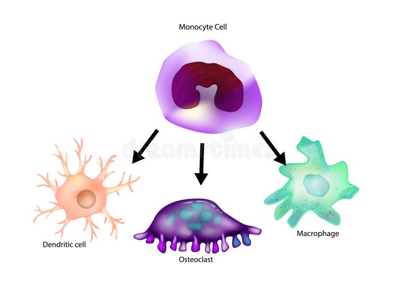
Free with trial Monocyte differentiation. Macrophage, Dendritic cell, Osteoclast. Type of leukocyte or white blood cell. Osteoblast cell vectors Monocyte differentiation. Macrophage, Dendritic cell, Osteoclast.
Free with trial Osteon development and structure. Bone tissue structure anatomy diagram. Haversian system. Labeled osteocyte, osteoblast, osteoclast, nerve, lamellae, lymphatic vessel, artery, central canal and vein. Osteoblast cell vectors Osteon development and structure. Bone tissue structure anatomy diagram. Haversian system. Labeled osteocyte, osteoblast
Free with trial This digital illustration showcases osteoblast activity involved in bone fracture healing, highlighting cellular structures and processes essential for tissue repair. Osteoblast cell illustrations Digital Illustration of Osteoblast Activity in Bone Fracture Healing. This digital illustration showcases osteoblast activity involved in bone fracture healing, highlighting cellular structures and processes essential for tissue repair
Free with trial This scientific illustration depicts the process of bone regeneration, showcasing osteoblast delivery and the crucial roles of growth factor delivery and mechanical stimulation. The diagram visualizes cellular processes leading to improved bone health and potential therapeutic applications. Osteoblast cell illustrations Bone Regeneration Osteoblast Delivery, Growth Factors, and Mechanical Stimulation. This scientific illustration depicts the process of bone regeneration, showcasing osteoblast delivery and the crucial roles of growth factor delivery and mechanical stimulation. The diagram visualizes cellular processes leading to improved bone health and potential therapeutic applications.
Free with trial Diagram illustrating the complex process of bone fracture healing, showcasing osteoblast differentiation, the delivery of growth factors via injection, and the effects of mechanical stimulation to promote new bone matrix formation and repair. Osteoblast cell illustrations Bone Fracture Healing Osteoblast Differentiation, Growth Factor Delivery Mechanical Stimulation. Diagram illustrating the complex process of bone fracture healing, showcasing osteoblast differentiation, the delivery of growth factors via injection, and the effects of mechanical stimulation to promote new bone matrix formation and repair.
Free with trial Light microscopy of osteoblast culture stained with Alizarin Red S showing calcium mineral deposition nodules during in vitro osteogenesis. Osteoblast cell illustrations Osteoblast Bone Matrix Mineralization Nodule ARS. Light microscopy of osteoblast culture stained with Alizarin Red S showing calcium mineral deposition nodules. Light microscopy of osteoblast culture stained with Alizarin Red S showing calcium mineral deposition nodules during in vitro osteogenesis
Free with trial Confocal fluorescence microscopy of bone marrow showing hematopoietic stem cell niche near sinusoidal blood vessels and endosteum. Osteoblast cell illustrations Stem Cell Niche Bone Marrow Sinusoid Architecture. Confocal fluorescence microscopy of bone marrow showing hematopoietic stem cell niche near sinusoidal blood vessels and endosteum
Free with trial This illustration depicts the process of osteoblast differentiation, highlighting the role of growth factors and mechanical stimulation in forming new bone matrix. It visually represents cellular development and bone tissue regeneration. Osteoblast cell illustrations Osteoblast Differentiation Growth Factors and Mechanical Stimulation leading to New Bone Matrix Formation. This illustration depicts the process of osteoblast differentiation, highlighting the role of growth factors and mechanical stimulation in forming new bone matrix. It visually represents cellular development and bone tissue regeneration.
Free with trial This illustrative diagram shows the key stages of bone regeneration. It depicts growth factor delivery via injection, mechanical stimulation applied to tissue, osteoblast differentiation into bone-forming cells, and subsequent bone matrix formation. Osteoblast cell illustrations Bone Regeneration Process Growth Factor Delivery, Mechanical Stimulation, Osteoblast Differentiation, Bone Matrix Format. This illustrative diagram shows the key stages of bone regeneration. It depicts growth factor delivery via injection, mechanical stimulation applied to tissue, osteoblast differentiation into bone-forming cells, and subsequent bone matrix formation.
Free with trial Explore a captivating digital illustration depicting osteoblast activity during the fracture healing process. Ideal for medical and scientific use. Osteoblast cell illustrations Digital Illustration of Osteoblast Activity in Fracture Healing Process. Explore a captivating digital illustration depicting osteoblast activity during the fracture healing process. Ideal for medical and scientific use
Free with trial This digital illustration depicts osteoblast activity during the fracture healing process, showcasing cellular dynamics and the intricate structure of bones in a vibrant visual representation. Osteoblast cell illustrations Digital Illustration of Osteoblast Activity in Fracture Healing Process. This digital illustration depicts osteoblast activity during the fracture healing process, showcasing cellular dynamics and the intricate structure of bones in a vibrant visual representation
Free with trial Osteogenic cells, also known as osteoprogenitor cells, are a type of stem cell found in bone tissue. They are responsible for the formation of new bone during the process of bone remodeling, growth, and repair. Osteogenic cells are derived from mesenchymal stem cells, which are multipotent stem cells found in various tissues, including bone marrow. These cells have the ability to differentiate into osteoblasts, the cells responsible for synthesizing and secreting the organic components of bone matrix. Osteoblast cell vectors Osteogenic cell
Free with trial Human bone cell color line icon. Microorganisms microbes, bacteria. Vector isolated element. Editable stroke. Osteoblast cell vectors Human bone cell color line icon. Microorganisms microbes, bacteria
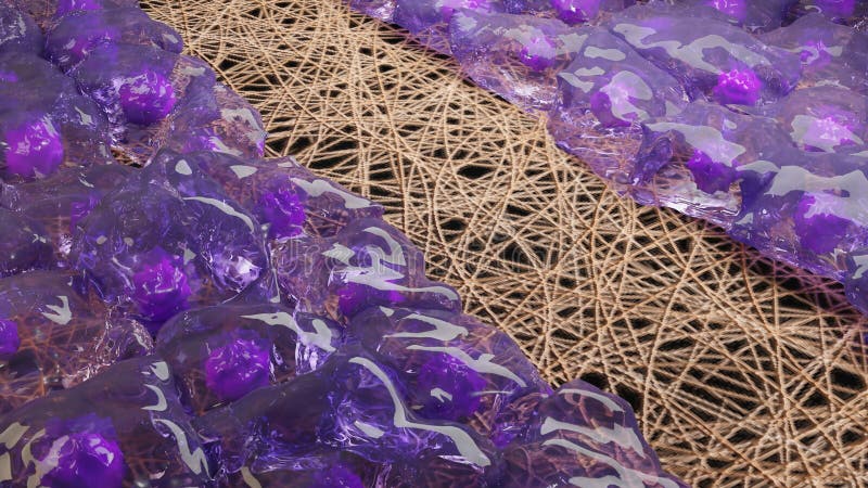
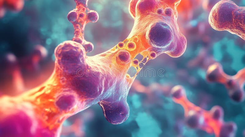
Free with trial Bone marrow cross-section shows cell development. Purple cells sprout from bone structure. Orange spheres float nearby. Internal bone cavity glows with bright, yellow and red light. Osteoblast cell illustrations Bone marrow cross-section shows cell development. Purple cells sprout from bone structure. Orange spheres float nearby. Internal
Free with trial This illustration depicts the dynamic process of bone remodeling, showing osteoclasts resorbing bone tissue and osteoblasts forming new bone. Cellular communication and signaling molecules are also visually represented. Osteoblast cell illustrations Osteoclast and Osteoblast Bone Remodeling Resorption - Cellular Biology Illustration. This illustration depicts the dynamic process of bone remodeling, showing osteoclasts resorbing bone tissue and osteoblasts forming new bone. Cellular communication and signaling molecules are also visually represented.
Free with trial This digital illustration showcases the activity of osteoblasts during the intricate process of fracture healing, emphasizing cellular structure and growth dynamics. Osteoblast cell illustrations Digital Illustration of Osteoblast Activity in Fracture Healing Process. This digital illustration showcases the activity of osteoblasts during the intricate process of fracture healing, emphasizing cellular structure and growth dynamics
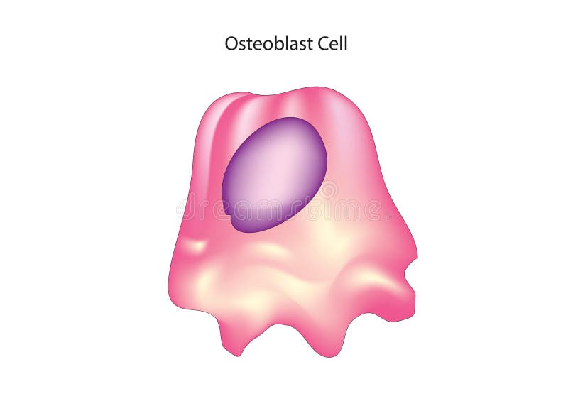
Free with trial Osteoblasts are specialized cells responsible for bone formation. They are derived from precursor cells called osteoprogenitor cells and play a crucial role in bone development, growth, and repair. Osteoblasts produce and secrete proteins, such as collagen, which form the organic matrix of bone, and also regulate the mineralization of bone tissue by depositing calcium and phosphate ions. Additionally, osteoblasts communicate with other cells, such as osteoclasts (which break down bone tissue) and osteocytes (mature bone cells embedded in the bone matrix), to maintain bone homeostasis. Osteoblast cell vectors Osteoblast
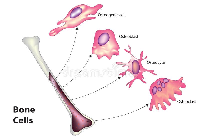
Free with trial Bone tissue is composed of several types of cells, each with specific functions in the growth, maintenance, and repair of bone. The main types of bone cells include osteoblasts, osteocytes, osteoclasts, and bone lining cells. Osteoblast cell vectors Bone cell classification. Bone tissue is composed of several types of cells, each with specific functions in the growth, maintenance, and repair of bone. The main types of bone cells include osteoblasts, osteocytes, osteoclasts, and bone lining cells.
Free with trial Dynamic Regeneration, Fracture Healing, Fibrocartilaginous Callus, Osteoblastogenesis, Formation and Bone Healing, Ossification Process, Scar Tissue, Chondrocyte Proliferation, Osteoblast, 3d render. Osteoblast cell illustrations Dynamic Regeneration, Fracture Healing, Fibrocartilaginous Callus, Osteoblastogenesis, Formation and Bone Healing, Ossification
Free with trial Dynamic Regeneration, Fracture Healing, Fibrocartilaginous Callus, Osteoblastogenesis, Formation and Bone Healing, Ossification Process, Scar Tissue, Chondrocyte Proliferation, Osteoblast, 3d render. Osteoblast cell illustrations Dynamic Regeneration, Fracture Healing, Fibrocartilaginous Callus, Osteoblastogenesis, Formation and Bone Healing, Ossification
Free with trial Homeostasis. Bone remodeling. Osteoclasts remove bone. Osteoblasts form bone. Hormonal regulation of tissue formation and resorption. Parathyroid glands synthesize PTH parathyroid hormone. Thyroid gland secretes Calcitonin. Role RANKL and OPG proteins. Vector illustration. Medical diagram. Osteoblast cell vectors Homeostasis. Hormonal regulation of Bone remodeling. Osteoclasts remove bone. Osteoblasts form bone. Homeostasis. Bone remodeling. Osteoclasts remove bone. Osteoblasts form bone. Hormonal regulation of tissue formation and resorption. Parathyroid glands synthesize PTH parathyroid hormone. Thyroid gland secretes Calcitonin. Role RANKL and OPG proteins. Vector illustration. Medical diagram
Free with trial Differentiation of Primary Human Osteoblasts cells. Osteoblasts and their applications in bone tissue. Osteoblast cell illustrations Illustration showing a Osteoblasts cells. Osteoblasts and their applications in bone tissue. Differentiation of Primary Human Osteoblasts cells. Osteoblasts and their applications in bone tissue
Free with trial Osteoblast Cells Design Vector Illustration. Osteoblast cell vectors Osteoblasts Cells Design. Osteoblast Cells Design Vector Illustration
Free with trial Illustration detailing the complex bone healing process. It highlights key anatomical structures like the periosteum and endosteum, and cellular activity involving growth factors and bone matrix regeneration. The diagram also shows external influences such as mechanical stimulation and laboratory research elements. Osteoblast cell illustrations Bone Healing Process Periosteum, Endosteum, Cells, Growth Factors, Mechanical Stimulation. Illustration detailing the complex bone healing process. It highlights key anatomical structures like the periosteum and endosteum, and cellular activity involving growth factors and bone matrix regeneration. The diagram also shows external influences such as mechanical stimulation and laboratory research elements.