Free with trial Skin is the soft outer covering of vertebrates. Other animal coverings such as the arthropod exoskeleton have different developmental origin, structure and chemical composition. The adjective cutaneous means of the skin (from Latin cutis, skin). In mammals, the skin is an organ of the integumentary system made up of multiple layers of ectodermal tissue, and guards the underlying muscles, bones, ligaments and internal organs. Outer covering illustrations Skin label. Skin is the soft outer covering of vertebrates. Other animal coverings such as the arthropod exoskeleton have different developmental origin, structure and chemical composition. The adjective cutaneous means of the skin (from Latin cutis, skin). In mammals, the skin is an organ of the integumentary system made up of multiple layers of ectodermal tissue, and guards the underlying muscles, bones, ligaments and internal organs.
Free with trial Skin is the soft outer covering of vertebrates. Other animal coverings such as the arthropod exoskeleton have different developmental origin, structure and chemical composition. The adjective cutaneous means of the skin (from Latin cutis, skin). Outer covering illustrations Skin anatomy labelled. Skin is the soft outer covering of vertebrates. Other animal coverings such as the arthropod exoskeleton have different developmental origin, structure and chemical composition. The adjective cutaneous means of the skin (from Latin cutis, skin).
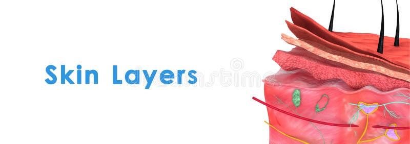
Free with trial Skin is the soft outer covering of vertebrates. Other animal coverings such as the arthropod exoskeleton have different developmental origin, structure and chemical composition. Outer covering illustrations Skin layers. Skin is the soft outer covering of vertebrates. Other animal coverings such as the arthropod exoskeleton have different developmental origin, structure and chemical composition.
Free with trial Skin is the soft outer covering of vertebrates. Other animal coverings such as the arthropod exoskeleton have different developmental origin, structure and chemical composition. Outer covering illustrations Skin Anatomy. Skin is the soft outer covering of vertebrates. Other animal coverings such as the arthropod exoskeleton have different developmental origin, structure and chemical composition.
Free with trial Lost Bird Outer Element Feather Sketch Vector. Fluffy Feather Bird Detail Covering Varmint Body Arise From Certain Well-defined Tracts On Skin. Designed In Retro Style Monochrome Illustration. Outer covering vectors Lost Bird Outer Element Feather Sketch Vector
Free with trial Skin is the soft outer covering of vertebrates. Other animal coverings such as the arthropod exoskeleton have different developmental origin, structure and chemical composition. Outer covering illustrations Skin anatomy. Skin is the soft outer covering of vertebrates. Other animal coverings such as the arthropod exoskeleton have different developmental origin, structure and chemical composition.
Free with trial Futuristic 3d rendering of an outer space plazma looking big bang. It is sparkling white in the center, with yellow protunerances and nebulas covering its contours in the blue background. Outer covering illustrations Abstract outer space illustration. Futuristic 3d rendering of an outer space plazma looking big bang. It is sparkling white in the center, with yellow protunerances and nebulas covering its contours in the blue background
Free with trial Collection of Different Feathers Set Ink Vector. Standing, Flying And Lying Fluffy Bird Feathers. Epidermal Growths Form Distinctive Outer Covering Or Plumage. Monochrome Hand Drawn Illustrations. Outer covering vectors Collection of Different Feathers Set Ink Vector
Free with trial Digitally generated full grey moon against outer space. Outer covering illustrations Composite image of digitally generated full grey moon. Digitally generated full grey moon against outer space
Free with trial Digitally generated full grey moon against outer space. Outer covering illustrations Composite image of digitally generated full grey moon. Digitally generated full grey moon against outer space
Free with trial Saturn Planet with ring in outer space. 3D rendered illustration. Elements of this image furnished by NASA. Outer covering illustrations Saturn Planet with ring in outer space
Free with trial Igloo on snow field against outer space. Outer covering illustrations Composite image of igloo on snow field. Igloo on snow field against outer space
Free with trial Digitally generated image of gift boxes on park bench against outer space. Outer covering illustrations Composite image of digitally generated image of gift boxes on park bench. Digitally generated image of gift boxes on park bench against outer space
Free with trial Science fiction exploading Earth planet with atmosphere and ring in outer space closeup. 3d rendering illustration. Elements of this image furnished by NASA. Outer covering illustrations Fiction exploading exoplanet with ring in outer space. Science fiction exploading Earth planet with atmosphere and ring in outer space closeup. 3d rendering illustration. Elements of this image furnished by NASA.
Free with trial A powerful and divine presence emerges as Jesus Christ, the Son of God, is depicted in full view. He is the Messiah, the Lamb of God, and the Savior who embodies the eternal Word. As the Good Shepherd, He offers solace and guidance, and as the King of Kings, He reigns with peace. Dressed in a tunic, chiton, and himation, His attire reflects ancient times, and His long hair and head covering exude reverence. Walking away, His silhouette carries the weight of His teachings�like the Sermon on the Mount�and His triumph over temptation. He is the embodiment of resurrection and the eternal truth, a beacon of hope and salvation for all. Outer covering illustrations Full view of the Messiah the Son of Man. A powerful and divine presence emerges as Jesus Christ, the Son of God, is depicted in full view. He is the Messiah, the Lamb of God, and the Savior who embodies the eternal Word. As the Good Shepherd, He offers solace and guidance, and as the King of Kings, He reigns with peace. Dressed in a tunic, chiton, and himation, His attire reflects ancient times, and His long hair and head covering exude reverence. Walking away, His silhouette carries the weight of His teachings�like the Sermon on the Mount�and His triumph over temptation. He is the embodiment of resurrection and the eternal truth, a beacon of hope and salvation for all.
Free with trial Child with Autism covering his ears from outer sounds and has issues with communication, flat vector illustration isolated on white. Autism disorder symptoms and signs. Outer covering vectors Child autist covers his ears from outer sounds flat vector illustration isolated. Child with Autism covering his ears from outer sounds and has issues with communication, flat vector illustration isolated on white. Autism disorder symptoms and signs.
Free with trial Computer generated illustration of wrinkled green lizard skin. Outer covering illustrations Reptile skin. Computer generated illustration of wrinkled green lizard skin
Free with trial A seed is an embryonic plant enclosed in a protective outer covering called the seed coat, usually with some stored food. It is a characteristic of spermatophytes (gymnosperm and angiosperm plants) and the product of the ripened ovule which occurs after fertilization and some growth within the mother plant. Outer covering illustrations Dry seed. A seed is an embryonic plant enclosed in a protective outer covering called the seed coat, usually with some stored food. It is a characteristic of spermatophytes (gymnosperm and angiosperm plants) and the product of the ripened ovule which occurs after fertilization and some growth within the mother plant.
Free with trial A seed is an embryonic plant enclosed in a protective outer covering called the seed coat, usually with some stored food. It is a characteristic of spermatophytes (gymnosperm and angiosperm plants) and the product of the ripened ovule which occurs after fertilization and some growth within the mother plant. Outer covering illustrations Dry seed. A seed is an embryonic plant enclosed in a protective outer covering called the seed coat, usually with some stored food. It is a characteristic of spermatophytes (gymnosperm and angiosperm plants) and the product of the ripened ovule which occurs after fertilization and some growth within the mother plant.
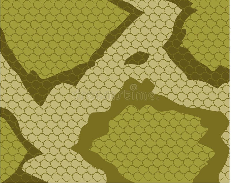
Free with trial An illustration of a snake's skin with scales. Outer covering illustrations Snake Skin Pattern. An illustration of a snake's skin with scales.
Free with trial The Earth surface is covered by clouds. Incredible image of Earth covered in clouds. Clouds covering a immense area of the planet and shaping the Earth's climate. Elements of this image furnished by NASA. Outer covering illustrations Cloudy Earth in space. The Earth surface is covered by clouds. Incredible image of Earth covered in clouds. Clouds covering a immense area of the planet and shaping the Earth's climate. Elements of this image furnished by NASA
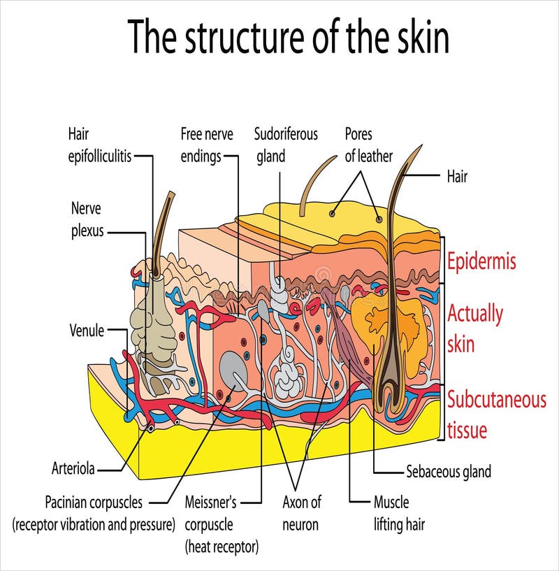
Free with trial Human skin is the outer layer of the body in humans. It is the largest organ of the body system covering up. The skin has several layers of ectodermal tissue and guards muscles, bones, ligaments and internal organs underneath. Outer covering vectors Skin organ and its function against a white background. Human skin is the outer layer of the body in humans. It is the largest organ of the body system covering up. The skin has several layers of ectodermal tissue and guards muscles, bones, ligaments and internal organs underneath.
Free with trial A slipped disc occurs when the outer covering of the disc tears and the internal gel filling (nucleus pulposus) gets pushed out of the covering (herniated), causing the gel to press on the nerve root. Outer covering vectors A slipped disc occurs when the outer covering of the disc tears
Free with trial This image showcases a whole head of Romanesco broccoli, known for its striking fractal pattern. The outer leaves are still attached, providing a vibrant green hue and a protective covering. The unique geometric shape and pale green florets are characteristic of this variety, which is both visually appealing and nutritious, often used in gourmet dishes for its distinct appearance and mild flavor. Outer covering illustrations Fresh green romanesco broccoli with outer leaves intact. This image showcases a whole head of Romanesco broccoli, known for its striking fractal pattern. The. This image showcases a whole head of Romanesco broccoli, known for its striking fractal pattern. The outer leaves are still attached, providing a vibrant green hue and a protective covering. The unique geometric shape and pale green florets are characteristic of this variety, which is both visually appealing and nutritious, often used in gourmet dishes for its distinct appearance and mild flavor
Free with trial White cauliflower (Brassica oleracea) with green leaves rests against a smooth blue fabric backdrop. The cauliflower is round with tightly packed curds and vibrant green outer leaves partially covering it. The texture of the cauliflower's surface is coarse and bumpy, contrasting with the smoothness of the background. The lighting highlights the natural color variations between the white florets and green leaves, creating an artistic composition. Outer covering illustrations Tightly packed curds and vibrant green outer leaves partially
Free with trial A solar eclipse is depicted, with the moon covering the sun, leaving a fiery, orange corona. The bright halo contrasts sharply against the dark sky, creating a dramatic visual. The eclipse showcases the alignment of the sun, moon, and Earth, an astronomical event that briefly darkens the day and reveals the sun's outer atmosphere. Outer covering illustrations A solar eclipse is depicted, with the moon covering the sun, leaving a fiery, orange
Free with trial This captivating image showcases a detailed close-up of a tree trunk's outer layer. The texture is truly remarkable, revealing a complex interplay of fibrous, ridged, and scaled patterns. This protective covering, often referred to as bark, is more than just an exterior it's a vital component of the tree's survival strategy against environmental stressors. Notice the rough, rugged surface, a. Outer covering illustrations Closeup View of a Rugged Tree Trunk Exploring the Protective Outer Layers of a Woody Stem. This captivating image showcases a detailed close-up of a tree trunk's outer layer. The texture is truly remarkable, revealing a complex interplay of fibrous, ridged, and scaled patterns. This protective covering, often referred to as bark, is more than just an exterior it's a vital component of the tree's survival strategy against environmental stressors. Notice the rough, rugged surface, a
Free with trial The image shows a single coconut with its fibrous husk fully covering the hard, brown outer shell. The husk appears slightly weathered and frayed at the edges, showcasing the natural texture and appearance of a mature coconut. Outer covering illustrations A whole coconut with its fibrous husk intact and brown outer shell. The image shows a single coconut with its fibrous husk fully covering the hard, brown outer shell. The husk appears slightly weathered and frayed at the edges, showcasing the natural texture and appearance of a mature coconut
Free with trial This image depicts a lightbulb with a unique design, featuring a gear-shaped outer shell instead of the traditional glass covering. The lightbulb is illuminated with a soft blue glow, symbolizing creativity and innovation. Outer covering illustrations A creative representation of a lightbulb with a gear-shaped outer shell. This image depicts a lightbulb with a unique design, featuring a gear-shaped outer shell instead of the traditional glass covering. The lightbulb is illuminated with a soft blue glow, symbolizing creativity and innovation
Free with trial This image presents a vivid and detailed view of the Earth from outer space, showcasing the continents of North America, South America, Africa, Europe, and Asia. The oceans are depicted in deep blue, with swirling white clouds covering parts of the planet. The image highlights the beauty and complexity of our home planet. Outer covering illustrations A vibrant and detailed view of the earth from outer space showcasing continents and oceans. This image presents a vivid and detailed view of the Earth from outer space, showcasing the continents of North America, South America, Africa, Europe, and Asia. The oceans are depicted in deep blue, with swirling white clouds covering parts of the planet. The image highlights the beauty and complexity of our home planet
Free with trial 3D render depicting epithelial tissue structure. The image shows a layered arrangement with outer cells connected, featuring prominent oval nuclei. Cells are organized in a uniform, stacked pattern, reflecting their role in covering surfaces. The background is dark green, enhancing the vibrant orange and pink hues of the tissue. This visualization helps illustrate the protective and absorptive functions of epithelial tissue, commonly found lining organs and cavities in the body. Outer covering illustrations Structure of Epithelial Tissue anatyomy with colourful background. 3d render. 3D render depicting epithelial tissue structure. The image shows a layered arrangement with outer cells connected, featuring prominent oval nuclei. Cells are organized in a uniform, stacked pattern, reflecting their role in covering surfaces. The background is dark green, enhancing the vibrant orange and pink hues of the tissue. This visualization helps illustrate the protective and absorptive functions of epithelial tissue, commonly found lining organs and cavities in the body.
Free with trial 3D render depicting epithelial tissue structure. The image shows a layered arrangement with outer cells connected, featuring prominent oval nuclei. Cells are organized in a uniform, stacked pattern, reflecting their role in covering surfaces. The background is dark green, enhancing the vibrant orange and pink hues of the tissue. This visualization helps illustrate the protective and absorptive functions of epithelial tissue, commonly found lining organs and cavities in the body. Outer covering illustrations Structure of Epithelial Tissue anatyomy with colourful background. 3d render. 3D render depicting epithelial tissue structure. The image shows a layered arrangement with outer cells connected, featuring prominent oval nuclei. Cells are organized in a uniform, stacked pattern, reflecting their role in covering surfaces. The background is dark green, enhancing the vibrant orange and pink hues of the tissue. This visualization helps illustrate the protective and absorptive functions of epithelial tissue, commonly found lining organs and cavities in the body.
Free with trial 3D render depicting epithelial tissue structure. The image shows a layered arrangement with outer cells connected, featuring prominent oval nuclei. Cells are organized in a uniform, stacked pattern, reflecting their role in covering surfaces. The background is dark green, enhancing the vibrant orange and pink hues of the tissue. This visualization helps illustrate the protective and absorptive functions of epithelial tissue, commonly found lining organs and cavities in the body. Outer covering illustrations Structure of Epithelial Tissue anatyomy with colourful background. 3d render. 3D render depicting epithelial tissue structure. The image shows a layered arrangement with outer cells connected, featuring prominent oval nuclei. Cells are organized in a uniform, stacked pattern, reflecting their role in covering surfaces. The background is dark green, enhancing the vibrant orange and pink hues of the tissue. This visualization helps illustrate the protective and absorptive functions of epithelial tissue, commonly found lining organs and cavities in the body.
Free with trial 3D render depicting epithelial tissue structure. The image shows a layered arrangement with outer cells connected, featuring prominent oval nuclei. Cells are organized in a uniform, stacked pattern, reflecting their role in covering surfaces. The background is dark green, enhancing the vibrant orange and pink hues of the tissue. This visualization helps illustrate the protective and absorptive functions of epithelial tissue, commonly found lining organs and cavities in the body. Outer covering illustrations Structure of Epithelial Tissue anatyomy with colourful background. 3d render. 3D render depicting epithelial tissue structure. The image shows a layered arrangement with outer cells connected, featuring prominent oval nuclei. Cells are organized in a uniform, stacked pattern, reflecting their role in covering surfaces. The background is dark green, enhancing the vibrant orange and pink hues of the tissue. This visualization helps illustrate the protective and absorptive functions of epithelial tissue, commonly found lining organs and cavities in the body.
Free with trial 3D render depicting epithelial tissue structure. The image shows a layered arrangement with outer cells connected, featuring prominent oval nuclei. Cells are organized in a uniform, stacked pattern, reflecting their role in covering surfaces. The background is dark green, enhancing the vibrant orange and pink hues of the tissue. This visualization helps illustrate the protective and absorptive functions of epithelial tissue, commonly found lining organs and cavities in the body. Outer covering illustrations Structure of Epithelial Tissue anatyomy with colourful background. 3d render. 3D render depicting epithelial tissue structure. The image shows a layered arrangement with outer cells connected, featuring prominent oval nuclei. Cells are organized in a uniform, stacked pattern, reflecting their role in covering surfaces. The background is dark green, enhancing the vibrant orange and pink hues of the tissue. This visualization helps illustrate the protective and absorptive functions of epithelial tissue, commonly found lining organs and cavities in the body.
Free with trial 3D render depicting epithelial tissue structure. The image shows a layered arrangement with outer cells connected, featuring prominent oval nuclei. Cells are organized in a uniform, stacked pattern, reflecting their role in covering surfaces. The background is dark green, enhancing the vibrant orange and pink hues of the tissue. This visualization helps illustrate the protective and absorptive functions of epithelial tissue, commonly found lining organs and cavities in the body. Outer covering illustrations Structure of Epithelial Tissue anatyomy with colourful background. 3d render. 3D render depicting epithelial tissue structure. The image shows a layered arrangement with outer cells connected, featuring prominent oval nuclei. Cells are organized in a uniform, stacked pattern, reflecting their role in covering surfaces. The background is dark green, enhancing the vibrant orange and pink hues of the tissue. This visualization helps illustrate the protective and absorptive functions of epithelial tissue, commonly found lining organs and cavities in the body.
Free with trial 3D render depicting epithelial tissue structure. The image shows a layered arrangement with outer cells connected, featuring prominent oval nuclei. Cells are organized in a uniform, stacked pattern, reflecting their role in covering surfaces. The background is dark green, enhancing the vibrant orange and pink hues of the tissue. This visualization helps illustrate the protective and absorptive functions of epithelial tissue, commonly found lining organs and cavities in the body. Outer covering illustrations Structure of Epithelial Tissue anatyomy with colourful background. 3d render. 3D render depicting epithelial tissue structure. The image shows a layered arrangement with outer cells connected, featuring prominent oval nuclei. Cells are organized in a uniform, stacked pattern, reflecting their role in covering surfaces. The background is dark green, enhancing the vibrant orange and pink hues of the tissue. This visualization helps illustrate the protective and absorptive functions of epithelial tissue, commonly found lining organs and cavities in the body.
Free with trial 3D render depicting epithelial tissue structure. The image shows a layered arrangement with outer cells connected, featuring prominent oval nuclei. Cells are organized in a uniform, stacked pattern, reflecting their role in covering surfaces. The background is dark green, enhancing the vibrant orange and pink hues of the tissue. This visualization helps illustrate the protective and absorptive functions of epithelial tissue, commonly found lining organs and cavities in the body. Outer covering illustrations Structure of Epithelial Tissue anatyomy with colourful background. 3d render. 3D render depicting epithelial tissue structure. The image shows a layered arrangement with outer cells connected, featuring prominent oval nuclei. Cells are organized in a uniform, stacked pattern, reflecting their role in covering surfaces. The background is dark green, enhancing the vibrant orange and pink hues of the tissue. This visualization helps illustrate the protective and absorptive functions of epithelial tissue, commonly found lining organs and cavities in the body.
Free with trial 3D render depicting epithelial tissue structure. The image shows a layered arrangement with outer cells connected, featuring prominent oval nuclei. Cells are organized in a uniform, stacked pattern, reflecting their role in covering surfaces. The background is dark green, enhancing the vibrant orange and pink hues of the tissue. This visualization helps illustrate the protective and absorptive functions of epithelial tissue, commonly found lining organs and cavities in the body. Outer covering illustrations Structure of Epithelial Tissue anatyomy with colourful background. 3d render. 3D render depicting epithelial tissue structure. The image shows a layered arrangement with outer cells connected, featuring prominent oval nuclei. Cells are organized in a uniform, stacked pattern, reflecting their role in covering surfaces. The background is dark green, enhancing the vibrant orange and pink hues of the tissue. This visualization helps illustrate the protective and absorptive functions of epithelial tissue, commonly found lining organs and cavities in the body.
Free with trial 3D render depicting epithelial tissue structure. The image shows a layered arrangement with outer cells connected, featuring prominent oval nuclei. Cells are organized in a uniform, stacked pattern, reflecting their role in covering surfaces. The background is dark green, enhancing the vibrant orange and pink hues of the tissue. This visualization helps illustrate the protective and absorptive functions of epithelial tissue, commonly found lining organs and cavities in the body. Outer covering illustrations Structure of Epithelial Tissue anatyomy with colourful background. 3d render. 3D render depicting epithelial tissue structure. The image shows a layered arrangement with outer cells connected, featuring prominent oval nuclei. Cells are organized in a uniform, stacked pattern, reflecting their role in covering surfaces. The background is dark green, enhancing the vibrant orange and pink hues of the tissue. This visualization helps illustrate the protective and absorptive functions of epithelial tissue, commonly found lining organs and cavities in the body.
Free with trial 3D render depicting epithelial tissue structure. The image shows a layered arrangement with outer cells connected, featuring prominent oval nuclei. Cells are organized in a uniform, stacked pattern, reflecting their role in covering surfaces. The background is dark green, enhancing the vibrant orange and pink hues of the tissue. This visualization helps illustrate the protective and absorptive functions of epithelial tissue, commonly found lining organs and cavities in the body. Outer covering illustrations Structure of Epithelial Tissue anatyomy with colourful background. 3d render. 3D render depicting epithelial tissue structure. The image shows a layered arrangement with outer cells connected, featuring prominent oval nuclei. Cells are organized in a uniform, stacked pattern, reflecting their role in covering surfaces. The background is dark green, enhancing the vibrant orange and pink hues of the tissue. This visualization helps illustrate the protective and absorptive functions of epithelial tissue, commonly found lining organs and cavities in the body.
Free with trial 3D render depicting epithelial tissue structure. The image shows a layered arrangement with outer cells connected, featuring prominent oval nuclei. Cells are organized in a uniform, stacked pattern, reflecting their role in covering surfaces. The background is dark green, enhancing the vibrant orange and pink hues of the tissue. This visualization helps illustrate the protective and absorptive functions of epithelial tissue, commonly found lining organs and cavities in the body. Outer covering illustrations Structure of Epithelial Tissue anatyomy with colourful background. 3d render. 3D render depicting epithelial tissue structure. The image shows a layered arrangement with outer cells connected, featuring prominent oval nuclei. Cells are organized in a uniform, stacked pattern, reflecting their role in covering surfaces. The background is dark green, enhancing the vibrant orange and pink hues of the tissue. This visualization helps illustrate the protective and absorptive functions of epithelial tissue, commonly found lining organs and cavities in the body.
Free with trial 3D render depicting epithelial tissue structure. The image shows a layered arrangement with outer cells connected, featuring prominent oval nuclei. Cells are organized in a uniform, stacked pattern, reflecting their role in covering surfaces. The background is dark green, enhancing the vibrant orange and pink hues of the tissue. This visualization helps illustrate the protective and absorptive functions of epithelial tissue, commonly found lining organs and cavities in the body. Outer covering illustrations Structure of Epithelial Tissue anatyomy with colourful background. 3d render. 3D render depicting epithelial tissue structure. The image shows a layered arrangement with outer cells connected, featuring prominent oval nuclei. Cells are organized in a uniform, stacked pattern, reflecting their role in covering surfaces. The background is dark green, enhancing the vibrant orange and pink hues of the tissue. This visualization helps illustrate the protective and absorptive functions of epithelial tissue, commonly found lining organs and cavities in the body.
Free with trial 3D render depicting epithelial tissue structure. The image shows a layered arrangement with outer cells connected, featuring prominent oval nuclei. Cells are organized in a uniform, stacked pattern, reflecting their role in covering surfaces. The background is dark green, enhancing the vibrant orange and pink hues of the tissue. This visualization helps illustrate the protective and absorptive functions of epithelial tissue, commonly found lining organs and cavities in the body. Outer covering illustrations Structure of Epithelial Tissue anatyomy with colourful background. 3d render. 3D render depicting epithelial tissue structure. The image shows a layered arrangement with outer cells connected, featuring prominent oval nuclei. Cells are organized in a uniform, stacked pattern, reflecting their role in covering surfaces. The background is dark green, enhancing the vibrant orange and pink hues of the tissue. This visualization helps illustrate the protective and absorptive functions of epithelial tissue, commonly found lining organs and cavities in the body.
Free with trial 3D render depicting epithelial tissue structure. The image shows a layered arrangement with outer cells connected, featuring prominent oval nuclei. Cells are organized in a uniform, stacked pattern, reflecting their role in covering surfaces. The background is dark green, enhancing the vibrant orange and pink hues of the tissue. This visualization helps illustrate the protective and absorptive functions of epithelial tissue, commonly found lining organs and cavities in the body. Outer covering illustrations Structure of Epithelial Tissue anatyomy with colourful background. 3d render. 3D render depicting epithelial tissue structure. The image shows a layered arrangement with outer cells connected, featuring prominent oval nuclei. Cells are organized in a uniform, stacked pattern, reflecting their role in covering surfaces. The background is dark green, enhancing the vibrant orange and pink hues of the tissue. This visualization helps illustrate the protective and absorptive functions of epithelial tissue, commonly found lining organs and cavities in the body.
Free with trial 3D render depicting epithelial tissue structure. The image shows a layered arrangement with outer cells connected, featuring prominent oval nuclei. Cells are organized in a uniform, stacked pattern, reflecting their role in covering surfaces. The background is dark green, enhancing the vibrant orange and pink hues of the tissue. This visualization helps illustrate the protective and absorptive functions of epithelial tissue, commonly found lining organs and cavities in the body. Outer covering illustrations Structure of Epithelial Tissue anatyomy with colourful background. 3d render. 3D render depicting epithelial tissue structure. The image shows a layered arrangement with outer cells connected, featuring prominent oval nuclei. Cells are organized in a uniform, stacked pattern, reflecting their role in covering surfaces. The background is dark green, enhancing the vibrant orange and pink hues of the tissue. This visualization helps illustrate the protective and absorptive functions of epithelial tissue, commonly found lining organs and cavities in the body.
Free with trial 3D render depicting epithelial tissue structure. The image shows a layered arrangement with outer cells connected, featuring prominent oval nuclei. Cells are organized in a uniform, stacked pattern, reflecting their role in covering surfaces. The background is dark green, enhancing the vibrant orange and pink hues of the tissue. This visualization helps illustrate the protective and absorptive functions of epithelial tissue, commonly found lining organs and cavities in the body. Outer covering illustrations Structure of Epithelial Tissue anatyomy with colourful background. 3d render. 3D render depicting epithelial tissue structure. The image shows a layered arrangement with outer cells connected, featuring prominent oval nuclei. Cells are organized in a uniform, stacked pattern, reflecting their role in covering surfaces. The background is dark green, enhancing the vibrant orange and pink hues of the tissue. This visualization helps illustrate the protective and absorptive functions of epithelial tissue, commonly found lining organs and cavities in the body.
Free with trial 3D render depicting epithelial tissue structure. The image shows a layered arrangement with outer cells connected, featuring prominent oval nuclei. Cells are organized in a uniform, stacked pattern, reflecting their role in covering surfaces. The background is dark green, enhancing the vibrant orange and pink hues of the tissue. This visualization helps illustrate the protective and absorptive functions of epithelial tissue, commonly found lining organs and cavities in the body. Outer covering illustrations Structure of Epithelial Tissue anatyomy with colourful background. 3d render. 3D render depicting epithelial tissue structure. The image shows a layered arrangement with outer cells connected, featuring prominent oval nuclei. Cells are organized in a uniform, stacked pattern, reflecting their role in covering surfaces. The background is dark green, enhancing the vibrant orange and pink hues of the tissue. This visualization helps illustrate the protective and absorptive functions of epithelial tissue, commonly found lining organs and cavities in the body.
Free with trial 3D render depicting epithelial tissue structure. The image shows a layered arrangement with outer cells connected, featuring prominent oval nuclei. Cells are organized in a uniform, stacked pattern, reflecting their role in covering surfaces. The background is dark green, enhancing the vibrant orange and pink hues of the tissue. This visualization helps illustrate the protective and absorptive functions of epithelial tissue, commonly found lining organs and cavities in the body. Outer covering illustrations Structure of Epithelial Tissue anatyomy with colourful background. 3d render. 3D render depicting epithelial tissue structure. The image shows a layered arrangement with outer cells connected, featuring prominent oval nuclei. Cells are organized in a uniform, stacked pattern, reflecting their role in covering surfaces. The background is dark green, enhancing the vibrant orange and pink hues of the tissue. This visualization helps illustrate the protective and absorptive functions of epithelial tissue, commonly found lining organs and cavities in the body.
Free with trial 3D render depicting epithelial tissue structure. The image shows a layered arrangement with outer cells connected, featuring prominent oval nuclei. Cells are organized in a uniform, stacked pattern, reflecting their role in covering surfaces. The background is dark green, enhancing the vibrant orange and pink hues of the tissue. This visualization helps illustrate the protective and absorptive functions of epithelial tissue, commonly found lining organs and cavities in the body. Outer covering illustrations Structure of Epithelial Tissue anatyomy with colourful background. 3d render. 3D render depicting epithelial tissue structure. The image shows a layered arrangement with outer cells connected, featuring prominent oval nuclei. Cells are organized in a uniform, stacked pattern, reflecting their role in covering surfaces. The background is dark green, enhancing the vibrant orange and pink hues of the tissue. This visualization helps illustrate the protective and absorptive functions of epithelial tissue, commonly found lining organs and cavities in the body.
Free with trial 3D render depicting epithelial tissue structure. The image shows a layered arrangement with outer cells connected, featuring prominent oval nuclei. Cells are organized in a uniform, stacked pattern, reflecting their role in covering surfaces. The background is dark green, enhancing the vibrant orange and pink hues of the tissue. This visualization helps illustrate the protective and absorptive functions of epithelial tissue, commonly found lining organs and cavities in the body. Outer covering illustrations Structure of Epithelial Tissue anatyomy with colourful background. 3d render. 3D render depicting epithelial tissue structure. The image shows a layered arrangement with outer cells connected, featuring prominent oval nuclei. Cells are organized in a uniform, stacked pattern, reflecting their role in covering surfaces. The background is dark green, enhancing the vibrant orange and pink hues of the tissue. This visualization helps illustrate the protective and absorptive functions of epithelial tissue, commonly found lining organs and cavities in the body.
Free with trial 3D render depicting epithelial tissue structure. The image shows a layered arrangement with outer cells connected, featuring prominent oval nuclei. Cells are organized in a uniform, stacked pattern, reflecting their role in covering surfaces. The background is dark green, enhancing the vibrant orange and pink hues of the tissue. This visualization helps illustrate the protective and absorptive functions of epithelial tissue, commonly found lining organs and cavities in the body. Outer covering illustrations Structure of Epithelial Tissue anatyomy with colourful background. 3d render. 3D render depicting epithelial tissue structure. The image shows a layered arrangement with outer cells connected, featuring prominent oval nuclei. Cells are organized in a uniform, stacked pattern, reflecting their role in covering surfaces. The background is dark green, enhancing the vibrant orange and pink hues of the tissue. This visualization helps illustrate the protective and absorptive functions of epithelial tissue, commonly found lining organs and cavities in the body.
Free with trial 3D render depicting epithelial tissue structure. The image shows a layered arrangement with outer cells connected, featuring prominent oval nuclei. Cells are organized in a uniform, stacked pattern, reflecting their role in covering surfaces. The background is dark green, enhancing the vibrant orange and pink hues of the tissue. This visualization helps illustrate the protective and absorptive functions of epithelial tissue, commonly found lining organs and cavities in the body. Outer covering illustrations Structure of Epithelial Tissue anatyomy with colourful background. 3d render. 3D render depicting epithelial tissue structure. The image shows a layered arrangement with outer cells connected, featuring prominent oval nuclei. Cells are organized in a uniform, stacked pattern, reflecting their role in covering surfaces. The background is dark green, enhancing the vibrant orange and pink hues of the tissue. This visualization helps illustrate the protective and absorptive functions of epithelial tissue, commonly found lining organs and cavities in the body.
Free with trial 3D render depicting epithelial tissue structure. The image shows a layered arrangement with outer cells connected, featuring prominent oval nuclei. Cells are organized in a uniform, stacked pattern, reflecting their role in covering surfaces. The background is dark green, enhancing the vibrant orange and pink hues of the tissue. This visualization helps illustrate the protective and absorptive functions of epithelial tissue, commonly found lining organs and cavities in the body. Outer covering illustrations Structure of Epithelial Tissue anatyomy with colourful background. 3d render. 3D render depicting epithelial tissue structure. The image shows a layered arrangement with outer cells connected, featuring prominent oval nuclei. Cells are organized in a uniform, stacked pattern, reflecting their role in covering surfaces. The background is dark green, enhancing the vibrant orange and pink hues of the tissue. This visualization helps illustrate the protective and absorptive functions of epithelial tissue, commonly found lining organs and cavities in the body.
Free with trial 3D render depicting epithelial tissue structure. The image shows a layered arrangement with outer cells connected, featuring prominent oval nuclei. Cells are organized in a uniform, stacked pattern, reflecting their role in covering surfaces. The background is dark green, enhancing the vibrant orange and pink hues of the tissue. This visualization helps illustrate the protective and absorptive functions of epithelial tissue, commonly found lining organs and cavities in the body. Outer covering illustrations Structure of Epithelial Tissue anatyomy with colourful background. 3d render. 3D render depicting epithelial tissue structure. The image shows a layered arrangement with outer cells connected, featuring prominent oval nuclei. Cells are organized in a uniform, stacked pattern, reflecting their role in covering surfaces. The background is dark green, enhancing the vibrant orange and pink hues of the tissue. This visualization helps illustrate the protective and absorptive functions of epithelial tissue, commonly found lining organs and cavities in the body.
Free with trial 3D render depicting epithelial tissue structure. The image shows a layered arrangement with outer cells connected, featuring prominent oval nuclei. Cells are organized in a uniform, stacked pattern, reflecting their role in covering surfaces. The background is dark green, enhancing the vibrant orange and pink hues of the tissue. This visualization helps illustrate the protective and absorptive functions of epithelial tissue, commonly found lining organs and cavities in the body. Outer covering illustrations Structure of Epithelial Tissue anatyomy with colourful background. 3d render. 3D render depicting epithelial tissue structure. The image shows a layered arrangement with outer cells connected, featuring prominent oval nuclei. Cells are organized in a uniform, stacked pattern, reflecting their role in covering surfaces. The background is dark green, enhancing the vibrant orange and pink hues of the tissue. This visualization helps illustrate the protective and absorptive functions of epithelial tissue, commonly found lining organs and cavities in the body.
Free with trial 3D render depicting epithelial tissue structure. The image shows a layered arrangement with outer cells connected, featuring prominent oval nuclei. Cells are organized in a uniform, stacked pattern, reflecting their role in covering surfaces. The background is dark green, enhancing the vibrant orange and pink hues of the tissue. This visualization helps illustrate the protective and absorptive functions of epithelial tissue, commonly found lining organs and cavities in the body. Outer covering illustrations Structure of Epithelial Tissue anatyomy with colourful background. 3d render. 3D render depicting epithelial tissue structure. The image shows a layered arrangement with outer cells connected, featuring prominent oval nuclei. Cells are organized in a uniform, stacked pattern, reflecting their role in covering surfaces. The background is dark green, enhancing the vibrant orange and pink hues of the tissue. This visualization helps illustrate the protective and absorptive functions of epithelial tissue, commonly found lining organs and cavities in the body.
Free with trial 3D render depicting epithelial tissue structure. The image shows a layered arrangement with outer cells connected, featuring prominent oval nuclei. Cells are organized in a uniform, stacked pattern, reflecting their role in covering surfaces. The background is dark green, enhancing the vibrant orange and pink hues of the tissue. This visualization helps illustrate the protective and absorptive functions of epithelial tissue, commonly found lining organs and cavities in the body. Outer covering illustrations Structure of Epithelial Tissue anatyomy with colourful background. 3d render. 3D render depicting epithelial tissue structure. The image shows a layered arrangement with outer cells connected, featuring prominent oval nuclei. Cells are organized in a uniform, stacked pattern, reflecting their role in covering surfaces. The background is dark green, enhancing the vibrant orange and pink hues of the tissue. This visualization helps illustrate the protective and absorptive functions of epithelial tissue, commonly found lining organs and cavities in the body.
Free with trial 3D render depicting epithelial tissue structure. The image shows a layered arrangement with outer cells connected, featuring prominent oval nuclei. Cells are organized in a uniform, stacked pattern, reflecting their role in covering surfaces. The background is dark green, enhancing the vibrant orange and pink hues of the tissue. This visualization helps illustrate the protective and absorptive functions of epithelial tissue, commonly found lining organs and cavities in the body. Outer covering illustrations Structure of Epithelial Tissue anatyomy with colourful background. 3d render. 3D render depicting epithelial tissue structure. The image shows a layered arrangement with outer cells connected, featuring prominent oval nuclei. Cells are organized in a uniform, stacked pattern, reflecting their role in covering surfaces. The background is dark green, enhancing the vibrant orange and pink hues of the tissue. This visualization helps illustrate the protective and absorptive functions of epithelial tissue, commonly found lining organs and cavities in the body.
Free with trial 3D render depicting epithelial tissue structure. The image shows a layered arrangement with outer cells connected, featuring prominent oval nuclei. Cells are organized in a uniform, stacked pattern, reflecting their role in covering surfaces. The background is dark green, enhancing the vibrant orange and pink hues of the tissue. This visualization helps illustrate the protective and absorptive functions of epithelial tissue, commonly found lining organs and cavities in the body. Outer covering illustrations Structure of Epithelial Tissue anatyomy with colourful background. 3d render. 3D render depicting epithelial tissue structure. The image shows a layered arrangement with outer cells connected, featuring prominent oval nuclei. Cells are organized in a uniform, stacked pattern, reflecting their role in covering surfaces. The background is dark green, enhancing the vibrant orange and pink hues of the tissue. This visualization helps illustrate the protective and absorptive functions of epithelial tissue, commonly found lining organs and cavities in the body.
Free with trial 3D render depicting epithelial tissue structure. The image shows a layered arrangement with outer cells connected, featuring prominent oval nuclei. Cells are organized in a uniform, stacked pattern, reflecting their role in covering surfaces. The background is dark green, enhancing the vibrant orange and pink hues of the tissue. This visualization helps illustrate the protective and absorptive functions of epithelial tissue, commonly found lining organs and cavities in the body. Outer covering illustrations Structure of Epithelial Tissue anatyomy with colourful background. 3d render. 3D render depicting epithelial tissue structure. The image shows a layered arrangement with outer cells connected, featuring prominent oval nuclei. Cells are organized in a uniform, stacked pattern, reflecting their role in covering surfaces. The background is dark green, enhancing the vibrant orange and pink hues of the tissue. This visualization helps illustrate the protective and absorptive functions of epithelial tissue, commonly found lining organs and cavities in the body.
Free with trial 3D render depicting epithelial tissue structure. The image shows a layered arrangement with outer cells connected, featuring prominent oval nuclei. Cells are organized in a uniform, stacked pattern, reflecting their role in covering surfaces. The background is dark green, enhancing the vibrant orange and pink hues of the tissue. This visualization helps illustrate the protective and absorptive functions of epithelial tissue, commonly found lining organs and cavities in the body. Outer covering illustrations Structure of Epithelial Tissue anatyomy with colourful background. 3d render. 3D render depicting epithelial tissue structure. The image shows a layered arrangement with outer cells connected, featuring prominent oval nuclei. Cells are organized in a uniform, stacked pattern, reflecting their role in covering surfaces. The background is dark green, enhancing the vibrant orange and pink hues of the tissue. This visualization helps illustrate the protective and absorptive functions of epithelial tissue, commonly found lining organs and cavities in the body.
Free with trial 3D render depicting epithelial tissue structure. The image shows a layered arrangement with outer cells connected, featuring prominent oval nuclei. Cells are organized in a uniform, stacked pattern, reflecting their role in covering surfaces. The background is dark green, enhancing the vibrant orange and pink hues of the tissue. This visualization helps illustrate the protective and absorptive functions of epithelial tissue, commonly found lining organs and cavities in the body. Outer covering illustrations Structure of Epithelial Tissue anatyomy with colourful background. 3d render. 3D render depicting epithelial tissue structure. The image shows a layered arrangement with outer cells connected, featuring prominent oval nuclei. Cells are organized in a uniform, stacked pattern, reflecting their role in covering surfaces. The background is dark green, enhancing the vibrant orange and pink hues of the tissue. This visualization helps illustrate the protective and absorptive functions of epithelial tissue, commonly found lining organs and cavities in the body.