Free with trial Osteoarthritis of hip joint. With cartilage breakdown and bone spurring, eps8. Pelvis vectors Osteoarthritis of hip joint. With cartilage breakdown and bone spurring, eps8
Free with trial Bone marrow cells. Medical illustration of the composition of bone marrow cells. Pelvis vectors Bone marrow cells. Medical illustration of the composition of bone marrow cells
Free with trial Brain and spine on black. Human brain and spine on black background. Pelvis illustrations Brain and spine on black. Human brain and spine on black background
Free with trial Pelvis. Human Pelvis, isolated on the white. Pelvis vectors Pelvis. Human Pelvis, isolated on the white
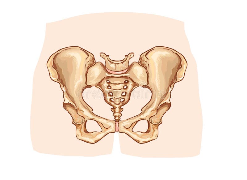
Free with trial Pelvis. Anterior view of the pelvic bones and the sacrum. Pelvis vectors Pelvis. Anterior view of the pelvic bones and the sacrum
Free with trial Man with heavy joint pain symptoms. Medical illustration of heavy joint pain symptoms in left shoulder elbow hand and pelvis. Pelvis illustrations Man with heavy joint pain symptoms. Medical illustration of heavy joint pain symptoms in left shoulder elbow hand and pelvis
Free with trial Spine and pelvis. 3d rendered anatomy illustration of a human spine with pelvis. Pelvis illustrations Spine and pelvis. 3d rendered anatomy illustration of a human spine with pelvis
Free with trial Medical infographic with orthopedic anatomy charts. Human silhouette in motion with marked spine, pelvis, knee, foot, shoulder, elbow, hand bones and joints. Orthopedics medical group design. Pelvis vectors Medical infographic with orthopedic anatomy charts. Human silhouette in motion with marked spine, pelvis, knee, foot, shoulder, elbow, hand bones and joints
Free with trial The hip. Medical accurate illustration of the hip. Pelvis illustrations The hip. Medical accurate illustration of the hip
Free with trial Spine Fracture. Spondylolysis (Spondylolisthesis) is a defect in the bony ring comprising the spinal column. displacement of a lumbar vertebra, most commonly occurring after a break or fracture. Pelvis vectors Spine Fracture. Spondylolysis (Spondylolisthesis) is a defect in the bony ring comprising the spinal column. displacement of a lumbar vertebra, most commonly
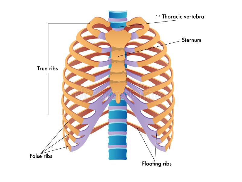
Free with trial Rib cage. Simplified illustration of the rib cage. Pelvis vectors Rib cage. Simplified illustration of the rib cage
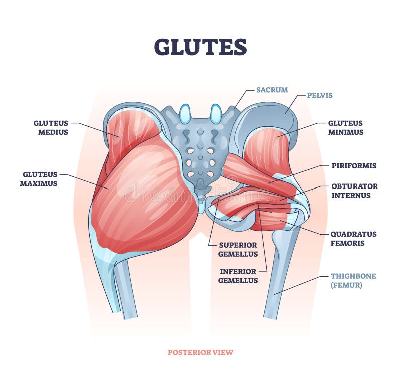
Free with trial Glutes as gluteal body muscles for human buttocks strength outline concept. Labeled educational anatomical scheme with physical skeletal and gluteus medius, maximus and minimus vector illustration. Pelvis vectors Glutes as gluteal body muscles for human buttocks strength outline concept. Labeled educational anatomical scheme with physical skeletal and gluteus medius
Free with trial Osteoporosis. Medical illustration healthy bone and unhealthy bone- osteoporosis. Osteoporosis may leads to bone fracture. Pelvis vectors Osteoporosis. Medical illustration healthy bone and unhealthy bone- . may leads to bone fracture. Osteoporosis. Medical illustration healthy bone and unhealthy bone- osteoporosis. Osteoporosis may leads to bone fracture.
Free with trial Anatomy of the kidney, vector illustration (for basic medical education, for clinics & Schools). Pelvis vectors Anatomy of the kidney. Vector illustration (for basic medical education, for clinics & Schools
Free with trial Hip joint structure. In a whole and in cross section, eps8. Pelvis vectors Hip joint structure. In a whole and in cross section, eps8
Free with trial Ankylosing spondylitis. Of the spine, early and late stages, eps8. Pelvis vectors Ankylosing spondylitis. Of the spine, early and late stages, eps8
Free with trial Ovary. Medical illustration of anatomy of ovary. Pelvis vectors Ovary. Medical illustration of anatomy of ovary
Free with trial Ovary. Female reproductive system. Female Ovary. Showing ovulation step by step. Pelvis vectors Ovary. Female reproductive system. Female Ovary. Showing ovulation step by step
Free with trial Fetus I. Digital illustration of Embryo inside a woman womb. Pelvis illustrations Fetus I. Digital illustration of Embryo inside a woman womb
Free with trial An illustration of a knee xray showing the injury. Pelvis illustrations Knee Pain. An illustration of a knee xray showing the injury. An illustration of a knee xray showing the injury.
Free with trial Human Hip, set, isolated on the white. Pelvis vectors Human Hip. Set, isolated on the white
Free with trial Hip fracture. Diagram showing the site of common hip injury as a result of osteoporosis or other bone diseases, eps8. Pelvis vectors Hip fracture. Diagram showing the site of common hip injury as a result of osteoporosis or other bone diseases, eps8
Free with trial Normal birth. Showing detail of fetal movement during delivery through the birth canal. Pelvis vectors Normal birth. Showing detail of fetal movement during delivery through the birth canal
Free with trial Human kidney. 3d rendered anatomy illustration from a profile of a human kidney. Pelvis illustrations Human kidney. 3d rendered anatomy illustration from a profile of a human kidney
Free with trial Pregnant woman and fetus. Colorful anatomy. Isolated on white. Pelvis illustrations Pregnant woman. Colorful anatomy. Isolated. Pregnant woman and fetus. Colorful anatomy. Isolated on white. Pregnant woman and fetus. Colorful anatomy. Isolated on white.
Free with trial Kidney Anatomy. Easy to edit vector illustration of kidney anatomy. Pelvis vectors Kidney Anatomy. Easy to edit vector illustration of kidney anatomy
Free with trial Uterus and Kidneys. Uterus, Fallopian Tubes and Ovaries, and Kidneys. Pelvis illustrations Uterus and Kidneys. Uterus, Fallopian Tubes and Ovaries, and Kidneys
Free with trial Knee - Anatomy Bones - Medical imaging. Pelvis illustrations Knee - Anatomy Bones. Medical imaging
Free with trial A 3d generated person posed like the famous Vitreous Man (Symbol of health) rendered in a x-ray style highlighting the spine. Pelvis illustrations Vatruvian Man & Spine. A 3d generated person posed like the famous Vitreous Man (Symbol of health) rendered in a x-ray style highlighting the spine. A 3d generated person posed like the famous Vitreous Man (Symbol of health) rendered in a x-ray style highlighting the spine.
Free with trial Kidney stone formation. Drawing to show the formation of a kidney stone, with detail of crystal formation in nephron and stone stuck in ureter. Pelvis illustrations Kidney stone formation. Drawing to show the formation of a kidney stone, with detail of crystal formation in nephron and stone stuck in ureter
Free with trial Human centre of gravity. Drawing to show the centre of gravity of a male adult human figure. Pelvis vectors Human centre of gravity. Drawing to show the centre of gravity of a male adult human figure
Free with trial Types of bone fractures. Illustrated with the femur (hip bone), eps8. Pelvis vectors Types of bone fractures. Illustrated with the femur (hip bone), eps8. Types of bone fractures illustrated with the femur (hip bone), eps8
Free with trial The pelvic floor muscles. Medically accurate illustration of the pelvic floor muscles. Pelvis illustrations The pelvic floor muscles. Medically accurate illustration of the pelvic floor muscles
Free with trial Abdominal aortic aneurysm, disease of the circulatory system, eps8. Pelvis vectors Abdominal aortic aneurysm. Disease of the circulatory system, eps8
Free with trial Arthritis of the hip joint, healthy joint and unhealthy joint with damaged cartilage and osteophytes. Pelvis vectors Arthritis of the hip joint. Healthy joint and unhealthy joint with damaged cartilage and osteophytes
Free with trial Osteoporosis in femur bone, human bone anatomy. Pelvis vectors Osteoporosis in femur bone. Human bone anatomy
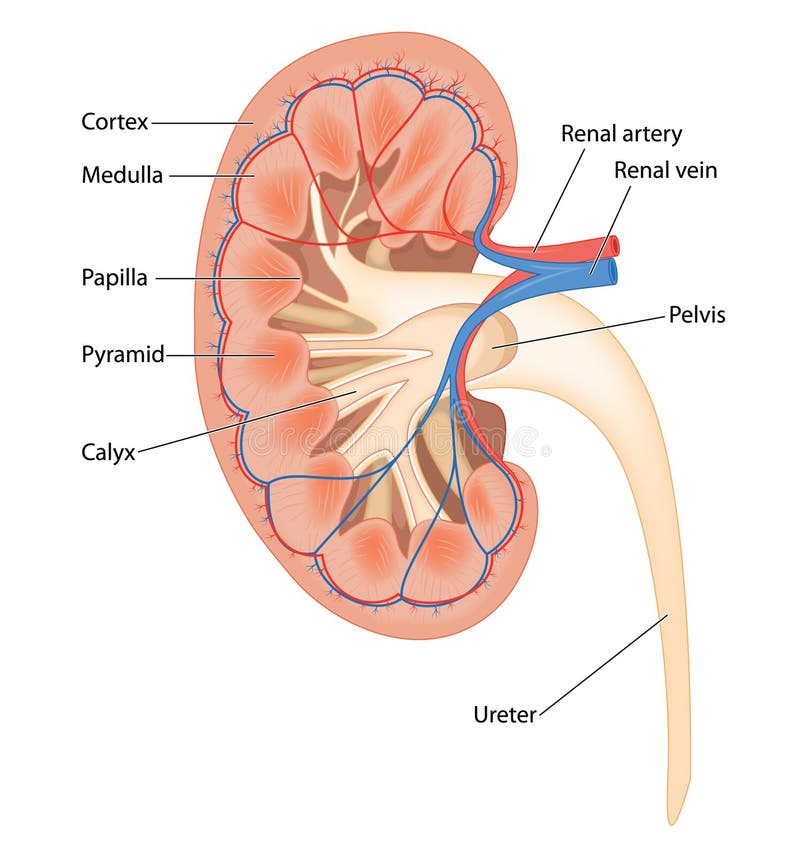
Free with trial Cross section of human kidney. Showing detail of blood supply, cortex, medulla, pyramids and calyx. Pelvis vectors Cross section of human kidney. Showing detail of blood supply, cortex, medulla, pyramids and calyx
Free with trial Kidney anatomy. Vector illustration of anatomy of human kidney. Pelvis vectors Kidney anatomy. Vector illustration of anatomy of human kidney
Free with trial Human hip joint and Osteoporosis. On a abstract blue background. Pelvis vectors Human hip joint and Osteoporosis. On a abstract blue background
Free with trial Rheumatoid arthritis of hip joint. With synovial membrane inflammation and cartilage and bones erosion, eps8. Pelvis vectors Rheumatoid arthritis of hip joint. With synovial membrane inflammation and cartilage and bones erosion, eps8
Free with trial Mother and child in human prenatal pregnancy. Pelvis vectors Mother and child in human prenatal pregnancy. A mother and child in human prenatal pregnancy
Free with trial Correct alignment of human body in standing posture for good personality and healthy of spine and bone. Health care and medical illustration. Pelvis vectors Correct alignment of body in standing posture. Correct alignment of human body in standing posture for good personality and healthy of spine and bone. Health. Correct alignment of human body in standing posture for good personality and healthy of spine and bone. Health care and medical illustration
Free with trial Vector illustration of a human pelvis and hip with pain in the hip joint and trochanteric bursitis. Front view. For advertising and medical publications. EPS 10. Pelvis vectors Pain in the hip joint_trochanteric bursitis. Vector illustration of a human pelvis and hip with pain in the hip joint and trochanteric bursitis. Front view. For. Vector illustration of a human pelvis and hip with pain in the hip joint and trochanteric bursitis. Front view. For advertising and medical publications. EPS 10.
Free with trial Rheumatism or rheumatic disorder medical set. Arthritis joint pain syndrome. Different body parts with pain rings on hand, leg and feet, spine, scull, pelvis. Rheumatology vector infographics elements. Pelvis vectors Rheumatism or rheumatic disorder medical set. Arthritis joint pain. Rheumatology vector infographics. Rheumatism or rheumatic disorder medical set. Arthritis. Rheumatism or rheumatic disorder medical set. Arthritis joint pain syndrome. Different body parts with pain rings on hand, leg and feet, spine, scull, pelvis. Rheumatology vector infographics elements
Free with trial Kidney. Detailed of kidney illustration computer generated on isolat background. Pelvis illustrations Kidney. Detailed of kidney illustration computer generated on isolat background
Free with trial Explore the intricate inner workings of the human kidney with our realistic cross-sectional illustration. This detailed artwork showcases the complex anatomy of the kidney, revealing its renal cortex, medulla, renal pelvis, and intricate network of nephrons. Ideal for educational materials, medical publications, and presentations, our illustration provides a comprehensive visual understanding of renal function and structure. Perfectly suited for enhancing medical textbooks, informative posters, and online resources, this artwork is meticulously crafted to accurately depict the essential components of the kidney in a visually engaging manner. Pelvis illustrations Medical illustration of a human kidney in cross section showing renal blood vessels, and internal anatomical structure. Explore the intricate inner workings of. Explore the intricate inner workings of the human kidney with our realistic cross-sectional illustration. This detailed artwork showcases the complex anatomy of the kidney, revealing its renal cortex, medulla, renal pelvis, and intricate network of nephrons. Ideal for educational materials, medical publications, and presentations, our illustration provides a comprehensive visual understanding of renal function and structure. Perfectly suited for enhancing medical textbooks, informative posters, and online resources, this artwork is meticulously crafted to accurately depict the essential components of the kidney in a visually engaging manner.
Free with trial Pelvis. Male pelvis under the X-rays. pelvis is highlighted in red. Pelvis illustrations Pelvis. Male pelvis under the X-rays. pelvis is highlighted in red
Free with trial Pelvis. Male pelvis under the X-rays. pelvis is highlighted in red. Pelvis illustrations Pelvis. Male pelvis under the X-rays. pelvis is highlighted in red
Free with trial Pelvis. Male pelvis under the X-rays. joints are highlighted in red. Pelvis illustrations Pelvis. Male pelvis under the X-rays. joints are highlighted in red
Free with trial Pelvis. Male pelvis under the X-rays. pelvis is highlighted in red. Pelvis illustrations Pelvis. Male pelvis under the X-rays. pelvis is highlighted in red
Free with trial Male pelvis. Bones of the lower extremity. Detailed medical illustration. Isolated on a white background. Bright and clean design. Pelvis vectors Male pelvis. Bones of the lower extremity. Detailed medical illustration. Isolated on a white background. Bright and clean design
Free with trial Female pelvis. Bones of the lower extremity. Detailed medical illustration. Isolated on a white background. Bright and clean design. Pelvis vectors Female pelvis. Bones of the lower extremity. Detailed medical illustration. Isolated on a white background. Bright and clean design
Free with trial A tilted pelvis. 3d rendered, medically accurate illustration of a tilted pelvis. Pelvis illustrations A tilted pelvis. 3d rendered, medically accurate illustration of a tilted pelvis
Free with trial Pelvis. Male pelvis under the X-rays. joints are highlighted in red. Pelvis illustrations Pelvis. Male pelvis under the X-rays. joints are highlighted in red
Free with trial Comparison of the skeleton of the male and female pelvis. Vector illustration of a comparison of the skeleton of the male and female pelvis. Pelvis vectors Comparison of the skeleton of the male and female pelvis. Vector illustration of a comparison of the skeleton of the male and female pelvis
Free with trial Horse Pelvis - Horse Equus Anatomy - isolated on white. Pelvis illustrations Horse Pelvis - Horse Equus Anatomy - isolated on white
Free with trial Human pelvis anatomy 3d medical illustration on white background. Eps 10. Pelvis illustrations Human pelvis anatomy 3d medical illustration on white background. Eps 10
Free with trial Child in womb - pelvis ischial spines - medical illustration. Pelvis vectors Child in womb - pelvis ischial spines - medical illustration. Child in womb w pelvis ischial spines medical illustration
Free with trial Male vs female pelvis. Main differences. Detailed vector illustration isolated on a white background. Medical and anatomical concept. Pelvis vectors Female Male Pelvis-09. Male vs female pelvis. Main differences. Detailed vector illustration isolated on a white background. Medical and anatomical concept. Male vs female pelvis. Main differences. Detailed vector illustration isolated on a white background. Medical and anatomical concept.
Free with trial The kidneys are a pair of organs located in the back of the abdomen. Each kidney is about 4 or 5 inches long -- about the size of a fist. The kidneys' function are to filter the blood. All the blood in our bodies passes through the kidneys several times a day. The kidneys remove wastes, control the body's fluid balance, and regulate the balance of electrolytes. As the kidneys filter blood, they create urine, which collects in the kidneys' pelvis -- funnel-shaped structures that drain down tubes called ureters to the bladder. Pelvis illustrations Kidneys. The kidneys are a pair of organs located in the back of the abdomen. Each kidney is about 4 or 5 inches long -- about the size of a fist. The kidneys'. The kidneys are a pair of organs located in the back of the abdomen. Each kidney is about 4 or 5 inches long -- about the size of a fist. The kidneys' function are to filter the blood. All the blood in our bodies passes through the kidneys several times a day. The kidneys remove wastes, control the body's fluid balance, and regulate the balance of electrolytes. As the kidneys filter blood, they create urine, which collects in the kidneys' pelvis -- funnel-shaped structures that drain down tubes called ureters to the bladder.
Free with trial Hip Pain. An illustration of an xray of the pelvis region, with a red spot showing the injured ankle. Pelvis illustrations Hip Pain. An illustration of an xray of the pelvis region, with a red spot showing the injured ankle
Free with trial Man with heavy joint pain symptoms. Medical illustration of heavy joint pain symptoms in left shoulder elbow hand and pelvis. Pelvis illustrations Man with heavy joint pain symptoms. Medical illustration of heavy joint pain symptoms in left shoulder elbow hand and pelvis
Free with trial Man with heavy joint pain symptoms. Medical illustration of heavy joint pain symptoms in left shoulder elbow hand and pelvis. Pelvis illustrations Man with heavy joint pain symptoms. Medical illustration of heavy joint pain symptoms in left shoulder elbow hand and pelvis
Free with trial The lumbar vertebrae are, in human anatomy, the five vertebrae between the rib cage and the pelvis. They are the largest segments of the vertebral column and are characterized by the absence of the foramen transversarium within the transverse process as it is only found in the cervical region, and by the absence of facets on the sides of the body. They are designated L1 to L5, starting at the top. The lumbar vertebrae help support the weight of the body, and permit movement. Pelvis illustrations Lumbar vertebrae L3. The lumbar vertebrae are, in human anatomy, the five vertebrae between the rib cage and the pelvis. They are the largest segments of the. The lumbar vertebrae are, in human anatomy, the five vertebrae between the rib cage and the pelvis. They are the largest segments of the vertebral column and are characterized by the absence of the foramen transversarium within the transverse process as it is only found in the cervical region, and by the absence of facets on the sides of the body. They are designated L1 to L5, starting at the top. The lumbar vertebrae help support the weight of the body, and permit movement.
Free with trial The kidneys are a pair of organs located in the back of the abdomen. Each kidney is about 4 or 5 inches long -- about the size of a fist. The kidneys' function are to filter the blood. All the blood in our bodies passes through the kidneys several times a day. The kidneys remove wastes, control the body's fluid balance, and regulate the balance of electrolytes. As the kidneys filter blood, they create urine, which collects in the kidneys' pelvis -- funnel-shaped structures that drain down tubes called ureters to the bladder. Each kidney contains around a million units called nephrons, each of which is a microscopic filter for blood. It's possible to lose as much as 90% of kidney function without experiencing any symptoms or problems. Pelvis illustrations Kidneys. The kidneys are a pair of organs located in the back of the abdomen. Each kidney is about 4 or 5 inches long -- about the size of a fist. The kidneys'. The kidneys are a pair of organs located in the back of the abdomen. Each kidney is about 4 or 5 inches long -- about the size of a fist. The kidneys' function are to filter the blood. All the blood in our bodies passes through the kidneys several times a day. The kidneys remove wastes, control the body's fluid balance, and regulate the balance of electrolytes. As the kidneys filter blood, they create urine, which collects in the kidneys' pelvis -- funnel-shaped structures that drain down tubes called ureters to the bladder. Each kidney contains around a million units called nephrons, each of which is a microscopic filter for blood. It's possible to lose as much as 90% of kidney function without experiencing any symptoms or problems.
Free with trial The kidneys are a pair of organs located in the back of the abdomen. Each kidney is about 4 or 5 inches long -- about the size of a fist. The kidneys' function are to filter the blood. All the blood in our bodies passes through the kidneys several times a day. The kidneys remove wastes, control the body's fluid balance, and regulate the balance of electrolytes. As the kidneys filter blood, they create urine, which collects in the kidneys' pelvis -- funnel-shaped structures that drain down tubes called ureters to the bladder. Pelvis illustrations Kidneys. The kidneys are a pair of organs located in the back of the abdomen. Each kidney is about 4 or 5 inches long -- about the size of a fist. The kidneys'. The kidneys are a pair of organs located in the back of the abdomen. Each kidney is about 4 or 5 inches long -- about the size of a fist. The kidneys' function are to filter the blood. All the blood in our bodies passes through the kidneys several times a day. The kidneys remove wastes, control the body's fluid balance, and regulate the balance of electrolytes. As the kidneys filter blood, they create urine, which collects in the kidneys' pelvis -- funnel-shaped structures that drain down tubes called ureters to the bladder.
Free with trial Human joints and body parts bones seamless pattern. Vector sketch spine pelvis, leg knee or shoulder scapula and elbow, arm and hand wrist with fingers or foot ankle for medical anatomy background. Pelvis vectors Vector seamless pattern human body bones sketch. Human joints and body parts bones seamless pattern. Vector sketch spine pelvis, leg knee or shoulder scapula and. Human joints and body parts bones seamless pattern. Vector sketch spine pelvis, leg knee or shoulder scapula and elbow, arm and hand wrist with fingers or foot ankle for medical anatomy background
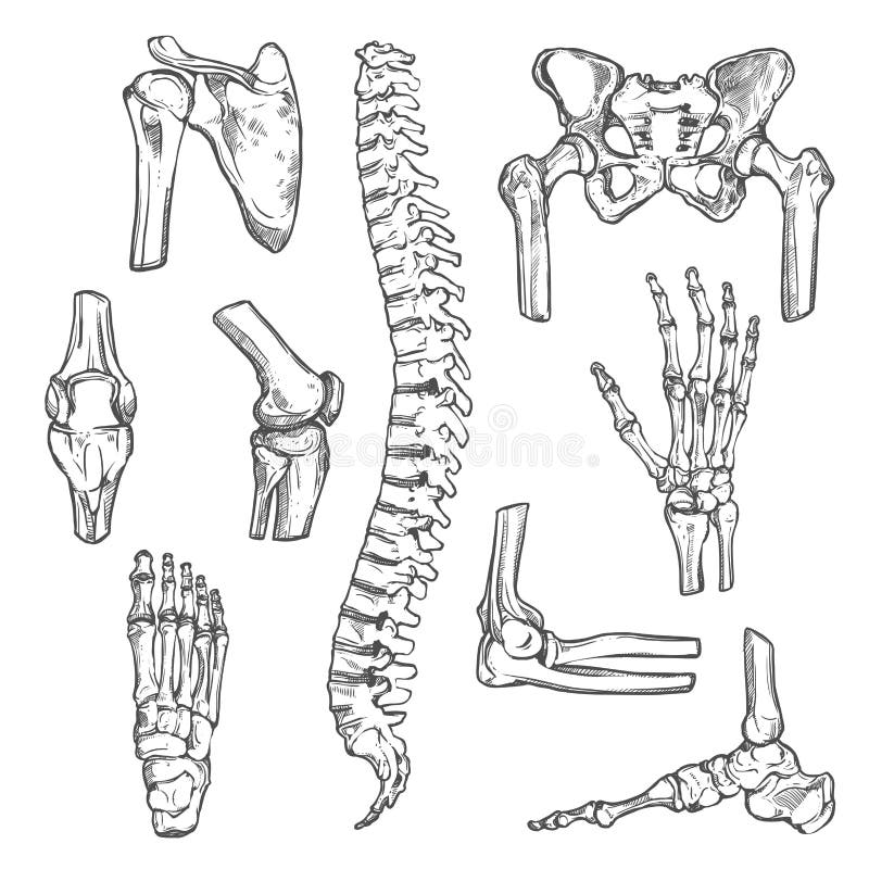
Free with trial Human joints and body parts bones sketch icons. Vector isolated set of spine pelvis, shoulder scapula or elbow, leg knee and foot ankle, arm and hand wrist with fingers for medical anatomy or surgery. Pelvis illustrations Vector sketch icons of human body bones and joints. Human joints and body parts bones sketch icons. Vector isolated set of spine pelvis, shoulder scapula or. Human joints and body parts bones sketch icons. Vector isolated set of spine pelvis, shoulder scapula or elbow, leg knee and foot ankle, arm and hand wrist with fingers for medical anatomy or surgery
Free with trial Medical banner template set with human skeleton bones. Running man silhouette with symbols of leg, hand, spine and pelvis joints supplemented by text layout. Orthopedic, rheumatology medicine design. Pelvis vectors Medical banner template with human skeleton bones. Medical banner template set with human skeleton bones. Running man silhouette with symbols of leg, hand, spine. Medical banner template set with human skeleton bones. Running man silhouette with symbols of leg, hand, spine and pelvis joints supplemented by text layout. Orthopedic, rheumatology medicine design
Free with trial Medical infographic of human skeletal system. Anatomy chart with leg, hand, foot, knee, spine, pelvis, shoulder, elbow bones and joints of human skeleton with text layout. Medicine, education design. Pelvis vectors Medical infographic of human skeletal system. Anatomy chart with leg, hand, foot, knee, spine, pelvis, shoulder, elbow bones and joints of human skeleton with
Free with trial Kidney. Vector medical illustration of Kidney. Pelvis vectors Kidney. Vector medical illustration of Kidney
Free with trial Diagram of the pelvic girdle labeled. A vector illustration of Diagram of the pelvic girdle labeled. Pelvis vectors Diagram of the pelvic girdle labeled. A vector illustration of Diagram of the pelvic girdle labeled
Free with trial The kidneys are located in the abdominal cavity, more specifically in the paravertebral gutter and lie in a retroperitoneal position at a slightly oblique angle. There are two kidneys. One is on each side of the spine. Pelvis illustrations Kidneys. The kidneys are located in the abdominal cavity, more specifically in the paravertebral gutter and lie in a retroperitoneal position at a slightly. The kidneys are located in the abdominal cavity, more specifically in the paravertebral gutter and lie in a retroperitoneal position at a slightly oblique angle. There are two kidneys. One is on each side of the spine.
Free with trial Hip joint. Hip fracture. Femoral Neck Fracture. Osteoarthritis of hip joint. Arthritic Hip. Osteoarthritis Hip Replacement on a white background. Pelvis vectors Hip joint. Hip fracture. Femoral Neck Fracture. Osteoarthritis of . Arthritic Hip. Osteoarthritis Hip Replacement on a white background
Free with trial Kidney. Detailed of kidney illustration computer generated on white background. Pelvis illustrations Kidney. Detailed of kidney illustration computer generated on white background
Free with trial Diastasis recti also known as abdominal separation, it is common among pregnant women. There is a gap between the rectus abdominis muscles. Pelvis vectors Diastasis recti. Also known as abdominal separation, it is common among pregnant women. There is a gap between the rectus abdominis muscles
Free with trial Anatomy Of Kidney. internal and external. human anatomy. Pelvis vectors Anatomy Of Kidney. Internal and external. human anatomy
Free with trial The urinary system. The human urinary system medical illustration with internal organs. Pelvis vectors The urinary system. The human urinary system medical illustration with internal organs
Free with trial Bone medical health questions and osteoporosis illustration concept as a close up diagram of the inside of human skeletal hip bones with a magnification glass showing a normal healthy condition degrading to abnormal unhealthy anatomy as a question mark. Pelvis illustrations Bone Medical Questions. Bone medical health questions and osteoporosis illustration concept as a close up diagram of the inside of human skeletal hip bones with. Bone medical health questions and osteoporosis illustration concept as a close up diagram of the inside of human skeletal hip bones with a magnification glass showing a normal healthy condition degrading to abnormal unhealthy anatomy as a question mark.
Free with trial Retro woman laundress to wash clothes, pop art vector illustration. Dirty and clean. Homework. Pelvis vectors Retro woman laundress to wash clothes. Pop art vector illustration. Dirty and clean. Homework
 Sign in