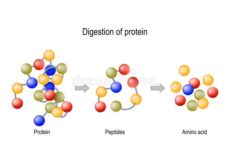
Free with trial Digestion of Protein. Enzymes proteases and peptidases are digestion breaks the protein into smaller peptide chains and into single amino acids, which are absorbed into the blood. Peptidase vectors Digestion of Protein. Enzymes proteases and peptidases, peptides and amino acids. Digestion of Protein. Enzymes proteases and peptidases are digestion breaks the protein into smaller peptide chains and into single amino acids, which are absorbed into the blood
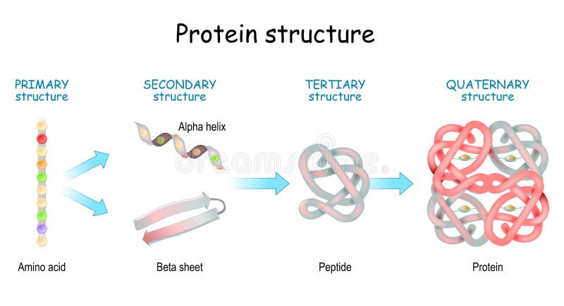
Free with trial Protein structure levels: Primary, Secondary, Tertiary, and Quaternary. From Amino acid to Alpha helix, Beta sheet, peptide, and protein molecule. concept. Vector illustration. Peptidase vectors Protein structure levels. From Amino acid to Alpha helix, Beta sheet, peptide, and protein molecule. Protein structure levels: Primary, Secondary, Tertiary, and Quaternary. From Amino acid to Alpha helix, Beta sheet, peptide, and protein molecule. concept. Vector illustration
Free with trial Digestion of protein. Enzymes proteases and peptidases are digestion breaks the protein into smaller peptide chains and into single amino acids, which are absorbed into the blood. Peptidase vectors Digestion of protein
Free with trial Structure of Protein from amino acid to peptide, and protein. Close-up of protein molecule. Vector illustration for medical, biological, science, and education use. Peptidase vectors Structure of Protein from amino acid to peptide, and protein. Close-up of protein molecule
Free with trial Functions of Glucagon-like peptide-1. weight loss. GLP-1. Treatment of diabetes. physiological properties of peptide hormone. Humans internal organs. vector diagram. Peptidase illustrations GLP-1. Functions of Glucagon-like peptide-1. Functions of Glucagon-like peptide-1. weight loss. GLP-1. Treatment of diabetes. physiological properties of peptide hormone. Humans internal organs. vector diagram
Free with trial Peptide synthesis. Two amino acids combined into a peptide to form a water molecule and a peptide bond. Vector illustration for medical, educational and science use. Peptidase vectors Peptide synthesis. Two amino acids combined into a peptide to form a water molecule and a peptide bond
Free with trial Alogliptin diabetes drug molecule. Belongs to dipeptidyl peptidase 4 DPP-4 or gliptin class of antidiabetic medicines. Skeletal formula. Peptidase illustrations Alogliptin diabetes drug molecule. Belongs to dipeptidyl peptidase 4 DPP-4 or gliptin class of antidiabetic medicines. Skeletal.
Free with trial Papain, also known as papaya proteinase I, a cysteine protease enzyme present in papaya Carica papaya and mountain papaya Vasconcellea cundinamarcensis. 3d structure. Peptidase illustrations Papain, also known as papaya proteinase I, a cysteine protease
Free with trial Glucagon-like peptide-1. Close-up of Cell membrane lipid bilayer with Receptor GLP1R. Vector illustration. Peptidase vectors Glucagon-like peptide-1 receptor. Glucagon-like peptide-1. Close-up of Cell membrane lipid bilayer with Receptor GLP1R. Vector illustration
Free with trial Protein digestion. Enzymes proteases are digestion breaks the protein into single amino acids, which are absorbed into the blood. Vector illustration. Peptidase vectors Protein digestion. Enzymes proteases are digestion breaks the protein into single amino acids
Free with trial Protein Digestion. Enzymes proteases and peptidases are digestion breaks the protein into smaller peptide chains and into single amino acids, which are absorbed into the blood. Vector illustration. Peptidase vectors Protein Digestion
Free with trial Vector illustration of protein digestion. Pepsin, trypsin and erepsin enzymes effect on protein molecule. Peptidase vectors Vector illustration of protein digestion
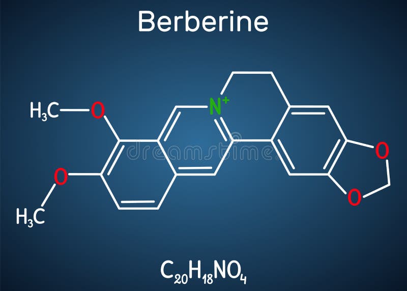
Free with trial Berberine C20H18NO4, herbal alkaloid molecule. Skeletal chemical formula. Illustration. Peptidase illustrations Berberine C20H18NO4, herbal alkaloid molecule. Skeletal chemical formula
Free with trial Berberine herbal medicine molecule. Skeletal formula. Peptidase illustrations Berberine herbal medicine molecule. Skeletal formula.
Free with trial The pepsins are enzymes secreted by the stomach that breaks down proteins. Vector illustration. Peptidase vectors Digestion of protein. Breaking the complex molecule first into peptides then into individual amino acids. The pepsins are enzymes secreted by the stomach that breaks down proteins. Vector illustration
Free with trial Glucagon-like Peptide 1 GLP-1 prevents macrovascular complications,coronary artery disease, , lipid metabolism, blood pressure inflammation, nitric oxide ROS, oxygenmolecule, 2d 3d graphic rendering. Peptidase illustrations Glucagon-like Peptide 1 GLP-1 prevents macrovascular complications,coronary artery disease, , lipid metabolism, blood pressure inf
Free with trial Berberine C20H18NO4, herbal alkaloid molecule. Structural chemical formula and molecule model. Vector illustration. Peptidase vectors Berberine C20H18NO4, herbal alkaloid molecule. Structural chemical formula and molecule model
Free with trial Berberine C20H18NO4, herbal alkaloid molecule. Structural chemical formula on the dark blue background. Vector illustration. Peptidase vectors Berberine C20H18NO4, herbal alkaloid molecule. Structural chemical formula on the dark blue background
Free with trial Glucagon-like Peptide 1 GLP-1 prevents macrovascular complications,coronary artery disease, , lipid metabolism, blood pressure inflammation, nitric oxide ROS, oxygenmolecule, 2d 3d graphic rendering. Peptidase illustrations Glucagon-like Peptide 1 GLP-1 molecule, chemical structure. GLP-1 is being investigated for the treatment of diabetes mellitus. Glucagon-like Peptide 1 GLP-1 prevents macrovascular complications,coronary artery disease, , lipid metabolism, blood pressure inflammation, nitric oxide ROS, oxygenmolecule, 2d 3d graphic rendering
Free with trial Berberine C20H18NO4, herbal alkaloid molecule. Molecule model. Illustration. Peptidase illustrations Berberine C20H18NO4, herbal alkaloid molecule. Molecule model
Free with trial Alogliptin is an orally administered anti-diabetic drug in the DPP-4 inhibitor class. 3d illustration. Peptidase illustrations Alogliptin molecular structure isolated on white. Alogliptin is an orally administered anti-diabetic drug in the DPP-4 inhibitor class. 3d illustration
Free with trial Glucagon-like peptide 1 (GLP1, 7-36) molecule, 3D rendering. Atoms are represented as spheres with conventional color coding: hydrogen (white), carbon (grey), nitrogen (blue), oxygen (red), sulfur (yellow. Peptidase illustrations Glucagon-like peptide 1 (GLP1, 7-36) molecule, 3D rendering. Atoms are represented as spheres with conventional color coding:
Free with trial Glucagon-like peptide 1 GLP1, 7-36 molecule, 3D rendering. Cartoon representation combined with semi-transparent surfaces. Peptidase illustrations Glucagon-like peptide 1 GLP1, 7-36 molecule, 3D rendering. Cartoon representation combined with semi-transparent surfaces.
Free with trial 3D rendering of Glucagon-like peptide 1 (GLP1, 7-36) molecule, a potent antihyperglycemic hormone. A neuropeptide and an incretin, chemical structure. treatment of diabetes,. Peptidase illustrations 3D rendering of Glucagon-like peptide 1 (GLP1, 7-36) molecule, a potent antihyperglycemic hormone.
Free with trial Dipeptidyl peptidase iv dpp iv also known as cd dpp iv processes numerous substrates such as. Peptidase illustrations Dipeptidyl peptidase iv dpp iv also known as cd dpp iv processs. dipeptidyl peptidase iv dpp iv also known as cd dpp iv processes numerous substrates such as
Free with trial Dipeptidyl Peptidase IV (DPP IV) Also known as CD, DPP IV processes numerous substrates, such as glucagon like peptides Our label distinguishes DPP IV. Peptidase illustrations Dipeptidyl Peptidase IV (DPP IV) Also known as CD, DPP IV proes. Dipeptidyl Peptidase IV (DPP IV) Also known as CD, DPP IV processes numerous substrates, such as glucagon like peptides Our label distinguishes DPP IV
Free with trial Structure of collagen with amino acid sequence for biology lessons. Peptidase vectors Structure of collagen with amino acid sequence
Free with trial Structure of collagen with amino acid sequence for biology lessons. Peptidase vectors Structure of collagen with amino acid sequence
Free with trial The connecting peptide, or C-peptide, is a short 31-amino-acid polypeptide that connects insulin's A-chain to its B-chain in the proinsulin molecule. In the context of diabetes or hypoglycemia, a measurement of C-peptide blood serum levels can be used to distinguish between different conditions with similar clinical features. Peptidase illustrations C-peptide amino acid - closeup view 3d illustration. The connecting peptide, or C-peptide, is a short 31-amino-acid polypeptide that connects insulin's A-chain to its B-chain in the proinsulin molecule. In the context of diabetes or hypoglycemia, a measurement of C-peptide blood serum levels can be used to distinguish between different conditions with similar clinical features.
Free with trial The connecting peptide, or C-peptide, is a short 31-amino-acid polypeptide that connects insulin's A-chain to its B-chain in the proinsulin molecule. In the context of diabetes or hypoglycemia, a measurement of C-peptide blood serum levels can be used to distinguish between different conditions with similar clinical features. Peptidase illustrations C-peptide amino acid - section view 3d illustration. The connecting peptide, or C-peptide, is a short 31-amino-acid polypeptide that connects insulin's A-chain. The connecting peptide, or C-peptide, is a short 31-amino-acid polypeptide that connects insulin's A-chain to its B-chain in the proinsulin molecule. In the context of diabetes or hypoglycemia, a measurement of C-peptide blood serum levels can be used to distinguish between different conditions with similar clinical features.
Free with trial The connecting peptide, or C-peptide, is a short 31-amino-acid polypeptide that connects insulin's A-chain to its B-chain in the proinsulin molecule. In the context of diabetes or hypoglycemia, a measurement of C-peptide blood serum levels can be used to distinguish between different conditions with similar clinical features. Peptidase illustrations C-peptide amino acid - isometric view 3d illustration. The connecting peptide, or C-peptide, is a short 31-amino-acid polypeptide that connects insulin's A-chain to its B-chain in the proinsulin molecule. In the context of diabetes or hypoglycemia, a measurement of C-peptide blood serum levels can be used to distinguish between different conditions with similar clinical features.
Free with trial Peptide Bond Chemical Structure Diagram - Basic R and Acidic Group. Peptidase vectors Peptide Bond Chemical Structure Diagram
Free with trial Vector Illustration of pepsin enzyme effect on protein molecule. Peptidase vectors Vector Illustration of pepsin enzyme effect on protein moleculet
Free with trial Vector illustration of protein digestion. Protease enzyme effect on protein molecule. Peptidase vectors Vector illustration of protein digestion. Protease enzyme effect on protein molecule
Free with trial Structure of tropocollagen for biology lessons. Peptidase vectors Structure of tropocollagen
Free with trial Black silhouette of structure of collagen. Peptidase vectors Silhouette of structure of collagen
Free with trial Coloring page with structure of procollagen for biology lessons. Peptidase vectors Coloring page with structure of procollagen
Free with trial Coloring page with structure of procollagen for biology lessons. Peptidase vectors Coloring page with structure of procollagen
Free with trial Structure of procollagen for biology lessons. Peptidase vectors Structure of procollagen
Free with trial Structure of procollagen for biology lessons. Peptidase illustrations Structure of procollagen
Free with trial Structure of procollagen for biology lessons. Peptidase vectors Structure of procollagen
Free with trial Structure of procollagen for biology lessons. Peptidase vectors Structure of procollagen
Free with trial Structure of procollagen for biology lessons. Peptidase illustrations Structure of procollagen
Free with trial Grampositive bacterial cell wall illustration gramnegative lipopolysaccharide, teichoic mycolic, periplasm murein grampositive bacterial cell wall. Peptidase illustrations Grampositive bacterial cell wall
Free with trial Saxagliptin hydrochloride molecule, structural chemical formula, ball-and-stick model, isolated image anti-diabetic drug. Peptidase illustrations Saxagliptin hydrochloride molecule, structural chemical formula, ball-and-stick model, isolated image anti-diabetic drug
Free with trial 'GLP-1 Therapy for Obesity: Metabolism, Appetite Regulation, Insulin Sensitivity, and Weight Management'. 'GLP-1 and Obesity: Mechanisms, Metabolic Effects, Appetite Control, and Therapeutic Potential'. Peptidase illustrations \'GLP-1 Therapy for Obesity: Metabolism, Appetite Regulation, Insulin Sensitivity, and Weight Management\'. 'GLP-1 Therapy for Obesity: Metabolism, Appetite Regulation, Insulin Sensitivity, and Weight Management'. 'GLP-1 and Obesity: Mechanisms, Metabolic Effects, Appetite Control, and Therapeutic Potential'
Free with trial Saxagliptin hydrochloride molecular structure 3d, anti-diabetic drug, structural chemical formula view from a microscope. Peptidase illustrations Saxagliptin hydrochloride molecular structure 3d, anti-diabetic drug, structural chemical formula view from a microscope
Free with trial Saxagliptin hydrochloride molecule 3d, molecular structure, ball and stick model, structural chemical formula anti-diabetic drug. Peptidase illustrations Saxagliptin hydrochloride molecule 3d, molecular structure, ball and stick model, structural chemical formula anti-diabetic drug
Free with trial The novel coronavirus, SARS-CoV-2, emerged in Wuhan, China, in late 2019, rapidly escalating into a global pandemic. This highly contagious pathogen causes severe respiratory illness, impacting individuals worldwide. Understanding its transmission pathways, genetic structure, and interaction with the human host is crucial for developing effective preventative measures and treatments. The virus. Peptidase illustrations Understanding the SARSCoV2 Virus Global Outbreak Transmission and Immune Response. The novel coronavirus, SARS-CoV-2, emerged in Wuhan, China, in late 2019, rapidly escalating into a global pandemic. This highly contagious pathogen causes severe respiratory illness, impacting individuals worldwide. Understanding its transmission pathways, genetic structure, and interaction with the human host is crucial for developing effective preventative measures and treatments. The virus
Free with trial Linagliptin diabetes drug molecule dipeptidyl peptidase 4 or DPP4 inhibitor. Skeletal formula. Peptidase illustrations Linagliptin diabetes drug molecule dipeptidyl peptidase 4 or DPP4 inhibitor. Skeletal formula.
Free with trial Saxagliptin diabetes drug molecule. Inhibitor of dipeptidyl peptidase-4 DPP4. Atoms are represented as spheres with conventional color coding: hydrogen white, carbon grey, oxygen red, nitrogen blue. Peptidase illustrations Saxagliptin diabetes drug molecule. Inhibitor of dipeptidyl peptidase-4 DPP4.
Free with trial Alogliptin diabetes drug molecule. Belongs to dipeptidyl peptidase 4 DPP-4 or gliptin class of antidiabetic medicines. Skeletal formula. Peptidase vectors Alogliptin diabetes drug molecule. Belongs to dipeptidyl peptidase 4 DPP-4 or gliptin class of antidiabetic medicines. Skeletal.
Free with trial Alogliptin diabetes drug molecule. Belongs to dipeptidyl peptidase 4 DPP-4 or gliptin class of antidiabetic medicines. Atoms are represented as spheres with conventional color coding: hydrogen white, carbon grey, oxygen red, nitrogen blue. Peptidase illustrations Alogliptin diabetes drug molecule. Belongs to dipeptidyl peptidase 4 DPP-4 or gliptin class of antidiabetic medicines.
Free with trial Linagliptin diabetes drug molecule (dipeptidyl peptidase 4 or DPP4 inhibitor). Atoms are represented as spheres with conventional color coding: hydrogen (white), carbon (grey), oxygen (red), nitrogen (blue. Peptidase illustrations Linagliptin diabetes drug molecule (dipeptidyl peptidase 4 or DPP4 inhibitor). Atoms are represented as spheres with conventional
Free with trial Alogliptin diabetes drug molecule. Belongs to dipeptidyl peptidase 4 (DPP-4) or gliptin class of antidiabetic medicines. Stylized 2D rendering and conventional skeletal formula. Peptidase illustrations Alogliptin diabetes drug molecule. Belongs to dipeptidyl peptidase 4 (DPP-4) or gliptin class of antidiabetic medicines
Free with trial Alogliptin diabetes drug molecule. Belongs to dipeptidyl peptidase 4 (DPP-4) or gliptin class of antidiabetic medicines. Atoms are represented as spheres with conventional color coding: hydrogen (white), carbon (grey), oxygen (red), nitrogen (blue. Peptidase illustrations Alogliptin diabetes drug molecule. Belongs to dipeptidyl peptidase 4 (DPP-4) or gliptin class of antidiabetic medicines
Free with trial Alogliptin diabetes drug molecule. Belongs to dipeptidyl peptidase 4 (DPP-4) or gliptin class of antidiabetic medicines. Conventional skeletal formula and stylized representations. Peptidase vectors Alogliptin diabetes drug molecule. Belongs to dipeptidyl peptidase 4 (DPP-4) or gliptin class of antidiabetic medicines
Free with trial Alogliptin diabetes drug molecule. Belongs to dipeptidyl peptidase 4 DPP-4 or gliptin class of antidiabetic medicines. Atoms are represented as spheres with conventional color coding: hydrogen white, carbon grey, oxygen red, nitrogen blue. Peptidase illustrations Alogliptin diabetes drug molecule. Belongs to dipeptidyl peptidase 4 DPP-4 or gliptin class of antidiabetic medicines.
Free with trial Tripeptidyl-peptidase I enzyme. Mutations in corresponding TPP1 gene lead to late infantile neuronal ceroid lipofuscinosis. Cerliponase alpha is a recombinant form of the enzyme. 3D rendering based on protein data bank entry 3ee6. Atoms shown as color-coded spheres. Per chain coloring. Peptidase illustrations Tripeptidyl-peptidase I enzyme. Mutations in corresponding TPP1 gene lead to late infantile neuronal ceroid lipofuscinosis.
Free with trial Tripeptidyl-peptidase I enzyme. Mutations in corresponding TPP1 gene lead to late infantile neuronal ceroid lipofuscinosis. Cerliponase alpha is a recombinant form of the enzyme. 3D rendering based on protein data bank entry 3ee6. Cartoon representation with secondary structure coloring (green sheets, red helices. Peptidase illustrations Tripeptidyl-peptidase I enzyme. Mutations in corresponding TPP1 gene lead to late infantile neuronal ceroid lipofuscinosis.
Free with trial Alogliptin diabetes drug molecule. Belongs to dipeptidyl peptidase 4 (DPP-4) or gliptin class of antidiabetic medicines. Atoms are represented as spheres with conventional color coding: hydrogen (white), carbon (grey), oxygen (red), nitrogen (blue. Peptidase illustrations Alogliptin diabetes drug molecule. Belongs to dipeptidyl peptidase 4 (DPP-4) or gliptin class of antidiabetic medicines
Free with trial Saxagliptin diabetes drug molecule. Inhibitor of dipeptidyl peptidase-4 DPP4. Skeletal formula. Peptidase illustrations Saxagliptin diabetes drug molecule. Inhibitor of dipeptidyl peptidase-4 DPP4. Skeletal formula.
Free with trial Linagliptin diabetes drug molecule dipeptidyl peptidase 4 or DPP4 inhibitor. Skeletal formula. Peptidase vectors Linagliptin diabetes drug molecule dipeptidyl peptidase 4 or DPP4 inhibitor. Skeletal formula.
Free with trial Saxagliptin diabetes drug molecule. Inhibitor of dipeptidyl peptidase-4 DPP4. Skeletal formula. Peptidase vectors Saxagliptin diabetes drug molecule. Inhibitor of dipeptidyl peptidase-4 DPP4. Skeletal formula.
Free with trial Saxagliptin diabetes drug molecule. Inhibitor of dipeptidyl peptidase-4 DPP4. Atoms are represented as spheres with conventional color coding: hydrogen white, carbon grey, oxygen red, nitrogen blue. Peptidase illustrations Saxagliptin diabetes drug molecule. Inhibitor of dipeptidyl peptidase-4 DPP4.
Free with trial Saxagliptin diabetes drug molecule. Inhibitor of dipeptidyl peptidase-4 DPP4. Atoms are represented as spheres with conventional color coding: hydrogen white, carbon grey, oxygen red, nitrogen blue. Peptidase illustrations Saxagliptin diabetes drug molecule. Inhibitor of dipeptidyl peptidase-4 DPP4.
Free with trial Saxagliptin diabetes drug molecule. Inhibitor of dipeptidyl peptidase-4 (DPP4). Stylized 2D rendering and conventional skeletal formula. Peptidase vectors Saxagliptin diabetes drug molecule. Inhibitor of dipeptidyl peptidase-4 (DPP4
Free with trial Crystal structure and molecular model of human dipeptidyl peptidase-4, a serine protease, a member of the prolyl oligopeptidase family that has been implicated in several diseases. 3d illustration. Peptidase illustrations Crystal structure and space-filling molecular model of human dipeptidyl peptidase-4, a member of the prolyl. Crystal structure and molecular model of human dipeptidyl peptidase-4, a serine protease, a member of the prolyl oligopeptidase family that has been implicated in several diseases. 3d illustration
Free with trial Crystal structure of human dipeptidyl peptidase-4, a serine protease, a member of the prolyl oligopeptidase family that has been implicated in several diseases. Scientific background. 3d illustration. Peptidase illustrations Crystal structure of human dipeptidyl peptidase-4, a serine protease, a member of the prolyl oligopeptidase family that
Free with trial Space-filling molecular model of human dipeptidyl peptidase-4. Rendering with differently colored protein chains based on protein data bank entry 1j2e. Scientific background. 3d illustration. Peptidase illustrations Space-filling molecular model of human dipeptidyl peptidase-4. Rendering with differently colored protein chains based
Free with trial Space-filling molecular model of human dipeptidyl peptidase-4. Atoms are represented as spheres with color coding: carbon (grey), oxygen (red), nitrogen (blue), sulfur (yellow). 3d illustration. Peptidase illustrations Molecular model of human dipeptidyl peptidase-4. Atoms are represented as spheres with conventional color coding: carbon. Space-filling molecular model of human dipeptidyl peptidase-4. Atoms are represented as spheres with color coding: carbon (grey), oxygen (red), nitrogen (blue), sulfur (yellow). 3d illustration
Free with trial Alogliptin diabetes drug molecule. Belongs to dipeptidyl peptidase 4 DPP-4 or gliptin class of antidiabetic medicines. Atoms are represented as spheres with conventional color coding: hydrogen white, carbon grey, oxygen red, nitrogen blue. Peptidase illustrations Alogliptin diabetes drug molecule. Belongs to dipeptidyl peptidase 4 DPP-4 or gliptin class of antidiabetic medicines.
Free with trial Tripeptidyl-peptidase I enzyme. Mutations in corresponding TPP1 gene lead to late infantile neuronal ceroid lipofuscinosis. Cerliponase alpha is a recombinant form of the enzyme. 3D rendering based on protein data bank entry 3ee6. Cartoon representation combined with semi-transparent surfaces. Dark background. Peptidase illustrations Tripeptidyl-peptidase I enzyme. Mutations in corresponding TPP1 gene lead to late infantile neuronal ceroid lipofuscinosis.
Free with trial 3d rendered Alogliptin diabetes drug molecule. dipeptidyl peptidase 4 (DPP-4) or gliptin class of antidiabetic medicines. anti-diabetic drug in the DPP-4 inhibitor class, 3d illustration. Peptidase illustrations Alogliptin diabetes drug molecule. dipeptidyl peptidase 4 (DPP-4) or gliptin class of antidiabetic
Free with trial Tripeptidyl-peptidase I enzyme. Mutations in corresponding TPP1 gene lead to late infantile neuronal ceroid lipofuscinosis. Cerliponase alpha is a recombinant form of the enzyme. 3D rendering based on protein data bank entry 3ee6. Combined cartoon and stick representation with backbone gradient coloring. Background black. Peptidase illustrations Tripeptidyl-peptidase I enzyme. Mutations in corresponding TPP1 gene lead to late infantile neuronal ceroid lipofuscinosis.
Free with trial Tripeptidyl-peptidase I enzyme. Mutations in corresponding TPP1 gene lead to late infantile neuronal ceroid lipofuscinosis. Cerliponase alpha is a recombinant form of the enzyme. 3D rendering based on protein data bank entry 3ee6. Cartoon representation combined with semi-transparent surfaces. Dark background. Peptidase illustrations Tripeptidyl-peptidase I enzyme. Mutations in corresponding TPP1 gene lead to late infantile neuronal ceroid lipofuscinosis.
Free with trial 3d rendered Alogliptin diabetes drug molecule. dipeptidyl peptidase 4 (DPP-4) or gliptin class of antidiabetic medicines. anti-diabetic drug in the DPP-4 inhibitor class, 3d illustration. Peptidase illustrations Alogliptin diabetes drug molecule. dipeptidyl peptidase 4 (DPP-4) or gliptin class of antidiabetic
Free with trial Linagliptin diabetes drug molecule (dipeptidyl peptidase 4 or DPP4 inhibitor). Atoms are represented as spheres with conventional color coding: hydrogen (white), carbon (grey), oxygen (red), nitrogen (blue. Peptidase illustrations Linagliptin diabetes drug molecule (dipeptidyl peptidase 4 or DPP4 inhibitor). Atoms are represented as spheres with conventional
Free with trial Linagliptin diabetes drug molecule (dipeptidyl peptidase 4 or DPP4 inhibitor). Atoms are represented as spheres with conventional color coding: hydrogen (white), carbon (grey), oxygen (red), nitrogen (blue. Peptidase illustrations Linagliptin diabetes drug molecule (dipeptidyl peptidase 4 or DPP4 inhibitor). Atoms are represented as spheres with conventional
Free with trial Alogliptin diabetes drug molecule. Belongs to dipeptidyl peptidase 4 (DPP-4) or gliptin class of antidiabetic medicines. Atoms are represented as spheres with conventional color coding: hydrogen (white), carbon (grey), oxygen (red), nitrogen (blue. Peptidase illustrations Alogliptin diabetes drug molecule. Belongs to dipeptidyl peptidase 4 (DPP-4) or gliptin class of antidiabetic medicines