Free with trial Blood cell formation from differentiation of hematopoietic stem cells in red bone marrow. Platelet bone marrow vectors Hematopoietic stem cell
Free with trial Blood cells Formation ( bone marrow produce blood cells series : erythrocytes , lymphocytes , neutrophils , monocytes , eosinophils , basophils , thrombocytes ) Haematology concept and infographics. Platelet bone marrow vectors Blood cells Formation
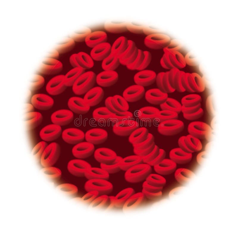
Free with trial Red Blood Cells computer generated illustration. Platelet bone marrow illustrations RBC [Red Blood Cells]. Red Blood Cells computer generated illustration
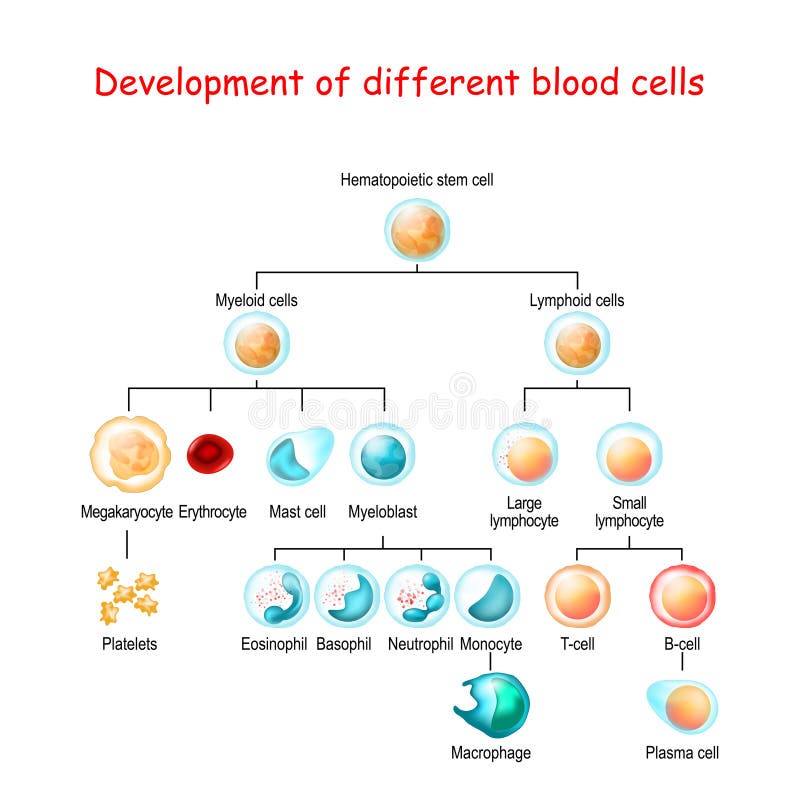
Free with trial Development of different blood cells from haematopoietic stem cell to mature cells. Vector illustration. Platelet bone marrow vectors Development of different blood cells from haematopoietic stem cell to mature cells
Free with trial Red bone marrow and Yellow marrow. Hematopoiesis. Platelets thrombocytes, White blood cells or leukocytes, Red blood cells or erythrocytes. Vector illustration. Platelet bone marrow vectors Bone marrow. Hematopoiesis. Red bone marrow and Yellow marrow. Hematopoiesis. Platelets thrombocytes, White blood cells or leukocytes, Red blood cells or erythrocytes. Vector illustration
Free with trial Illustration showing the structure of a megakaryocyte, a large nucleated cell in the bone marrow which is the precursor for the production of platelets or thrombocytes. Platelet bone marrow illustrations Megakaryocyte
Free with trial Limbo blood cells in bone marrow. Infographics. Vector illustration. Platelet bone marrow vectors Limbo blood cells in bone marrow. Infographics. Vector illustration
Free with trial Leukemia, the nasty blood cancer. Platelet bone marrow vectors Leukemia Bone. Leukemia, the nasty blood cancer
Free with trial Medical tests for patients with leukemia and blood disorders, cancer diagram illustration. Platelet bone marrow vectors Leukemia
Free with trial Limbo platelets in the bone marrow. Dieback of platelets in the spleen, the liver. The life of the platelet. Infographics. Vector illustration on isolated background. Platelet bone marrow vectors Limbo platelets in the bone marrow. Dieback of platelets in the spleen, the liver. The life of the platelet. Infographics.
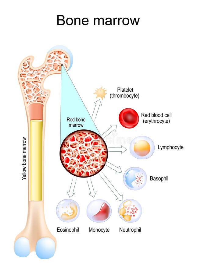
Free with trial Bone marrow. Difference between Yellow and Red bone marrow. Blood cells develop in bone marrow from stem cells. White blood cells Eosinophil, Neutrophil, Basophil, Lymphocyte, Monocyte. Platelet or thrombocyte, and Red blood cell or erythrocyte. Platelet bone marrow vectors Yellow bone marrow and Red bone marrow. Blood cells develop. Bone marrow. Difference between Yellow and Red bone marrow. Blood cells develop in bone marrow from stem cells. White blood cells Eosinophil, Neutrophil, Basophil, Lymphocyte, Monocyte. Platelet or thrombocyte, and Red blood cell or erythrocyte
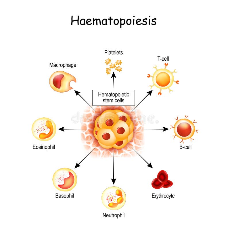
Free with trial Haematopoiesis is the formation of blood cells. All cellular blood components are derived from hematopoietic stem cells. hemocytoblast in red bone marrow, white and red blood cells, Macrophage and Platelets. Platelet bone marrow vectors Haematopoiesis is the formation of blood cells. hemocytoblast in red bone marrow, white and red blood cells, Macrophage and. Haematopoiesis is the formation of blood cells. All cellular blood components are derived from hematopoietic stem cells. hemocytoblast in red bone marrow, white and red blood cells, Macrophage and Platelets
Free with trial Bone marrow. Stem cell. Red blood cell thrombocytes neutrophil basophil monocyte lymphocyte eosinophils. Platelet bone marrow vectors Bone marrow. Stem cell.
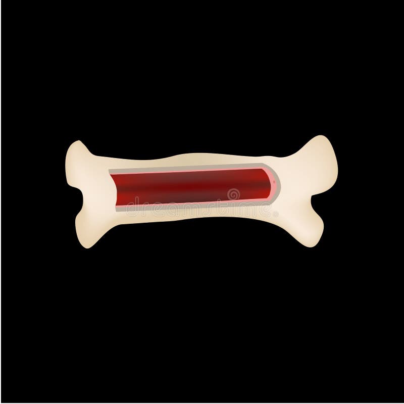
Free with trial Bone marrow and blood cells formation diagram. Hematopoiesis. Femur bone with type of blood cell. Erythrocyte Lymphocyte Neutrophil Eosinophil Basophil Monocyte Thrombocyte. Isolated. 3D render. Platelet bone marrow illustrations Bone marrow and blood cells formation diagram . Hematopoiesis . Femur bone with type of blood cell . Erythrocyte Lymphocyte
Free with trial Red bone marrow. Through the magnifying glass, Hematopoiesis from Stem cells to Platelets, Red and White blood cells is visible. Blood cell formation. Vector illustration. Medical poster. Schematic diagram. Platelet bone marrow vectors Cells in Red bone marrow.Through the magnifying glass. Red bone marrow. Through the magnifying glass, Hematopoiesis from Stem cells to Platelets, Red and White blood cells is visible. Blood cell formation. Vector illustration. Medical poster. Schematic diagram
Free with trial Bone stem cell. Bone marrow. Blood cells. Infographics. Vector illustration on isolated background. Platelet bone marrow vectors Bone stem cell. Bone marrow. Blood cells. Infographics. Vector illustration on isolated background.
Free with trial Thrombopoietin (TPO) is a hormone that regulates platelet production in neonates with thrombocytopenia, TPO levels usually rise to stimulate the bone marrow to produce more platelets, though the response may be limited in sick or premature infants. Platelet bone marrow vectors Thrombocytopenia in Neonates flashcard illustration. Thrombopoietin (TPO) is a hormone that regulates platelet production in neonates with thrombocytopenia, TPO levels usually rise to stimulate the bone marrow to produce more platelets, though the response may be limited in sick or premature infants.
Free with trial Platelets, also called thrombocytes `clot` and `cell`, are a component of blood whose function along with the coagulation factors is to react to bleeding from blood vessel injury by clumping, thereby initiating a blood clot. Platelets have no cell nucleus they are fragments of cytoplasm that are derived from the megakaryocytes of the bone marrow, which then enter the circulation. Platelet bone marrow illustrations Platelet thrombocyte with red and white blood cells 3d illustration. Platelets, also called thrombocytes `clot` and `cell`, are a component of blood whose function along with the coagulation factors is to react to bleeding from blood vessel injury by clumping, thereby initiating a blood clot. Platelets have no cell nucleus they are fragments of cytoplasm that are derived from the megakaryocytes of the bone marrow, which then enter the circulation.
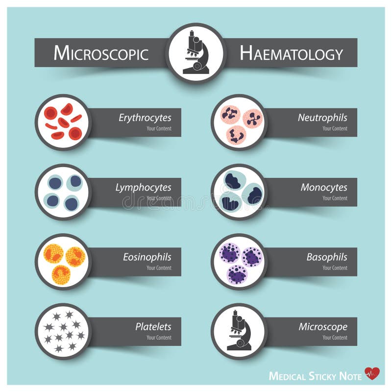
Free with trial Microscopic Haematology. Medical sticky note style Bone marrow produce blood cells series : erythrocytes. lymphocytes. neutrophils. monocytes. eosinophils. basophils. thrombocytes. Platelet bone marrow vectors Microscopic Haematology
Free with trial Thrombopoietin 3d structure, a glycoprotein hormone produced by the liver and kidney which regulates the production of platelets. It stimulates the production and differentiation of megakaryocytes. Platelet bone marrow illustrations Thrombopoietin 3d structure, a glycoprotein hormone
Free with trial Stem Cell Haematopoiesis Diagram illustration. Platelet bone marrow vectors Stem Cell Haematopoiesis Diagram
Free with trial Blood smear is often used as a follow-up test to abnormal results on a complete blood count (CBC) to evaluate the different types of blood cells. Platelet bone marrow illustrations Blood smear. Blood smear is often used as a follow-up test to abnormal results on a complete blood count (CBC) to evaluate the different types of blood cells
Free with trial Blood smear is often used as a follow-up test to abnormal results on a complete blood count (CBC) to evaluate the different types of blood cells. Platelet bone marrow illustrations Blood smear. Blood smear is often used as a follow-up test to abnormal results on a complete blood count (CBC) to evaluate the different types of blood cells
Free with trial Blood smear is often used as a follow-up test to abnormal results on a complete blood count (CBC) to evaluate the different types of blood cells. Platelet bone marrow illustrations Blood smear. Blood smear is often used as a follow-up test to abnormal results on a complete blood count (CBC) to evaluate the different types of blood cells
Free with trial Reticulocyte with red blood cells on blood smear. Platelet bone marrow illustrations Reticulocyte
Free with trial Transmission electron microscopy of bone marrow megakaryocyte showing platelet demarcation membrane system and platelet shedding into sinusoid. Platelet bone marrow illustrations Bone Marrow Megakaryocyte Platelet Shedding TEM. Transmission electron microscopy of bone marrow megakaryocyte showing platelet demarcation membrane system and. Transmission electron microscopy of bone marrow megakaryocyte showing platelet demarcation membrane system and platelet shedding into sinusoid
Free with trial Bone Marrow, the source of red blood cells or erythrocytes, white blood cells and platelets. Blood cells develop in bone marrow. Hematopoiesis, biology education. Platelet bone marrow vectors Bone Marrow, the source of red blood cells or erythrocytes, white blood cells and platelets. Blood cells develop in bone marrow.
Free with trial Bone marrow in a case of immune thrombocytopenic purpura. Note the hyperplasia of megakaryocytes, the platelet-producing cells. Wright x 1000. Platelet bone marrow illustrations Immune thrombocytopenic purpura. Bone marrow. Bone marrow in a case of immune thrombocytopenic purpura. Note the hyperplasia of megakaryocytes, the platelet-producing cells. Wright x 1000.
Free with trial Bone marrow in a case of immune thrombocytopenic purpura. Note the hyperplasia of megakaryocytes, the platelet-producing cells. Wright x 1000. Platelet bone marrow illustrations Immune thrombocytopenic purpura. Bone marrow. Bone marrow in a case of immune thrombocytopenic purpura. Note the hyperplasia of megakaryocytes, the platelet-producing cells. Wright x 1000.
Free with trial Bone marrow in a case of immune thrombocytopenic purpura. Note the hyperplasia of megakaryocytes, the platelet-producing cells. Wright x 1000. Platelet bone marrow illustrations Immune thrombocytopenic purpura. Bone marrow. Bone marrow in a case of immune thrombocytopenic purpura. Note the hyperplasia of megakaryocytes, the platelet-producing cells. Wright x 1000.
Free with trial Bone Marrow, the source of red blood cells or erythrocytes, white blood cells and platelets. Blood cells develop in bone marrow. Hematopoiesis, biology education. Platelet bone marrow vectors Bone Marrow, the source of red blood cells or erythrocytes, white blood cells and platelets. Blood cells develop in bone marrow.
Free with trial Detailed 3D microscopic view of blood cells flowing through vessels and bone marrow showcasing a white blood cell's function in the human body. Useful in medical illustrations for healthcare. Platelet bone marrow illustrations Blood Cells Flowing Through Vessels and Marrow with White Cell in Detailed Microscopic View Medical Concept. Detailed 3D microscopic view of blood cells flowing through vessels and bone marrow showcasing a white blood cell's function in the human body. Useful in medical illustrations for healthcare
Free with trial In oncology, polycythemia vera (PV) is an uncommon myeloproliferative neoplasm in which the bone marrow makes too many red blood cells. The majority of cases are caused by mutations in the JAK2 gene, most commonly resulting in a single amino acid change in its protein product from valine to phenylalanine at position 617. Platelet bone marrow illustrations High level of RBC and platelet in Polycythemia vera (PV) - closeup view 3d illustration. In oncology, polycythemia vera (PV) is an uncommon myeloproliferative neoplasm in which the bone marrow makes too many red blood cells. The majority of cases are caused by mutations in the JAK2 gene, most commonly resulting in a single amino acid change in its protein product from valine to phenylalanine at position 617.
Free with trial In oncology, polycythemia vera (PV) is an uncommon myeloproliferative neoplasm in which the bone marrow makes too many red blood cells. The majority of cases are caused by mutations in the JAK2 gene, most commonly resulting in a single amino acid change in its protein product from valine to phenylalanine at position 617. Platelet bone marrow illustrations High level of RBC and platelet in Polycythemia vera (PV) - isometric view 3d illustration. In oncology, polycythemia vera (PV) is an uncommon myeloproliferative neoplasm in which the bone marrow makes too many red blood cells. The majority of cases are caused by mutations in the JAK2 gene, most commonly resulting in a single amino acid change in its protein product from valine to phenylalanine at position 617.
Free with trial Blood cells produced in the bone marrow are divided into three main types: red blood cells, white blood cells, and platelets, which make up about 45% of the blood volume. They carry oxygen, fight infection, and clot the blood, respectively. Platelet bone marrow illustrations Types of Blood Cells in the Body. Blood cells produced in the bone marrow are divided into three main types: red blood cells, white blood cells, and platelets, which make up about 45% of the blood volume. They carry oxygen, fight infection, and clot the blood, respectively.
Free with trial Red blood cells tile able seamless, A type of blood cell that is made in the bone marrow and found in the blood. Red blood cells contain a protein called hemoglobin, which carries oxygen from the lungs to all parts of the body. Platelet bone marrow illustrations Red blood cells tile able seamless
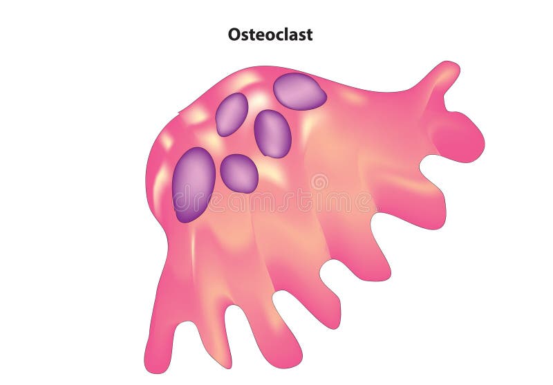
Free with trial Osteoclasts are specialized cells that are responsible for bone resorption, which is the process of breaking down bone tissue. They are multinucleated cells derived from hematopoietic stem cells in the bone marrow, specifically from the monocyte macrophage lineage. The primary function of osteoclasts is to remove old or damaged bone tissue during bone remodeling, a process essential for maintaining bone health and integrity. Bone remodeling involves a balance between bone resorption by osteoclasts and bone formation by osteoblasts. Platelet bone marrow vectors Osteoclast
Free with trial Mesenchymal stem cells (MSCs) are a type of multipotent stem cell found in various tissues throughout the body, including bone marrow, adipose tissue, umbilical cord tissue, and dental pulp. They possess the ability to differentiate into a variety of cell types, including osteoblasts (bone cells), chondrocytes (cartilage cells), adipocytes (fat cells), and other cell lineages of mesodermal origin. Platelet bone marrow vectors Mesenchymal stem cell
Free with trial 3d rendering of Thrombocytes, are pieces of very large cells in the bone marrow called megakaryocytes. Platelet bone marrow illustrations 3d rendering of Thrombocytes and red blood cells. 3d rendering of Thrombocytes, are pieces of very large cells in the bone marrow called megakaryocytes
Free with trial 3d rendering of Thrombocytes, are pieces of very large cells in the bone marrow called megakaryocytes. Platelet bone marrow illustrations 3d rendering of Thrombocytes and red blood cells. 3d rendering of Thrombocytes, are pieces of very large cells in the bone marrow called megakaryocytes
Free with trial 3d rendering of Thrombocytes, are pieces of very large cells in the bone marrow called megakaryocytes. Platelet bone marrow illustrations 3d rendering of Thrombocytes and red blood cells. 3d rendering of Thrombocytes, are pieces of very large cells in the bone marrow called megakaryocytes
Free with trial 3d rendering of Thrombocytes, are pieces of very large cells in the bone marrow called megakaryocytes. Platelet bone marrow illustrations 3d rendering of Thrombocytes and red blood cells. 3d rendering of Thrombocytes, are pieces of very large cells in the bone marrow called megakaryocytes
Free with trial 3d rendering of Thrombocytes, are pieces of very large cells in the bone marrow called megakaryocytes. Platelet bone marrow illustrations 3d rendering of Thrombocytes and red blood cells. 3d rendering of Thrombocytes, are pieces of very large cells in the bone marrow called megakaryocytes
Free with trial 3d rendering of Thrombocytes, are pieces of very large cells in the bone marrow called megakaryocytes. Platelet bone marrow illustrations 3d rendering of Thrombocytes and red blood cells. 3d rendering of Thrombocytes, are pieces of very large cells in the bone marrow called megakaryocytes
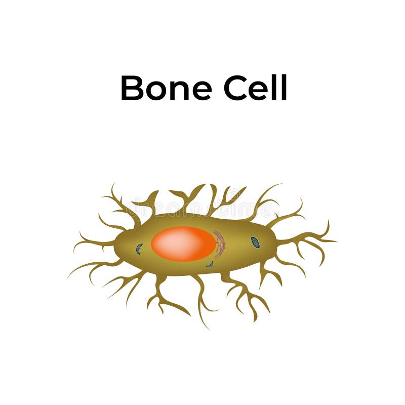
Free with trial Bone is composed of four main types of cells: osteoblasts, osteocytes, osteoclasts, and bone lining cells. Osteoblasts are responsible for bone formation, osteocytes are mature bone cells embedded in the matrix, osteoclasts break down bone tissue, and bone lining cells cover inactive bone surfaces. Platelet bone marrow vectors Bone Cell Structure Design Vector Illustration. Bone is composed of four main types of cells: osteoblasts, osteocytes, osteoclasts, and bone lining cells. Osteoblasts are responsible for bone formation, osteocytes are mature bone cells embedded in the matrix, osteoclasts break down bone tissue, and bone lining cells cover inactive bone surfaces.
Free with trial In oncology, polycythemia vera (PV) is an uncommon myeloproliferative neoplasm in which the bone marrow makes too many red blood cells. The majority of cases are caused by mutations in the JAK2 gene, most commonly resulting in a single amino acid change in its protein product from valine to phenylalanine at position 617. Platelet bone marrow illustrations High level of Red blood cell in Polycythemia vera (PV) - closeup view 3d illustration. In oncology, polycythemia vera (PV) is an uncommon myeloproliferative neoplasm in which the bone marrow makes too many red blood cells. The majority of cases are caused by mutations in the JAK2 gene, most commonly resulting in a single amino acid change in its protein product from valine to phenylalanine at position 617.
Free with trial In oncology, polycythemia vera (PV) is an uncommon myeloproliferative neoplasm in which the bone marrow makes too many red blood cells. The majority of cases are caused by mutations in the JAK2 gene, most commonly resulting in a single amino acid change in its protein product from valine to phenylalanine at position 617. Platelet bone marrow illustrations High level of Red blood cell in Polycythemia vera (PV) - isometric view 3d illustration. In oncology, polycythemia vera (PV) is an uncommon myeloproliferative neoplasm in which the bone marrow makes too many red blood cells. The majority of cases are caused by mutations in the JAK2 gene, most commonly resulting in a single amino acid change in its protein product from valine to phenylalanine at position 617.
Free with trial This microscopic image reveals the intricate composition of platelet-rich plasma (PRP). Suspended within a golden liquid are red and white blood cells, highlighting the rich biological components crucial for tissue regeneration. PRP therapy, a cutting-edge regenerative medicine treatment, offers a powerful approach to skin and tissue repair. The image showcases the potential of PRP injections. Platelet bone marrow illustrations PlateletRich Plasma PRP A Microscopic Look at Regenerative Medicine for Skin and Tissue Repair. This microscopic image reveals the intricate composition of platelet-rich plasma (PRP). Suspended within a golden liquid are red and white blood cells, highlighting the rich biological components crucial for tissue regeneration. PRP therapy, a cutting-edge regenerative medicine treatment, offers a powerful approach to skin and tissue repair. The image showcases the potential of PRP injections
Free with trial Blast cell anatomy overview featuring an immature blast cell with large nucleus and scant cytoplasm, context with red blood cell and neutrophil. Outline diagram. Platelet bone marrow vectors Blast cell anatomy overview featuring an immature blast cell with ... Blast cell anatomy overview featuring an immature blast cell with large nucleus and scant cytoplasm, context with red blood cell and neutrophil. Outline diagram
Free with trial Generative ai, A close-up scientific visualization shows red and white blood cells flowing, highlighting the complexity and vital functions within the human body. Platelet bone marrow illustrations Blood cells in vivid. generative ai, A close-up scientific visualization shows red and white blood cells flowing, highlighting the complexity and vital functions within the human body
Free with trial Red blood cell count with low and high and normal levels outline diagram. Labeled educational scheme with abnormal RBC test results with possible causes vector illustration. Hematological diseases. Platelet bone marrow vectors Red blood cell count with low and high and normal levels outline diagram
Free with trial Microscopic view of red blood cells, also known as platelets essential for blood clotting. These fragments help stop bleeding by forming clots. They are crucial for vascular health. Platelet bone marrow illustrations Microscopic view of red blood cells, also known as platelets, essential for blood clotting. These fragments help stop bleeding by. Microscopic view of red blood cells, also known as platelets essential for blood clotting. These fragments help stop bleeding by forming clots. They are crucial for vascular health.
Free with trial A 3D rendering of blood clot that travels to another part of the body is called an embolus. Platelet bone marrow illustrations A 3D rendering of embolus or blood clot. A 3D rendering of blood clot that travels to another part of the body is called an embolus
Free with trial A 3D rendering of blood clot that travels to another part of the body is called an embolus. Platelet bone marrow illustrations A 3D rendering of embolus or blood clot. A 3D rendering of blood clot that travels to another part of the body is called an embolus
Free with trial A 3D rendering of blood clot that travels to another part of the body is called an embolus. Platelet bone marrow illustrations A 3D rendering of embolus or blood clot. A 3D rendering of blood clot that travels to another part of the body is called an embolus
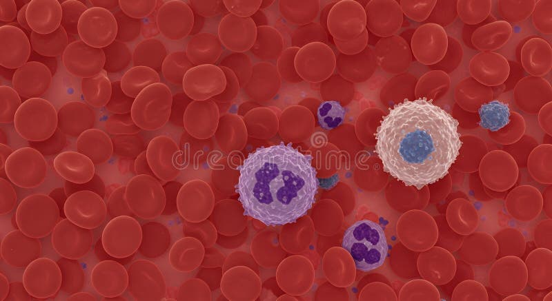
Free with trial A microscopic view showcasing red blood cells and different types of white blood cells within a blood sample. Platelet bone marrow illustrations Microscopic view shows red and white blood cells flowing. A microscopic view showcasing red blood cells and different types of white blood cells within a blood sample
Free with trial Under the microscope- background macro for scientific medical concept - platelets. Platelet bone marrow illustrations Under the microscope- background macro for scientific medical concept - platelets
Free with trial Blood picture of chronic lymphocytic leukemia or CLL, analyze by microscope, original magnification 400x. Platelet bone marrow illustrations Blood picture of chronic lymphocytic leukemia or CLL, analyze by microscope, original magnification 400x.
Free with trial WBC White Blood Cell - cellular component of blood that helps defend the body against infection, acronym text concept background. Platelet bone marrow illustrations WBC White Blood Cell - cellular component of blood that helps defend the body against infection, acronym text concept background
Free with trial Platelet life circle. The life circle of the thrombocyte from the birth in the bone marrow to the spleen. Platelet bone marrow vectors Platelet life circle. The life circle of the thrombocyte from bone marrow to spleen. Platelet life circle. The life circle of the thrombocyte from the birth in the bone marrow to the spleen
Free with trial Bone marrow Stem cell. Platelets, Red and White blood cells. Diagram. Platelet bone marrow vectors Bone marrow Stem cell. Platelets, Red and White blood cells.
Free with trial Bone stem cell. Bone marrow. Blood cells. Infographics. Vector illustration on isolated background. Platelet bone marrow vectors Bone stem cell. Bone marrow. Blood cells. Infographics. Vector illustration on isolated background.
Free with trial Activatet platelet cell, Thrombocyte are a component of blood whose function is to react to bleeding from blood vessel injury by clumping, thereby initiating a blood clot. 3d illustration. Platelet bone marrow illustrations Activatet platelet cell Thrombocyte. Activatet platelet cell, Thrombocyte are a component of blood whose function is to react to bleeding from blood vessel injury by clumping, thereby initiating a blood clot. 3d illustration
Free with trial Platelets, also called thrombocytes `clot` and `cell`, are a component of blood whose function along with the coagulation factors is to react to bleeding from blood vessel injury by clumping, thereby initiating a blood clot. Platelets have no cell nucleus they are fragments of cytoplasm that are derived from the megakaryocytes of the bone marrow, which then enter the circulation. Platelet bone marrow illustrations Platelet thrombocyte with red and white blood cells 3d illustration close-up. Platelets, also called thrombocytes `clot` and `cell`, are a component of blood whose function along with the coagulation factors is to react to bleeding from blood vessel injury by clumping, thereby initiating a blood clot. Platelets have no cell nucleus they are fragments of cytoplasm that are derived from the megakaryocytes of the bone marrow, which then enter the circulation.
Free with trial Stem cell transplant. Stem cells divide and change into the different types of blood cells. Vector illustration. Platelet bone marrow vectors Stem cell transplant
Free with trial A vector image illustration (lThalassemia trait infographic. ). Platelet bone marrow vectors Thalassemia trait infographic
Free with trial Acute promyelocytic leukemia cells or APL, analyze by microscope, original magnification 1000x. Platelet bone marrow illustrations Acute promyelocytic leukemia cells or APL
Free with trial Acute promyelocytic leukemia cells or APL, analyze by microscope, original magnification 1000x. Platelet bone marrow illustrations Acute promyelocytic leukemia cells or APL
Free with trial Factor XIII or fibrin stabilizing factor is a zymogen found from the blood of humans and some other animals. It is activated by thrombin to factor XIIIa. 3D cartoon and Gaussian surface models, chain id color scheme, based on PDB 1FIE, white background. Platelet bone marrow illustrations Structure of human factor XIII. Factor XIII or fibrin stabilizing factor is a zymogen found from the blood of humans and some other animals. It is activated by thrombin to factor XIIIa. 3D cartoon and Gaussian surface models, chain id color scheme, based on PDB 1FIE, white background.
Free with trial WBC - White Blood Cell acronym, medical concept background. Platelet bone marrow illustrations WBC - White Blood Cell acronym
Free with trial WBC - White Blood Cell acronym, medical concept background. Platelet bone marrow illustrations WBC - White Blood Cell acronym, medical concept background
Free with trial Microscopic view of red blood cells in motion, Microscopic view of red blood cells in motion. Platelet bone marrow illustrations Microscopic view of red blood cells in motion
Free with trial A special type of immune cell that is found in tissues, such as the skin, and boosts immune responses by showing antigens on its surface to other cells of the immune system. A dendritic cell is a type of phagocyte and a type of antigen-presenting cell APC. Platelet bone marrow illustrations Mature dendritic cell - super closeup view 3d illustration. A special type of immune cell that is found in tissues, such as the skin, and boosts immune responses by showing antigens on its surface to other cells of the immune system. A dendritic cell is a type of phagocyte and a type of antigen-presenting cell APC.