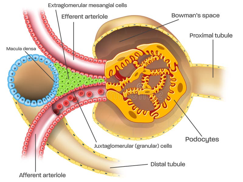
Free with trial Juxtaglomerular apparatus of kidney nephron and glomerulus medical illustration with caption. Podocyte illustrations Juxtaglomerular apparatus of kidney nephron illustration with captions. Juxtaglomerular apparatus of kidney nephron and glomerulus medical illustration with caption
Free with trial Juxtaglomerular apparatus of kidney nephron and glomerulus medical illustration without captions. Podocyte illustrations Juxtaglomerular apparatus of kidney nephron illustration without captions. Juxtaglomerular apparatus of kidney nephron and glomerulus medical illustration without captions
Free with trial Podocytes are cells in Bowman`s capsule in the kidneys that wrap around capillaries of the glomerulus. Podocytes make up the epithelial lining of Bowman`s capsule, the third layer through which filtration of blood takes place. Several proteins are required for the pedicels to wrap around the capillaries and function. Podocyte illustrations Kidney. Podocytes are cells in the Bowman`s capsule. Podocytes are cells in Bowman`s capsule in the kidneys that wrap around capillaries of the glomerulus. Podocytes make up the epithelial lining of Bowman`s capsule, the third layer through which filtration of blood takes place. Several proteins are required for the pedicels to wrap around the capillaries and function.
Free with trial Glomerulonephritis impaired kidney filtration contrasts healthy vs inflamed glomerulus, Bowmans capsule, and urine sample to show leakage, proteinuria, and hematuria. Outline diagram. Podocyte vectors Glomerulonephritis impaired kidney filtration contrasts healthy vs inflamed glomerulus, Bowmans ... Glomerulonephritis impaired kidney filtration contrasts healthy vs inflamed glomerulus, Bowmans capsule, and urine sample to show leakage, proteinuria, and hematuria. Outline diagram
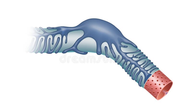
Free with trial 3d computer illustraton of podocytes on a glomerulus. Podocytes wrap around glomeruli in the kidney. They have long processes that interdigitate with the processes of other podocytes building up a fine filter for water, salts and small biomolecules, while proteins and other macromolecules are retained in the blood. Podocyte illustrations Podocytes on a glomerulus filtering the blood. 3d computer illustraton of podocytes on a glomerulus. Podocytes wrap around glomeruli in the kidney. They have long processes that interdigitate with the processes of other podocytes building up a fine filter for water, salts and small biomolecules, while proteins and other macromolecules are retained in the blood.
Free with trial Kidney filtration membrane diagram shows filtrate flow through podocyte slits around the glomerulus in Bowmans capsule, clarifying the barrier s function. Outline diagram. Podocyte vectors Kidney filtration membrane diagram shows filtrate flow through podocyte slits ... Kidney filtration membrane diagram shows filtrate flow through podocyte slits around the glomerulus in Bowmans capsule, clarifying the barrier s function. Outline diagram
Free with trial Explore a futuristic concept of podocyte regeneration, showcasing intricate cellular structures in a sci-fi environment enriched with advanced technology visuals. Podocyte illustrations Futuristic Concept of Podocyte Regeneration in Sci-Fi Setting. Explore a futuristic concept of podocyte regeneration, showcasing intricate cellular structures in a sci-fi environment enriched with advanced technology visuals
Free with trial A futuristic interpretation of podocyte regeneration, showcasing vibrant cellular structures within a kidney, blending science and imagination in a unique visual style. Podocyte illustrations Sci-Fi Inspired Concept of Podocyte Regeneration in Futuristic Design. A futuristic interpretation of podocyte regeneration, showcasing vibrant cellular structures within a kidney, blending science and imagination in a unique visual style
Free with trial This futuristic sci-fi image showcases the concept of podocyte regeneration, emphasizing innovative biology and technology in kidney health and research. Podocyte illustrations Futuristic Concept of Podocyte Regeneration in Sci-Fi Imagery. This futuristic sci-fi image showcases the concept of podocyte regeneration, emphasizing innovative biology and technology in kidney health and research
Free with trial Explore a captivating sci-fi concept of podocyte regeneration, blending biology and technology in a vivid and dynamic artwork that inspires curiosity about the future of life. Podocyte illustrations Futuristic Sci-Fi Concept Art of Podocyte Regeneration in Action. Explore a captivating sci-fi concept of podocyte regeneration, blending biology and technology in a vivid and dynamic artwork that inspires curiosity about the future of life
Free with trial Generative ai, The artistic rendering depicts the glomerulus with blue cells atop a yellow podocyte base, all encased in a defined blue capsule, showcasing inner structures connected by delicate strands. Podocyte illustrations This is a vibrant, digitally-rendered scanning electron micrograph of a nephron\'s. generative ai, The artistic rendering depicts the glomerulus with blue cells atop a yellow podocyte base, all encased in a defined blue capsule, showcasing inner structures connected by delicate strands
Free with trial Nephrotic syndrome overview, side-by-side glomeruli show healthy vs leaky filter leading to proteinuria, key objects, glomerulus, albumin leak, urine drop. Outline diagram. Podocyte vectors Nephrotic syndrome overview, side-by-side glomeruli show healthy vs
Free with trial This artistic illustration depicts podocytes in the human kidney, showcasing their complex structure in vibrant colors, perfect for educational and medical purposes. Podocyte illustrations Artistic Medical Illustration of Podocytes in Human Kidney Structure. This artistic illustration depicts podocytes in the human kidney, showcasing their complex structure in vibrant colors, perfect for educational and medical purposes
Free with trial This conceptual artwork showcases podocytes, crucial cells in the kidney that filter blood. The vivid colors and intricate details highlight the beauty of biological structures essential for human health. Podocyte illustrations Conceptual Artwork of Podocytes Filtering Blood in Kidneys. This conceptual artwork showcases podocytes, crucial cells in the kidney that filter blood. The vivid colors and intricate details highlight the beauty of biological structures essential for human health
Free with trial Detailed conceptual artwork showcasing podocytes, the specialized cells that filter blood in kidneys, highlighting their intricate structure and function. Podocyte illustrations Conceptual Artwork of Podocytes Filtering Blood in Human Body. Detailed conceptual artwork showcasing podocytes, the specialized cells that filter blood in kidneys, highlighting their intricate structure and function
Free with trial This vibrant conceptual artwork illustrates podocytes at work filtering blood in the human kidney, highlighting their intricate structure and function in renal health. Podocyte illustrations Conceptual Artwork of Podocytes Filtering Blood in Human Kidney. This vibrant conceptual artwork illustrates podocytes at work filtering blood in the human kidney, highlighting their intricate structure and function in renal health
Free with trial This detailed 3D rendering depicts podocytes interacting with capillaries, showcasing intricate cell architecture and biological processes essential for kidney function and vascular health. Podocyte illustrations 3D Rendering of Podocytes Interacting with Capillaries in Biological Study. This detailed 3D rendering depicts podocytes interacting with capillaries, showcasing intricate cell architecture and biological processes essential for kidney function and vascular health
Free with trial This artistic medical illustration depicts podocytes in the kidney, showcasing their vital role in filtration. A vibrant and detailed visual for educational purposes. Podocyte illustrations Artistic Medical Illustration of Podocytes in Kidney Structure. This artistic medical illustration depicts podocytes in the kidney, showcasing their vital role in filtration. A vibrant and detailed visual for educational purposes
Free with trial This 3D rendering captures the dynamic interaction of podocytes with capillaries, showcasing intricate cellular structures and their biological significance. Podocyte illustrations 3D Rendering of Podocytes Interacting with Capillaries in Detail. This 3D rendering captures the dynamic interaction of podocytes with capillaries, showcasing intricate cellular structures and their biological significance
Free with trial A high-magnification microscopic view of a kidney glomerulus, stained with Hematoxylin and Eosin (H&E). The image showcases the intricate structure of the glomerular capillaries, podocytes, and mesangial cells. This detailed histological sample is ideal for medical education, research, and scientific publications related to nephrology and renal pathology. Podocyte illustrations Microscopic View of Kidney Glomerulus - Histology Image. A high-magnification microscopic view of a kidney glomerulus, stained with Hematoxylin and Eosin (H&E). The image showcases the intricate structure of the glomerular capillaries, podocytes, and mesangial cells. This detailed histological sample is ideal for medical education, research, and scientific publications related to nephrology and renal pathology.
Free with trial Detailed structure of a kidney glomerulus filtration barrier. The image highlights the complex network of cells and fibers essential for the filtration process, with vibrant pink hues illustrating various tissue layers. The unique shapes and patterns are indicative of biological processes in action. Podocyte illustrations Close-up microscopic view of kidney glomerulus filtration barrier, showing intricate cellular structures involved in filtration. Detailed structure of a kidney glomerulus filtration barrier. The image highlights the complex network of cells and fibers essential for the filtration process, with vibrant pink hues illustrating various tissue layers. The unique shapes and patterns are indicative of biological processes in action.
Free with trial A renal corpuscle is composed of a glomerulus and the Bowman`s capsule. Water, ions and small molecules pass from the bloodstream into the Bowman`s capsule and are then processed to form urine. Podocyte illustrations Renal corpuscles filtering the blood in the kidney. A renal corpuscle is composed of a glomerulus and the Bowman`s capsule. Water, ions and small molecules pass from the bloodstream into the Bowman`s capsule and are then processed to form urine.
Free with trial Renal corpuscle podocytes can be identified using immunohistochemical techniques to detect vimentin protein. This image shows the marking of glomerular podocytes in a human kidney stained with a technique to detect vimentin. Podocyte illustrations Kidney. Podocytes, Vimentin. Renal corpuscle podocytes can be identified using immunohistochemical techniques to detect vimentin protein. This image shows the marking of glomerular podocytes in a human kidney stained with a technique to detect vimentin
Free with trial Pedicel disease, vintage engraved illustration. Podocyte illustrations Pedicel disease, vintage engraving. Pedicel disease, vintage engraved illustration