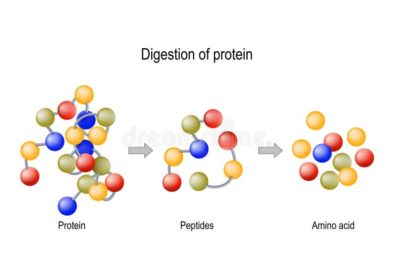
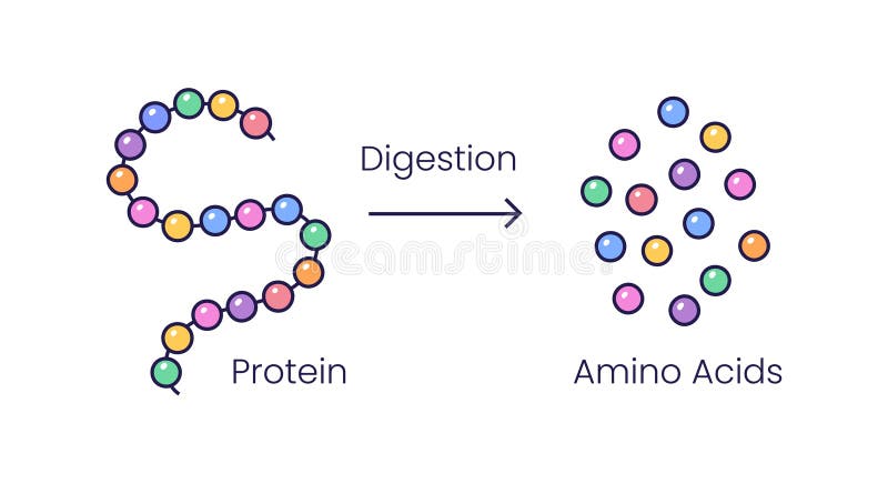
Free with trial Digestion of Protein. Enzymes proteases and peptidases are digestion breaks the protein into smaller peptide chains and into single amino acids, which are absorbed into the blood. Protease protein vectors Digestion of Protein. Enzymes proteases and peptidases, peptides and amino acids. Digestion of Protein. Enzymes proteases and peptidases are digestion breaks the protein into smaller peptide chains and into single amino acids, which are absorbed into the blood
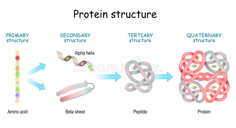
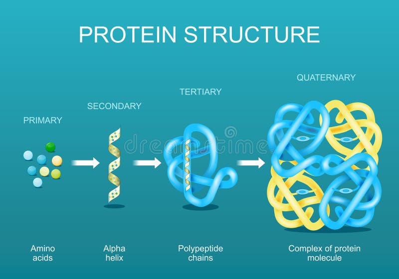
Free with trial Levels of protein structure from amino acids to Complex of protein molecule. Protein is a polymer polypeptide that formed from sequences of amino acids. Levels of protein structure: Primary, Secondary, Tertiary, and Quaternary. Protease protein vectors Levels of protein structure from amino acids to Complex of protein molecule
Free with trial Protein structure levels: Primary, Secondary, Tertiary, and Quaternary. From Amino acid to Alpha helix, Beta sheet, peptide, and protein molecule. concept. Vector illustration. Protease protein vectors Protein structure levels. From Amino acid to Alpha helix, Beta sheet, peptide, and protein molecule. Protein structure levels: Primary, Secondary, Tertiary, and Quaternary. From Amino acid to Alpha helix, Beta sheet, peptide, and protein molecule. concept. Vector illustration
Free with trial Enzyme pepsin 3D model on a white background. Protease protein illustrations Enzyme pepsin 3D model
Free with trial Molecular model of HIV protease protein, a target for anti-viral drugs to treat HIV and AIDS. Protease protein illustrations HIV protease protein
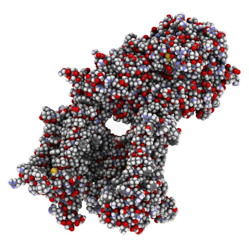
Free with trial Organic chemistry: model of the protein molecule - illustration of a biological particle. Protease protein illustrations Protein structure. Organic chemistry: model of the protein molecule - illustration of a biological particle
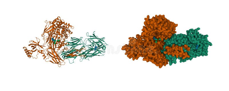
Free with trial Chemical structure of human alpha-thrombin, a key enzyme in the coagulation cascade. Thrombin is also used, in combination with fibrinogen, in so called 'meat glue'. Left image is a space-filling, all-atom representation; the right image is a ribbon + surface representation of the same protein. Protease protein illustrations Thrombin structure. Chemical structure of human alpha-thrombin, a key enzyme in the coagulation cascade. Thrombin is also used, in combination with fibrinogen, in so called 'meat glue'. Left image is a space-filling, all-atom representation; the right image is a ribbon + surface representation of the same protein.
Free with trial Chemical structure of a human trypsin enzyme molecule. Trypsin is an enzyme that contributes to the digestion of proteins in the digestive system. Protease protein illustrations Trypsin enzyme molecule, chemical structure. Chemical structure of a human trypsin enzyme molecule. Trypsin is an enzyme that contributes to the digestion of proteins in the digestive system.
Free with trial Protein Digestion. Enzymes proteases and peptidases are digestion breaks the protein into smaller peptide chains and into single amino acids, which are absorbed into the blood. Protease protein vectors Protein Digestion. Enzymes
Free with trial Digestion of protein. Enzymes proteases and peptidases are digestion breaks the protein into smaller peptide chains and into single amino acids, which are absorbed into the blood. Protease protein vectors Digestion of protein
Free with trial Structure of Protein from amino acid to peptide, and protein. Close-up of protein molecule. Vector illustration for medical, biological, science, and education use. Protease protein vectors Structure of Protein from amino acid to peptide, and protein. Close-up of protein molecule
Free with trial 3D Ribbon model of molecule: chemical structure of human keratin filaments, protein component of skin, hair and other tissues. Protease protein illustrations Molecular model of keratin. 3D Ribbon model of molecule: chemical structure of human keratin filaments, protein component of skin, hair and other tissues
Free with trial Organic chemistry: model of the proteine molecule - illustration of a biological particle. Protease protein illustrations Protein structure. Organic chemistry: model of the proteine molecule - illustration of a biological particle
Free with trial Activated coagulation factor VII (FVIIa), chemical structure. Plays role in blood clotting (coagulation). Recombinant protein used in hemophilia treatment. Cartoon model & semi-transparent surface. Coloring per chain. Protease protein illustrations Activated coagulation factor VII (FVIIa), chemical structure.
Free with trial From left to right. : spike protein, RNA polymerase, main protease. This proteins are the main targets for. drugs against COVID-19.. PDB entries: 6vsb, 7btf, 6lu7. Protease protein illustrations Potential drug target proteins of coronavirus SARS-CoV-2:spike protein, RNA polymerase, main protease. From left to right.: spike protein, RNA polymerase, main protease. This proteins are the main targets for .drugs against COVID-19.. PDB entries: 6vsb, 7btf, 6lu7
Free with trial Gamma secretase protein complex. Multi-subunit intramembrane protease that plays role in processing of proteins such as amyloid precursor protein and notch. 3D illustration. Atoms shown as spheres with conventional color coding. Protease protein illustrations Gamma secretase protein complex. Multi-subunit intramembrane protease that plays role in processing of proteins such as amyloid
Free with trial Gamma secretase protein complex. Multi-subunit intramembrane protease that plays role in processing of proteins such as amyloid precursor protein and notch. 3D illustration. Cartoon models with per chain coloring. Protease protein illustrations Gamma secretase protein complex. Multi-subunit intramembrane protease that plays role in processing of proteins such as amyloid
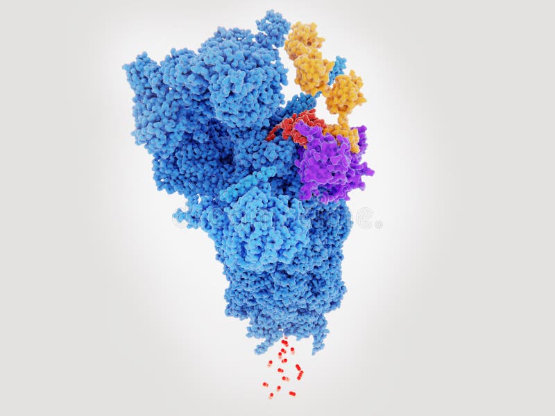
Free with trial Proteasomes are a large molecular machine that degrade unneeded or damaged proteins that have been tagged with polyubiquitin yellow. The ubiquitin hydrolase violet detaches ubiquitin from the protein, that is unfolded and degraded into small peptides bottom. Source: PDB entry 5GJQ. Protease protein illustrations Proteasome degrading a protein red tagged with polyubiquitin. Proteasomes are a large molecular machine that degrade unneeded or damaged proteins that have been tagged with polyubiquitin yellow. The ubiquitin hydrolase violet detaches ubiquitin from the protein, that is unfolded and degraded into small peptides bottom. Source: PDB entry 5GJQ
Free with trial Apoptosome is a large quaternary protein structure formed in the process of apoptosis (programmed cell death). Protease protein illustrations Apoptosome is a large quaternary protein structure formed in the
Free with trial Protease-activated receptor 4, a member of the large family of 7-transmembrane-region receptors that couple to guanosine-nucleotide-binding proteins. activated by thrombin and trypsin. 3d rendering. Protease protein illustrations Protease-activated receptor 4, a member of the large family of 7
Free with trial Protein digestion. Enzymes proteases are digestion breaks the protein into single amino acids, which are absorbed into the blood. Vector illustration. Protease protein vectors Protein digestion. Enzymes proteases are digestion breaks the protein into single amino acids
Free with trial Protein Digestion. Enzymes proteases and peptidases are digestion breaks the protein into smaller peptide chains and into single amino acids, which are absorbed into the blood. Vector illustration. Protease protein vectors Protein Digestion
Free with trial Protein kinase N1, an enzyme which may mediate the Rho-dependent signaling pathway. 3d rendering. Protease protein illustrations Protein kinase N1, an enzyme which may mediate the Rho-dependent signaling pathway. 3d rendering
Free with trial Vector illustration of protein digestion. Pepsin, trypsin and erepsin enzymes effect on protein molecule. Protease protein vectors Vector illustration of protein digestion
Free with trial Papain enzyme. Protease present in papaya fruit. Cartoon representation with secondary structure coloring (green sheets, red helices. Protease protein illustrations Papain enzyme. Protease present in papaya fruit
Free with trial Papain enzyme. Protease present in papaya fruit. Cartoon & stick representation with backbone gradient coloring. Protease protein illustrations Papain enzyme. Protease present in papaya fruit
Free with trial Caspase 3 apoptosis protein. Enzyme that plays important role in programmed cell death. Cartoon model, secondary structure coloring: alpha-helices green, beta sheets blue. Protease protein illustrations Caspase 3 apoptosis protein. Enzyme that plays important role in programmed cell death
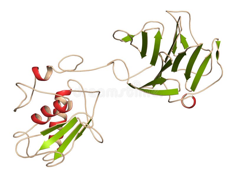
Free with trial A carboxypeptidase is a protease enzyme that hydrolyzes a peptide bond at the carboxy-terminal end of a protein or peptide. 3D cartoon model, secondary structure color scheme, PDB 2v77, white background. Protease protein illustrations Crystal structure of human carboxypeptidase A1. A carboxypeptidase is a protease enzyme that hydrolyzes a peptide bond at the carboxy-terminal end of a protein or peptide. 3D cartoon model, secondary structure color scheme, PDB 2v77, white background.
Free with trial Cathepsin K enzyme bound to the inhibitor odanacatib. 3D rendering based on protein data bank entry 5tdi. Atoms shown as color-coded spheres. Protein and inhibitor shown in different colors. Protease protein illustrations Cathepsin K enzyme bound to the inhibitor odanacatib. 3D rendering based on protein data bank entry 5tdi
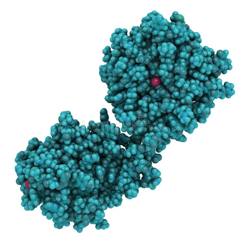
Free with trial Cystatin C (V57D mutant). Protein used as biomarker of kidney function. 3D rendering based on protein data bank entry 3sva. Atoms are represented as spheres with conventional color coding. Protease protein illustrations Cystatin C (V57D mutant). Protein used as biomarker of kidney function. 3D rendering based on protein data bank entry 3sva
Free with trial Human activated protein C (APC, drotrecogin alfa, without Gla-domain). Has anti-thrombotic and anti-inflammatory properties. Used to treat sepsis (obsolete). Atoms shown as spheres with conventional color coding: hydrogen (white), carbon (grey), oxygen (red), nitrogen (blue), sulfur (yellow. Protease protein illustrations Human activated protein C (APC, drotrecogin alfa, without Gla-domain). Has anti-thrombotic and anti-inflammatory properties. Used
Free with trial Cathepsin K enzyme bound to the inhibitor odanacatib. 3D rendering based on protein data bank entry 5tdi. Combined cartoon and stick representation with backbone gradient coloring. Background black. Protease protein illustrations Cathepsin K enzyme bound to the inhibitor odanacatib. 3D rendering based on protein data bank entry 5tdi
Free with trial Plasminogen plasmin precursor protein. Plasmin is an enzyme responsible for the breakdown of fibrin fibrinolysis. Cartoon representation. Secondary structure coloring. Protease protein illustrations Plasminogen plasmin precursor protein. Plasmin is an enzyme responsible for the breakdown of fibrin fibrinolysis.
Free with trial Activated coagulation factor VII (FVIIa), chemical structure. Plays role in blood clotting (coagulation). Recombinant protein used in hemophilia treatment. Atoms shown as spheres with conventional color coding: hydrogen (white), carbon (grey), oxygen (red), nitrogen (blue), sulfur (yellow. Protease protein illustrations Activated coagulation factor VII (FVIIa), chemical structure. Plays role in blood clotting (coagulation). Recombinant protein used
Free with trial Ribbon model of ubiquitin, protein that directs other proteins to various cellular compartments or marks them for destruction. Lysine side-chains and C-terminus depicted as ball-and-stick. Protease protein illustrations Ubiquitin
Free with trial Chemical structure of the yeast proteasome. The proteasome breaks apart unneeded and damaged proteins inside the cell. Two different views of the same protein complex from different angles. Protease protein illustrations Yeast proteasome
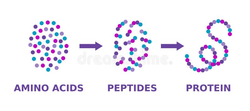
Free with trial Scientific diagram. Proteins molecule synthesis. Molecule made of long chains of polypeptides. Vector illustration. Protease protein vectors Protein structure. Amino acids, peptides, protein. Proteins formation model. Scientific diagram. Proteins molecule synthesis. Molecule made of long chains of polypeptides. Vector illustration.
Free with trial Complement component 3, often simply called C3, is a protein of the immune system. It plays a central role in the complement system and contributes to innate immunity. 3D cartoon and Gaussian surface models with differently colored protein chains, white background. Protease protein illustrations Structure of the complement component C3. Complement component 3, often simply called C3, is a protein of the immune system. It plays a central role in the complement system and contributes to innate immunity. 3D cartoon and Gaussian surface models with differently colored protein chains, white background.
Free with trial The pepsins are enzymes secreted by the stomach that breaks down proteins. Vector illustration. Protease protein vectors Digestion of protein. Breaking the complex molecule first into peptides then into individual amino acids. The pepsins are enzymes secreted by the stomach that breaks down proteins. Vector illustration
Free with trial Matrix metalloproteinase 12 MMP-12, macrophage elastase enzyme. MMPs are proteases involved in the breakdown of extracellular matrix. 3D rendering based on protein data bank entry 3ba0. Atoms shown as color-coded spheres. Protein and metal ions shown in different colors. Protease protein illustrations Matrix metalloproteinase 12 MMP-12, macrophage elastase enzyme. MMPs are proteases involved in the breakdown of extracellular. Matrix metalloproteinase 12 MMP-12, macrophage elastase enzyme. MMPs are proteases involved in the breakdown of extracellular matrix. 3D rendering based on protein data bank entry 3ba0. Atoms shown as color-coded spheres. Protein and metal ions shown in different colors
Free with trial Matrix metalloproteinase 12 MMP-12, macrophage elastase enzyme. MMPs are proteases involved in the breakdown of extracellular matrix. 3D rendering based on protein data bank entry 3ba0. Cartoon representation with secondary structure coloring green sheets, red helices. Protease protein illustrations Matrix metalloproteinase 12 MMP-12, macrophage elastase enzyme. MMPs are proteases involved in the breakdown of extracellular. Matrix metalloproteinase 12 MMP-12, macrophage elastase enzyme. MMPs are proteases involved in the breakdown of extracellular matrix. 3D rendering based on protein data bank entry 3ba0. Cartoon representation with secondary structure coloring green sheets, red helices
Free with trial Proteasomes are protein complexes which degrade unneeded or damaged proteins by proteolysis, a chemical reaction that breaks peptide bonds. 3D cartoon and Gaussian surface models, based on PDB 5le5, white background. Protease protein illustrations Structure of human 20S proteasome. Proteasomes are protein complexes which degrade unneeded or damaged proteins by proteolysis, a chemical reaction that breaks peptide bonds. 3D cartoon and Gaussian surface models, based on PDB 5le5, white background.
Free with trial Proteasome collaborate with ubiquitin. Protein degradation machineries in eukaryotic cells 3d rendering. Protease protein illustrations Isolated Proteasome collaborate with ubiquitin. Proteasome collaborate with ubiquitin. Protein degradation machineries in eukaryotic cells 3d rendering
Free with trial Proteasome collaborate with ubiquitin. Protein degradation machineries in eukaryotic cells 3d rendering. Protease protein illustrations Isolated Proteasome collaborate with ubiquitin. Proteasome collaborate with ubiquitin. Protein degradation machineries in eukaryotic cells 3d rendering
Free with trial DNA Replication and Transcription. Steps. double helix is unwound. Each separated strand acts as a template for replicating a new strand. Vector diagram for scientific, medical, and educational use. poster. Protease protein vectors DNA Replication and Transcription
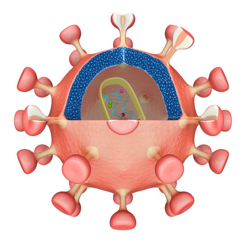
Free with trial The viral core (or capsid) is usually bullet-shaped and is made from the protein p24. Inside the core are three enzymes required for HIV replication called reverse transcriptase, integrase and protease. Also held within the core is HIV's genetic material, which consists of two identical strands of RNA. Protease protein illustrations HIV Virus. The viral core (or capsid) is usually bullet-shaped and is made from the protein p24. Inside the core are three enzymes required for HIV replication called reverse transcriptase, integrase and protease. Also held within the core is HIV's genetic material, which consists of two identical strands of RNA.
Free with trial The viral core (or capsid) is usually bullet-shaped and is made from the protein p24. Inside the core are three enzymes required for HIV replication called reverse transcriptase, integrase and protease. Also held within the core is HIV's genetic material, which consists of two identical strands of RNA. Protease protein illustrations HIV Virus. The viral core (or capsid) is usually bullet-shaped and is made from the protein p24. Inside the core are three enzymes required for HIV replication called reverse transcriptase, integrase and protease. Also held within the core is HIV's genetic material, which consists of two identical strands of RNA.
Free with trial P53 blue tagged with ubiquitin yellow is degraded into small peptides by a proteasome violett. p53 prevents cancer formation and acts as a guardian of the genome. Mutations in the p53 gene contribute to about half of the cases of human cancer. Protease protein illustrations The tumor suppressor p53 is degraded by a proteasome. P53 blue tagged with ubiquitin yellow is degraded into small peptides by a proteasome violett. p53 prevents cancer formation and acts as a guardian of the genome. Mutations in the p53 gene contribute to about half of the cases of human cancer
Free with trial Trypsin molecular chemical formula. Enzyme of the pancreas. Infographics. Vector illustration on isolated background. Protease protein vectors Trypsin molecular chemical formula. Enzyme of the pancreas. Infographics. Vector illustration on isolated background
Free with trial Pepsin is a molecular chemical formula. Enzyme of the stomach. Infographics. Vector illustration on an isolated background. Protease protein vectors Pepsin is a molecular chemical formula. Enzyme of the stomach. Infographics. Vector illustration on an isolated background.
Free with trial Gelatinase is a molecular chemical formula. Enzyme of the stomach. Infographics. Vector illustration on isolated background. Protease protein vectors Gelatinase is a molecular chemical formula. Enzyme of the stomach. Infographics. Vector illustration on isolated background
Free with trial Pepsin enzyme molecule. Digestive enzyme found in the stomach, where it breaks down food proteins into smaller fragments. Atoms are represented as spheres. Protease protein illustrations Pepsin enzyme molecule.
Free with trial The complement system, also known as complement cascade, is a part of the immune system that enhances complements the ability of antibodies and phagocytic cells to clear microbes and damaged cells from an organism, promote inflammation, and attack the pathogen`s cell membrane. 3D cartoon and Gaussian surface models with differently colored elements of the complex, white background. Protease protein illustrations Structure of the complement complex. The complement system, also known as complement cascade, is a part of the immune system that enhances complements the ability of antibodies and phagocytic cells to clear microbes and damaged cells from an organism, promote inflammation, and attack the pathogen`s cell membrane. 3D cartoon and Gaussian surface models with differently colored elements of the complex, white background.
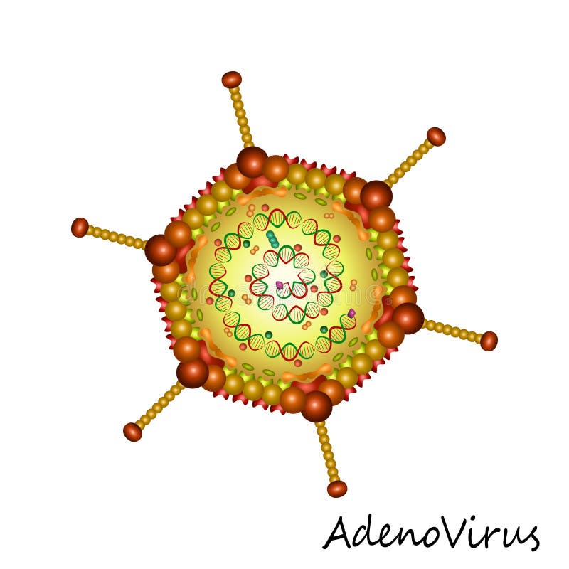
Free with trial Adenovirus virus particle structure. This is file of EPS10 format. Protease protein illustrations Adenovirus virus particle structure
Free with trial Gelatinase is a molecular chemical formula. Functions. Enzyme of the stomach. Infographics. Vector illustration on isolated background. Protease protein vectors Gelatinase is a molecular chemical formula. Functions. Enzyme of the stomach. Infographics. Vector illustration
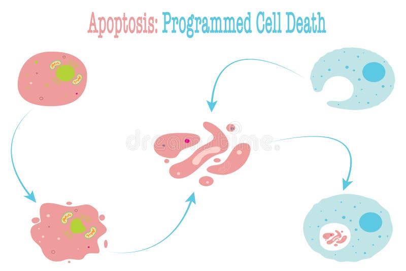
Free with trial Vector illustration infographic diagram of apoptosis, cell programmed death. Protease protein illustrations Apoptosis vector illustration diagram. vector illustration infographic diagram of apoptosis, cell programmed death
Free with trial Gelatinase is a molecular chemical formula. Enzyme of the stomach. Infographics. Vector illustration on black background. Protease protein vectors Gelatinase is a molecular chemical formula. Enzyme of the stomach. Infographics. Vector illustration on black background
Free with trial Trypsin IV is a proteinase expressed in the human brain. 3D cartoon model, white background. Protease protein illustrations Structure of human trypsin IV brain trypsin. Trypsin IV is a proteinase expressed in the human brain. 3D cartoon model, white background.
Free with trial 3d rendering of ubiquitin is attached to target proteins by a process called ubiquitination. Protease protein illustrations Ubiquitin is attached to target proteins by a process
Free with trial Pepsin is an endopeptidase that breaks down proteins into smaller peptides. A 3D cartoon model isolated, white background. Protease protein illustrations Structure of human pepsin complexed with inhibitor pepstatin. Pepsin is an endopeptidase that breaks down proteins into smaller peptides. A 3D cartoon model isolated, white background.
Free with trial Gastricsin is a molecular chemical formula. Enzyme of the stomach, gastric juice. Infographics. Vector illustration on isolated background. Protease protein vectors Gastricsin is a molecular chemical formula. Enzyme of the stomach, gastric juice. Infographics. Vector illustration
Free with trial Trypsin digestive enzyme molecule (human). 3D rendering. Enzyme that contributes to the digestion of proteins in the digestive system. Cartoon representation (red) combined with semi-transparent surfaces. Protease protein illustrations Trypsin digestive enzyme molecule (human). 3D rendering. Enzyme that contributes to the digestion of proteins in the digestive
Free with trial Thrombin EC 3. 4. 21. 5, fibrinogenase, thrombase, thrombofort, topical, thrombin-C, tropostasin, activated blood-coagulation factor II, blood-coagulation factor IIa, factor IIa, E thrombin, beta-thrombin, gamma-thrombin is a serine protease, an enzyme that, in humans, is encoded by the F2 gene. 3D cartoon and Gaussian surface models, secondary structure color scheme, based on PDB 2a0q, white background. Protease protein illustrations Structure of thrombin (factor II). Thrombin EC 3.4.21.5, fibrinogenase, thrombase, thrombofort, topical, thrombin-C, tropostasin, activated blood-coagulation factor II, blood-coagulation factor IIa, factor IIa, E thrombin, beta-thrombin, gamma-thrombin is a serine protease, an enzyme that, in humans, is encoded by the F2 gene. 3D cartoon and Gaussian surface models, secondary structure color scheme, based on PDB 2a0q, white background.
Free with trial Arginine amino acid capsules vitamins complex minerals. 3D Model of molecule brown isolated on white background. For food supplement ad package design. Science medic concept. Vector EPS10. Protease protein vectors Arginine amino acid capsules vitamins complex minerals
Free with trial Tryptophan amino acid capsules vitamins complex minerals. 3D Model of molecule green isolated on white background. For food supplement ad package design. Science medic concept. Vector EPS10. Protease protein vectors Tryptophan amino acid capsules vitamins complex minerals
Free with trial Methionine amino acid capsules vitamins complex minerals. 3D Model of molecule blue isolated on white background. For food supplement ad package design. Science medic concept. Vector EPS10. Protease protein vectors Methionine amino acid capsules vitamins complex minerals
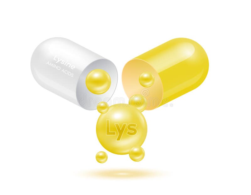
Free with trial Lysine amino acid float out of the capsule. Vitamins complex and minerals yellow isolated on white background. For food supplement ad package design. Science medic concept. 3D Vector EPS10. Protease protein vectors Lysine amino acid float out of the capsule. Vitamins complex and minerals yellow isolated on white background
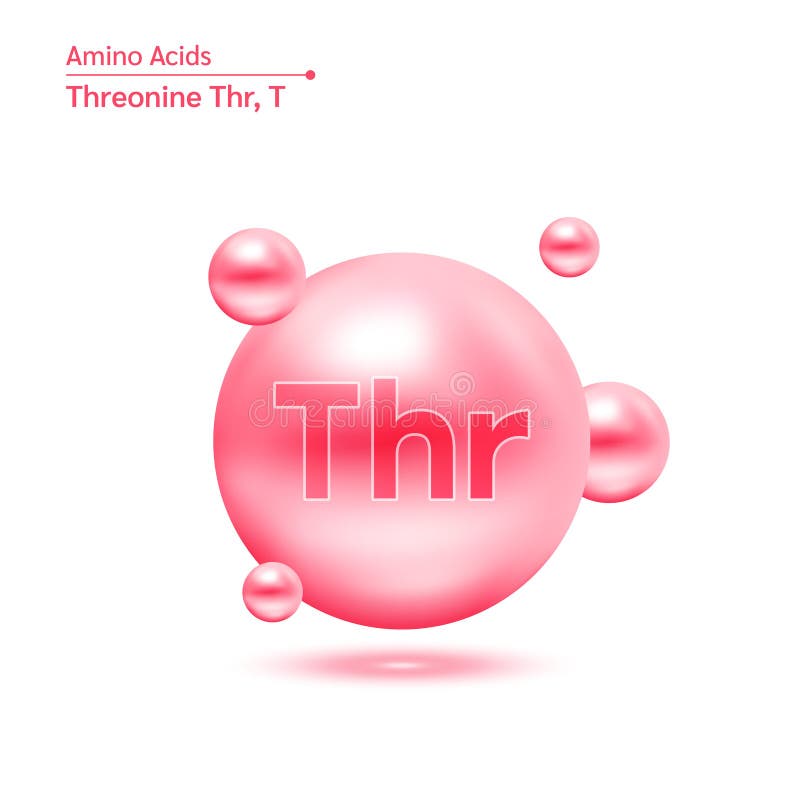
Free with trial Threonine (Thr) amino acid capsules vitamins complex minerals. 3D Model of molecule pink isolated on white background. For food supplement ad package design. Science medic concept. Vector EPS10. Protease protein vectors Threonine amino acid capsules vitamins complex minerals. 3D Model of molecule pink isolated on white background. For food su. Threonine (Thr) amino acid capsules vitamins complex minerals. 3D Model of molecule pink isolated on white background. For food supplement ad package design. Science medic concept. Vector EPS10
Free with trial Vector illustration of protein digestion. Protease enzyme effect on protein molecule. Protease protein vectors Vector illustration of protein digestion. Protease enzyme effect on protein molecule
Free with trial Close-up shot of Protease Extract powder in wooden bowl. Product rich protein content, health supplement, culinary ingredient for food preparation, cooking or baking nutritious meal. Protease protein illustrations Close-up shot of Protease Extract powder in wooden bowl. Product rich protein content, health supplement, culinary ingredient for
Free with trial This image depicts a 3D rendering of alpha-2 macroglobulin protein molecules, showcasing their intricate structure and complex interactions. The microscopic view provides a detailed look at the molecular arrangement, highlighting the protein's role in protease inhibition and its potential applications in pharmaceutical research and development. The image is relevant to fields such as biochemistry, molecular biology, and medicine, and could be used to illustrate scientific concepts, support educational materials, or promote research initiatives. Protease protein vectors A microscopic view of alpha-2 macroglobulin protein molecules in a 3D rendering. This image depicts a 3D rendering of alpha-2 macroglobulin protein molecules. This image depicts a 3D rendering of alpha-2 macroglobulin protein molecules, showcasing their intricate structure and complex interactions. The microscopic view provides a detailed look at the molecular arrangement, highlighting the protein's role in protease inhibition and its potential applications in pharmaceutical research and development. The image is relevant to fields such as biochemistry, molecular biology, and medicine, and could be used to illustrate scientific concepts, support educational materials, or promote research initiatives.
Free with trial Protein structure. Amino acids, Alpha helix, Polypeptide chains, and Complex of protein molecule. Protein is a polymer (polypeptide) that formed from sequences of amino acids. Isometric Flat vector illustration. Protease protein vectors Protein structure
Free with trial Vector Illustration of pepsin enzyme effect on protein molecule. Protease protein vectors Vector Illustration of pepsin enzyme effect on protein moleculet
Free with trial HIV virus structure diagram anatomy of virus labeled human immunodeficiency virus, capsid, glycoprotein, ssRNA, protease. Microbiology virology educational medical infographic healthcare illustration. Protease protein vectors HIV virus structure diagram anatomy of virus labeled human immunodeficiency virus, capsid, glycoprotein, ssRNA, protease.
Free with trial Proteasome structure anatomy diagram process. Labeled Immunoproteasome, Regulagory Cap. Protein complexes, degradation of proteins by proteolysis, a chemical reaction breaks peptide bonds. Protease protein vectors Proteasome structure anatomy diagram process. Labeled Immunoproteasome, Regulagory Cap. Protein complexes, degradation of proteins
Free with trial A metaphor showing Protein in equilibrium with Carbs, symbolizing a desired harmony between them. Stability. Harmonious and preferable setup. Protease protein illustrations Protein and carbs in balance. A metaphor showing Protein in equilibrium with Carbs, symbolizing a desired harmony between them. Stability. Harmonious and preferable setup
Free with trial Proteasomes are molecular machines for breaking down proteins called proteolysis, only target proteins that have been marked for destruction called ubiquitin to the target protein, 3d rendering. Protease protein illustrations Proteasomes is breaking down protein molecule. Proteasomes are molecular machines for breaking down proteins called proteolysis, only target proteins that have been marked for destruction called ubiquitin to the target protein, 3d rendering
Free with trial Proteasomes are molecular machines for breaking down proteins called proteolysis, only target proteins that have been marked for destruction called ubiquitin to the target protein, 3d rendering. Protease protein illustrations Proteasomes is breaking down protein molecule. Proteasomes are molecular machines for breaking down proteins called proteolysis, only target proteins that have been marked for destruction called ubiquitin to the target protein, 3d rendering
Free with trial Proteasomes are molecular machines for breaking down proteins called proteolysis, only target proteins that have been marked for destruction called ubiquitin to the target protein, 3d rendering. Protease protein illustrations Proteasomes is breaking down protein molecule. Proteasomes are molecular machines for breaking down proteins called proteolysis, only target proteins that have been marked for destruction called ubiquitin to the target protein, 3d rendering