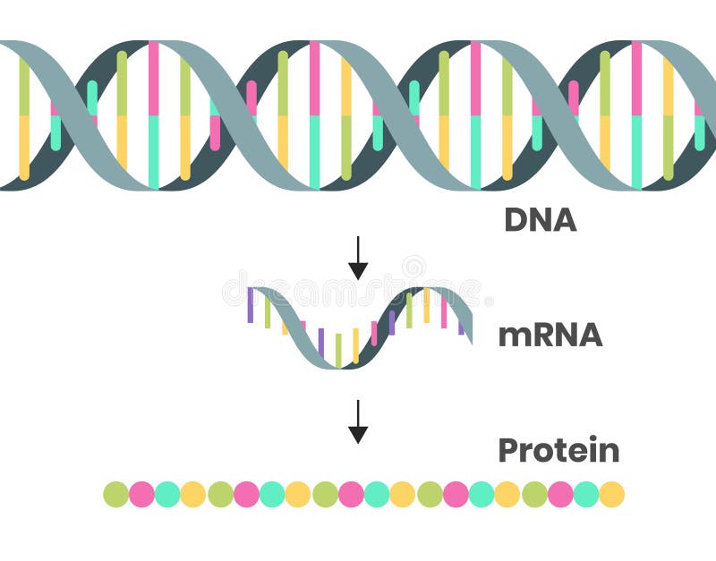
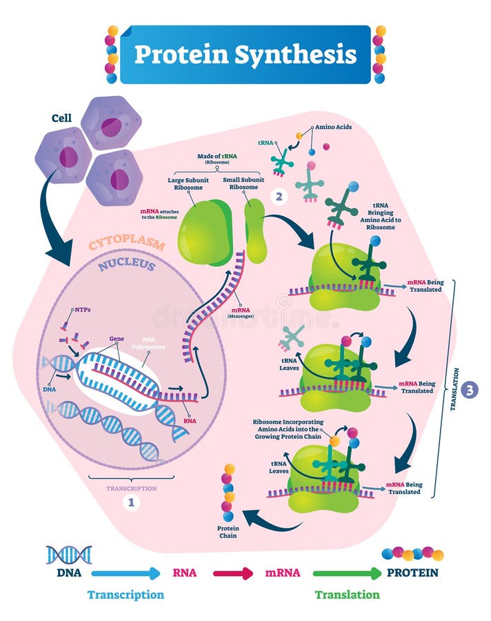
Free with trial Protein synthesis vector illustration. Labeled transcription and translation steps diagram with full cycle explanation. How body creates protein chain from cytoplasm. Protein chain vectors Protein synthesis vector illustration. Transcription and translation. Protein synthesis vector illustration. Labeled transcription and translation steps diagram with full cycle explanation. How body creates protein chain from cytoplasm.
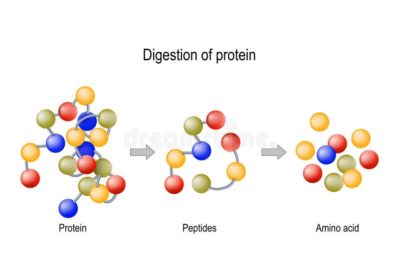
Free with trial Digestion of Protein. Enzymes proteases and peptidases are digestion breaks the protein into smaller peptide chains and into single amino acids, which are absorbed into the blood. Protein chain vectors Digestion of Protein. Enzymes proteases and peptidases, peptides and amino acids. Digestion of Protein. Enzymes proteases and peptidases are digestion breaks the protein into smaller peptide chains and into single amino acids, which are absorbed into the blood
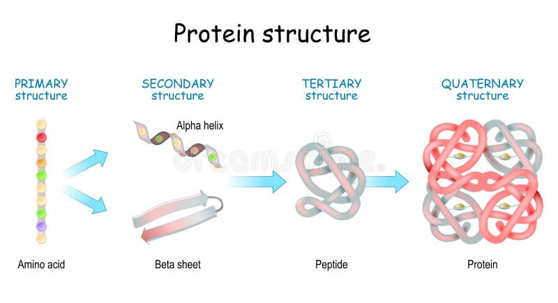
Free with trial Levels of protein structure from amino acids to Complex of protein molecule. Protein is a polymer polypeptide that formed from sequences of amino acids. Levels of protein structure: Primary, Secondary, Tertiary, and Quaternary. Protein chain vectors Levels of protein structure from amino acids to Complex of protein molecule
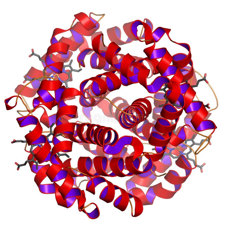
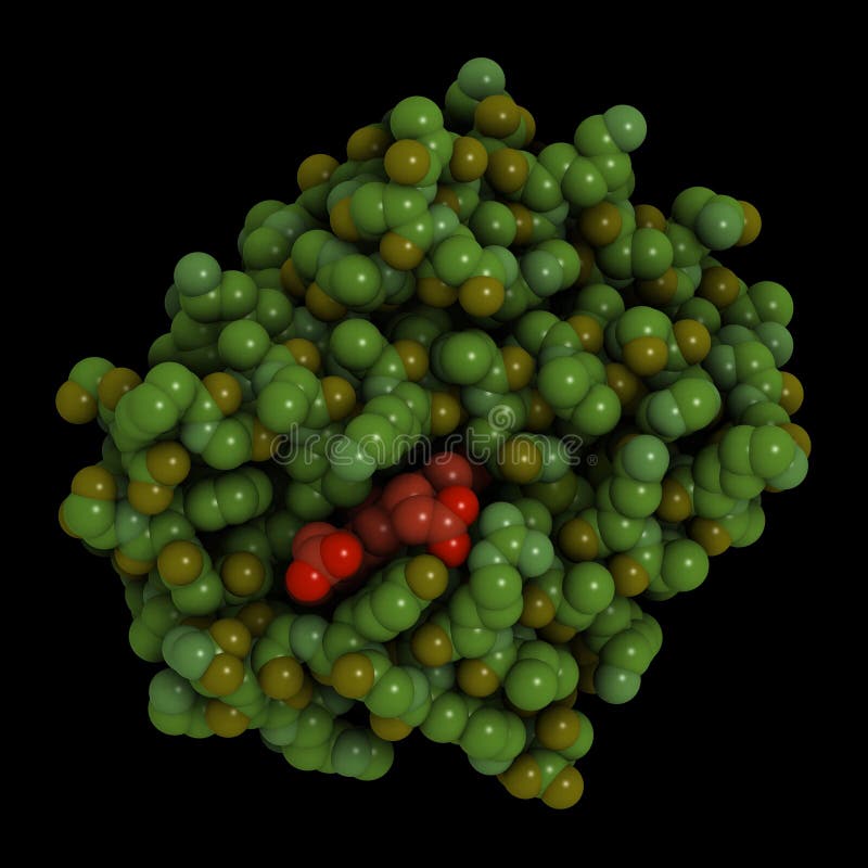
Free with trial Hemoglobin or haemoglobin (frequently abbreviated as Hb) is what makes our blood colored red. It transports oxygen from the lungs to the cells and CO2 back from the cells to the lung. Here, the globular shape of the protein (this is why it is called hemoGLOBIN) is visible. The heme groups (containing iron that can bind oxygen or CO2) are shown as ball-and-stick model. Rendered in great detail on white background. Fog simulates depth. Modifications such as other colorings, less fog, or perspecive views are available on request. Protein chain illustrations Globular protein. Hemoglobin or haemoglobin (frequently abbreviated as Hb) is what makes our blood colored red. It transports oxygen from the lungs to the cells and CO2 back from the cells to the lung. Here, the globular shape of the protein (this is why it is called hemoGLOBIN) is visible. The heme groups (containing iron that can bind oxygen or CO2) are shown as ball-and-stick model. Rendered in great detail on white background. Fog simulates depth. Modifications such as other colorings, less fog, or perspecive views are available on request.
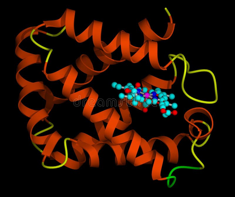
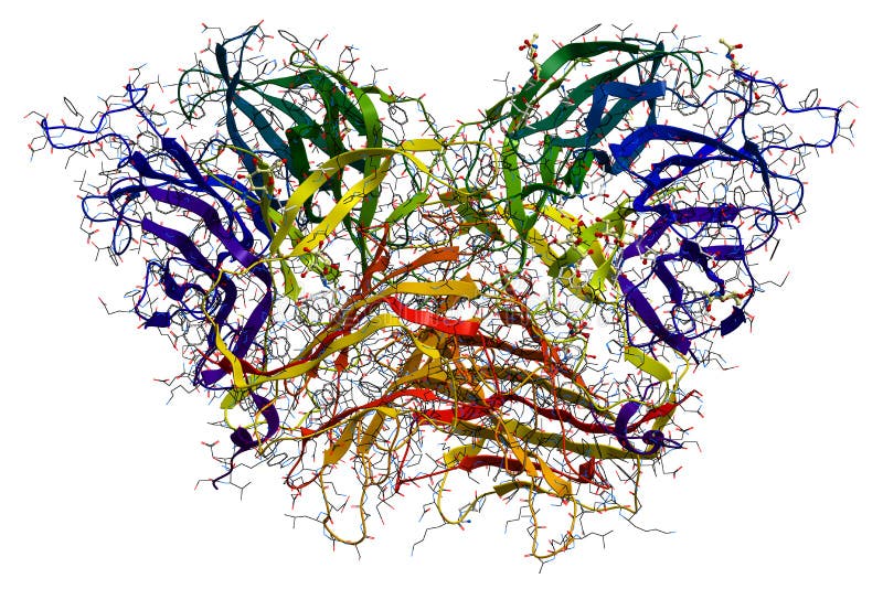
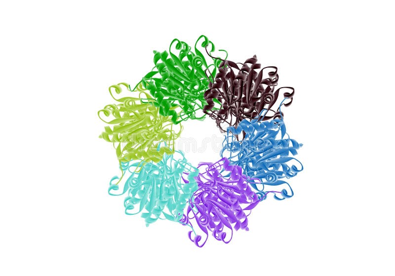
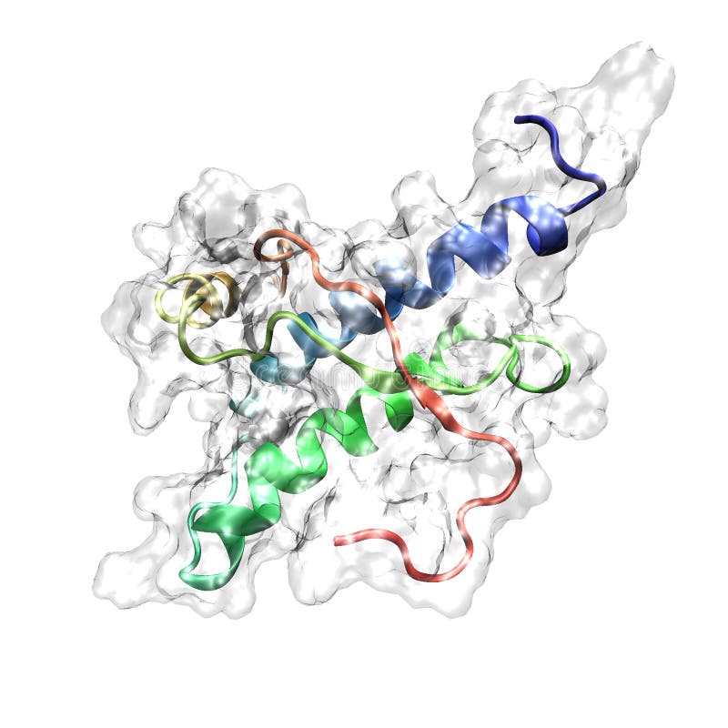
Free with trial Enzyme that produces fluorine compounds (Fluorinase). The protein consists of six identical chains, here colored from blue (N-terminus) to red (C-terminus), rainbow coloring. It is shown in cartoon representation: alpha-helices are represented by spirals, beta-sheets by arrows. The ball-and-stick molecules are the active sites. Detailed rendering. Protein chain illustrations Protein on white background. Enzyme that produces fluorine compounds (Fluorinase). The protein consists of six identical chains, here colored from blue (N-terminus) to red (C-terminus), rainbow coloring. It is shown in cartoon representation: alpha-helices are represented by spirals, beta-sheets by arrows. The ball-and-stick molecules are the active sites. Detailed rendering.
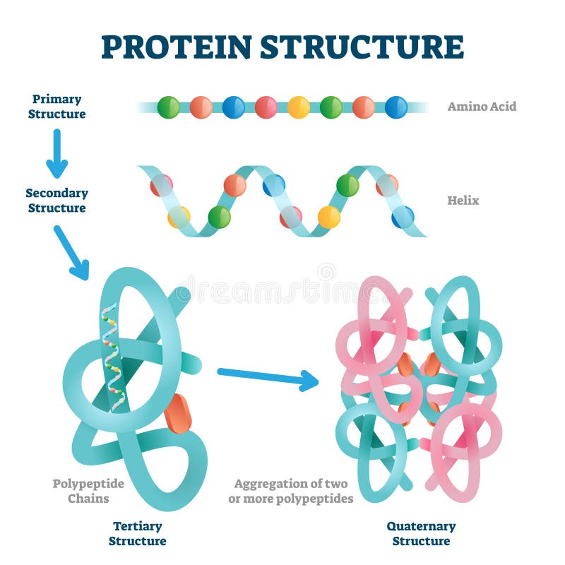
Free with trial Protein structure levels: Primary, Secondary, Tertiary, and Quaternary. From Amino acid to Alpha helix, Beta sheet, peptide, and protein molecule. concept. Vector illustration. Protein chain vectors Protein structure levels. From Amino acid to Alpha helix, Beta sheet, peptide, and protein molecule. Protein structure levels: Primary, Secondary, Tertiary, and Quaternary. From Amino acid to Alpha helix, Beta sheet, peptide, and protein molecule. concept. Vector illustration
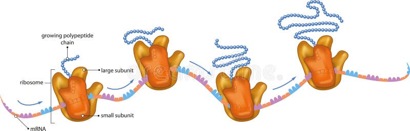
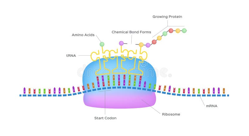
Free with trial Vector illustration of ribosomes, RNA and polypeptide chain. Protein chain vectors Ribosomes
Free with trial Vectored illustration of common farm animals as cow, pig and rooster. Protein chain vectors Farm animals vector silhouette. Vectored illustration of common farm animals as cow, pig and rooster
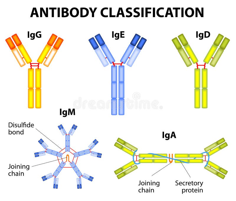
Free with trial Different types of immunoglobulins. IgG, IgA, IgD, IgE, and IgM. Protein chain vectors Antibody classification. Different types of immunoglobulins. IgG, IgA, IgD, IgE, and IgM
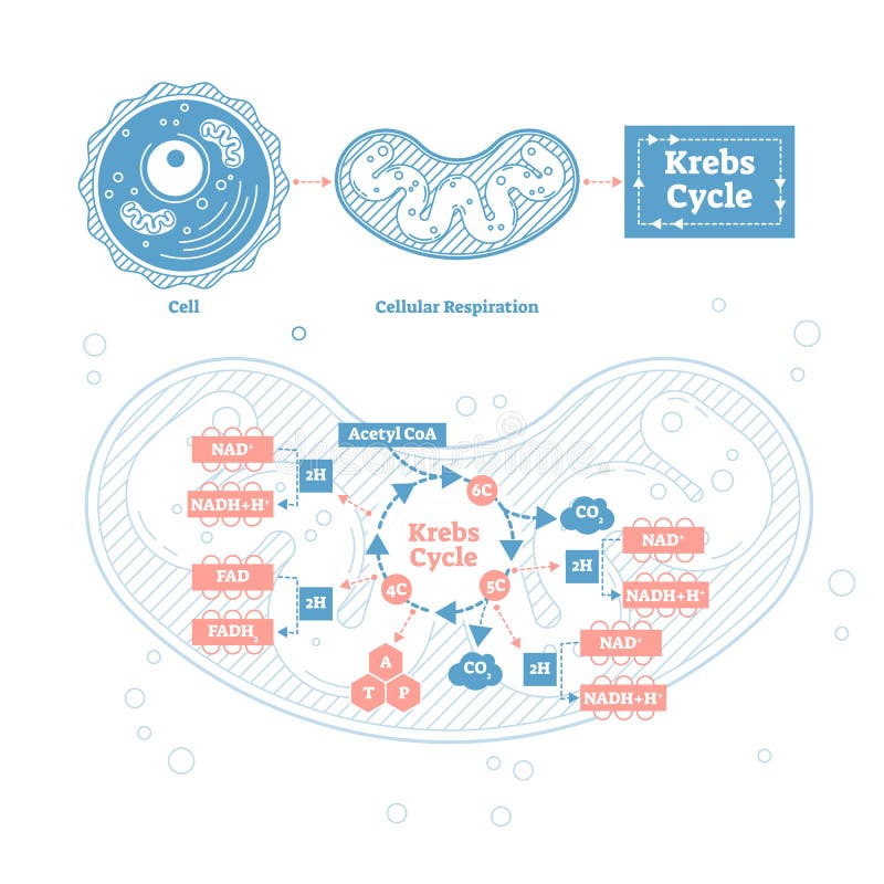
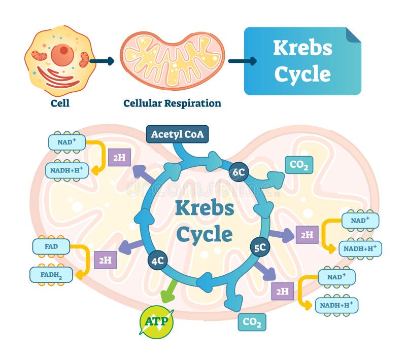
Free with trial Krebs cycle vector illustration. Citric tricarboxylic acid labeled scheme. Educational diagram with cell, cellular respiration and ATP. Human power molecular metabolism. Protein chain vectors Krebs cycle vector illustration. Citric tricarboxylic acid labeled scheme
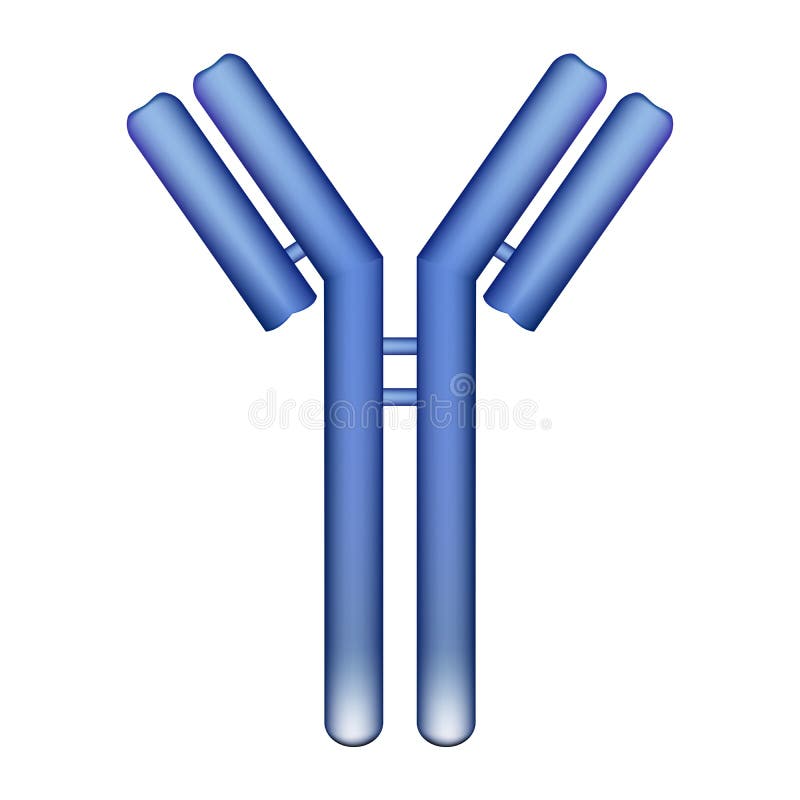
Free with trial Vector illustration of antibody molecule immunoglobulin. Protein chain vectors Antibody molecule
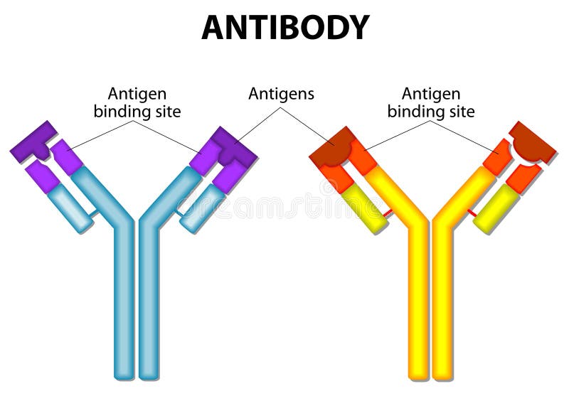
Free with trial Antibody molecule and Antigen. vector diagram. Protein chain vectors Antibody and Antigen. Antibody molecule and Antigen. vector diagram
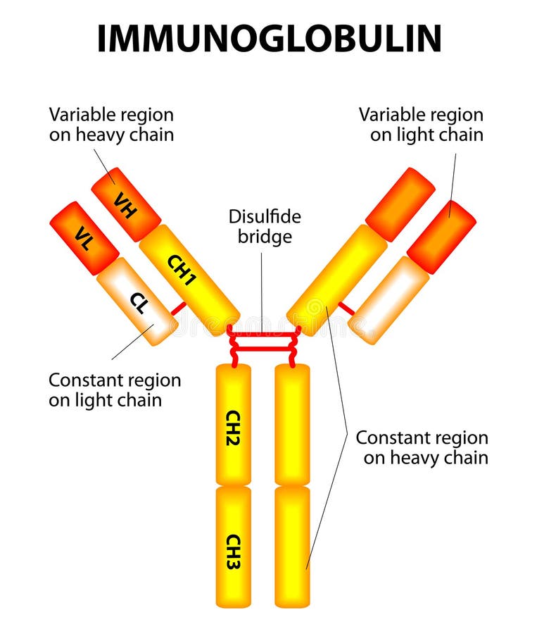
Free with trial Diagram showing the major components of a typical antibody molecule (IgE). Protein chain vectors Anatomy of an antibody. Diagram showing the major components of a typical antibody molecule (IgE)
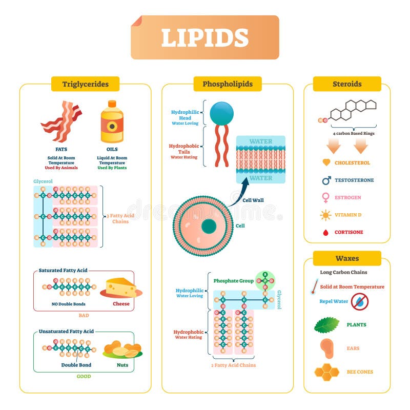
Free with trial Lipids vector illustration infographic. Triglycerides, waxes, phospholipids, and steroids diagram. Labeled structure with fatty chains, saturated bad acid example with cheese and unsaturated with nuts. Protein chain vectors Lipids vector illustration. Triglycerides, waxes and steroids diagram. Lipids vector illustration infographic. Triglycerides, waxes, phospholipids, and steroids diagram. Labeled structure with fatty chains, saturated bad acid example with cheese and unsaturated with nuts
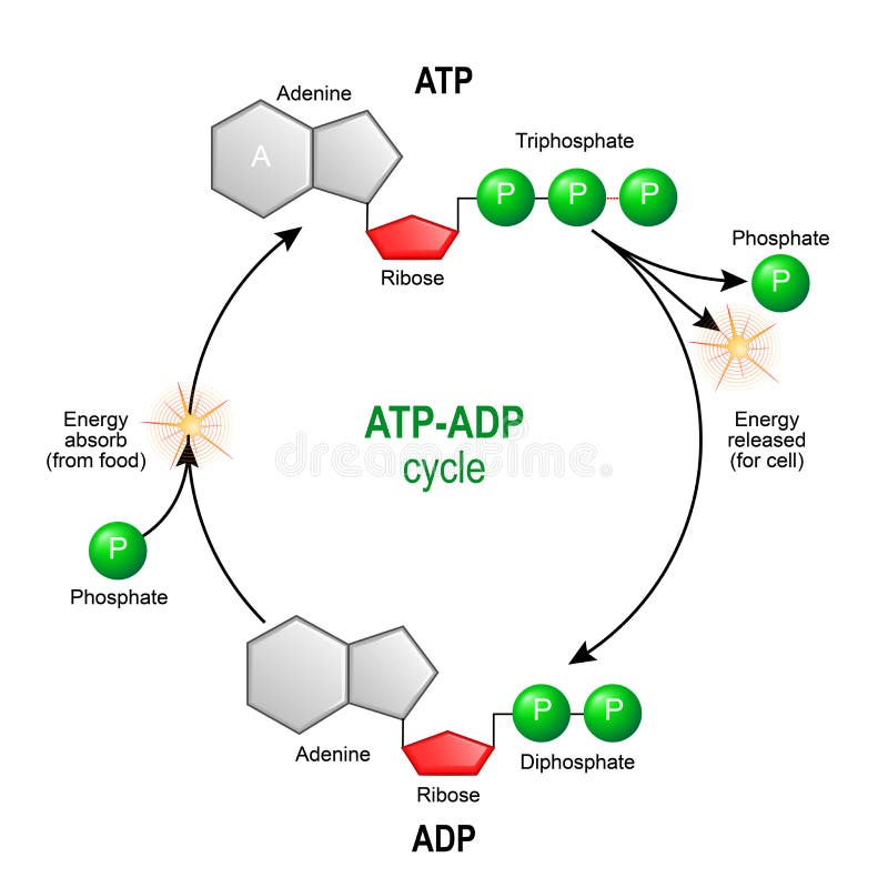
Free with trial ATP ADP cycle. Adenosine triphosphate ATP is a organic chemical that provides energy for cell. intracellular energy transfer. Adenosine diphosphate ADP is organic compound for metabolism in cell. Vector diagram for educational, biological, medical and science use. nmodel of molecule adenosine triphosphate, and Adenosine diphosphate. Protein chain vectors ATP ADP cycle. Adenosine triphosphate ATP is a organic chemica
Free with trial Structure of human insulin diagramed in two different ways, eps8. Protein chain vectors Structure of human insulin
Free with trial Antibody on a white background. Vector diagram of the basic unit of immunoglobulin. Protein chain vectors Antibody on a white background
Free with trial Illustration of rainbow DNA (deoxyribonucleic acid) with defocus on background. Protein chain illustrations Rainbow DNA (deoxyribonucleic acid)
Free with trial Enzyme pepsin 3D model on a white background. Protein chain illustrations Enzyme pepsin 3D model
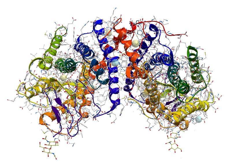
Free with trial Alpha-Amylase, an enzyme that hydrolyses polysaccharides, such as starch and glycogen, to glucose and maltose. Protein chain illustrations Enzyme Alpha-Amylase. Alpha-Amylase, an enzyme that hydrolyses polysaccharides, such as starch and glycogen, to glucose and maltose.
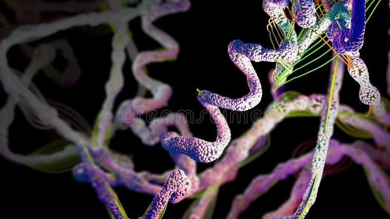
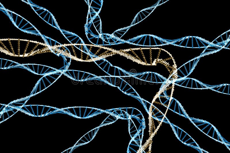
Free with trial Abstract bright protein chain on dark background. Protein chain illustrations Protein chain
Free with trial Protein structure vector illustration. Labeled amino acid chain molecules types scheme. Educational collection with various primary structure, helix, polypeptide tertiary and quaternary levels closeup. Protein chain vectors Protein structure vector illustration. Labeled amino acid chain molecules. Protein structure vector illustration. Labeled amino acid chain molecules types scheme. Educational collection with various primary structure, helix, polypeptide tertiary and quaternary levels closeup
Free with trial Chain of amino acid or bio molecules called protein - 3d illustration. Protein chain illustrations Chain of amino acid or bio molecules called protein
Free with trial Chain of amino acid or bio molecules called protein - 3d illustration. Protein chain illustrations Chain of amino acid or bio molecules called protein
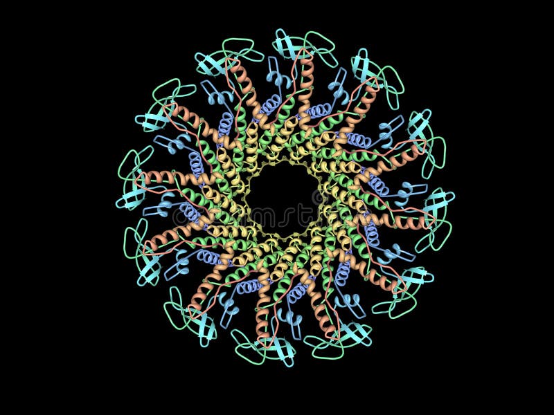
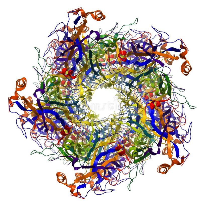
Free with trial Crystal Structure of Circadian Clock Protein KaiC at 2. 8 A resolution. KaiC is an essential circadian protein in cyanobacteria. The structure resembles a double doughnut with a central pore that is partially sealed at one end. The crystal structure reveals ATP binding, inter-subunit organization, a scaffold for Kai-protein complex formation, the location of critical KaiC mutations, and evolutionary relationships to other proteins. A key auto-phosphorylation site on KaiC (T432) is identified from the crystal structure, and mutation of this residue abolishes circadian rhythmicity. The crystal structure of KaiC will be essential for understanding this circadian clockwork and for establishing its links to global gene expression. Protein chain illustrations Protein
Free with trial Crystal Structure of Circadian Clock Protein KaiC. Protein chain illustrations Protein
Free with trial A molecular model of a prion protein. This protein is involved in diseases such as CJD and BSE (Mad cow disease). Protein chain illustrations Prion protein
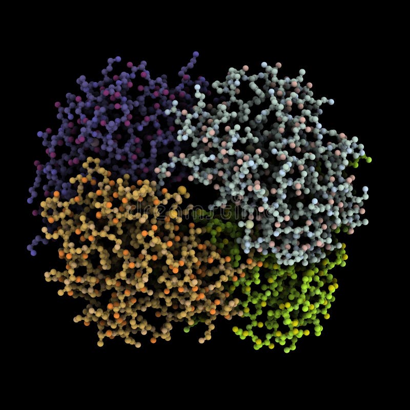
Free with trial Computer generated illustration protein complex with each subunit in different color. Protein chain illustrations Protein complex
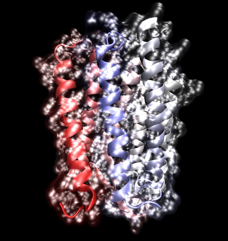
Free with trial Molecular model of the protein, rhodopsin, which is involved in vision. Protein chain illustrations Membrane protein. Molecular model of the protein, rhodopsin, which is involved in vision
Free with trial Electron transport chain as respiratory embedded transporters outline diagram. Labeled educational detailed protein complexes scheme with ATP synthase, NADH and FADH2 process vector illustration. Protein chain vectors Electron transport chain as respiratory embedded transporters outline diagram. Labeled educational detailed protein complexes scheme with ATP synthase, NADH
Free with trial Organic chemistry: model of the protein molecule - illustration of a biological particle. Protein chain illustrations Protein structure. Organic chemistry: model of the protein molecule - illustration of a biological particle
Free with trial Molecular model of HIV protease protein, a target for anti-viral drugs to treat HIV and AIDS. Protein chain illustrations HIV protease protein
Free with trial Protein synthesis / ribosome assemble protein molecules on white background. Protein chain vectors Protein synthesis / ribosome assemble protein molecules
Free with trial Amino acids structure vector illustration. Labeled example of Serine molecule diagram. Closeup with hydrogen, side chain and carboxyl group. Protein builders that are life sustaining macronutrients. Protein chain vectors Amino acids structure vector illustration. Serine molecule diagram. Amino acids structure vector illustration. Labeled example of Serine molecule diagram. Closeup with hydrogen, side chain and carboxyl group. Protein builders that are life sustaining macronutrients.
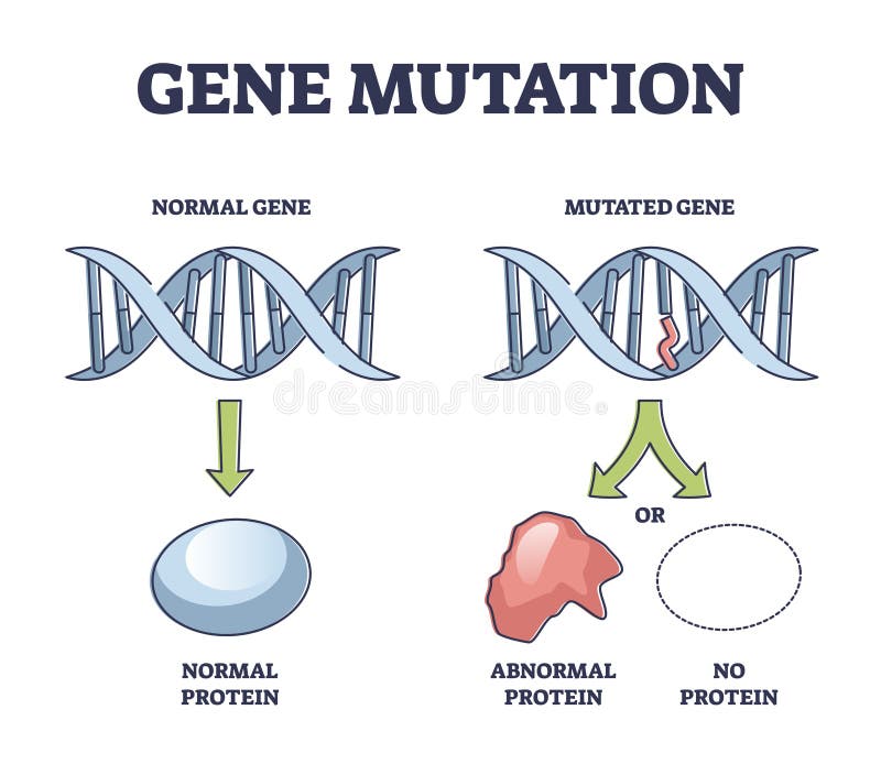
Free with trial Gene mutation models comparison with abnormal helix protein outline diagram. Labeled educational genetic DNA sequence scheme with artificial modification vector illustration. Biological manipulation. Protein chain vectors Gene mutation models comparison with abnormal helix protein outline diagram
Free with trial Rhodopsin (the extremely sensitive to light pigment involved in vision process) protein structure. Protein chain illustrations Rhodopsin protein structure. Rhodopsin (the extremely sensitive to light pigment involved in vision process) protein structure
Free with trial Major Capsid Protein L1 of Human Papilloma Virus type 16 molecular structure. Protein chain illustrations Major Capsid Protein of Human Papilloma Virus. Major Capsid Protein L1 of Human Papilloma Virus type 16 molecular structure
Free with trial Epigenetic mechanisms as DNA acid gene protein expression in outline diagram. Educational labeled scientific scheme with methylation, histone modification and marking process vector illustration. Protein chain vectors Epigenetic mechanisms as DNA acid gene protein expression in outline diagram
Free with trial DNA structure molecule 3D rendering. Exact representation of DNA from protein data bank. Protein chain illustrations DNA
Free with trial Collagen crystal structure molecule 3D rendering. Alpha channel added for easy extraction. Exact representation of collagen 1BKVA0 from protein data bank. Protein chain illustrations Collagen
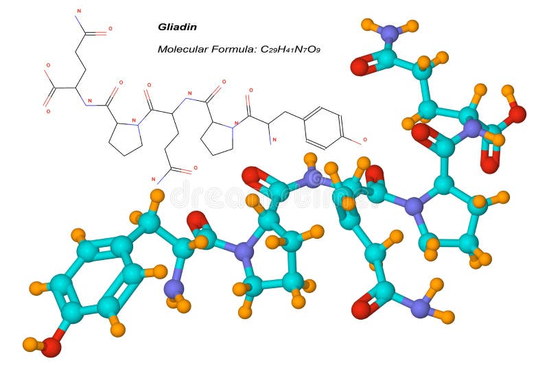
Free with trial Gliadin molecule, component of gluten, is a protein present in wheat and other cereals. It is the toxic factor associated with celiac disease. Protein chain illustrations Gliadin molecule, component of gluten
Free with trial Ribosomes vector illustration. Anatomical and medical labeled scheme with tRNA, Amino acid, protein, cell, small and large subunit, mRNA. Explained closeup Golgi apparatus diagram. Biology basics. Protein chain vectors Ribosomes vector illustration. Anatomical and medical labeled scheme. Explained closeup diagram. Ribosomes vector illustration. Anatomical and medical labeled scheme with tRNA, Amino acid, protein, cell, small and large subunit, mRNA. Explained closeup Golgi apparatus diagram. Biology basics.
Free with trial Organic molecules vector illustration. Labeled chemical educational scheme. Diagram description with monomer, polymer and cellular structure vs carbohydrate, nucleotide, protein and lipid infographic. Protein chain vectors Organic molecules vector illustration. Labeled chemical educational scheme.
Free with trial Computer generated illustration of hemoglobin molecule with cofactor atoms rendered in yellow. Protein chain illustrations Hemoglobin
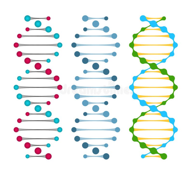
Free with trial Three variants of double strand DNA molecules showing the nucleotide pairs in a double helix vector illustration. Protein chain vectors Three variants of double strand DNA molecules
Free with trial Structure of the Antibody molecule. IgE and Antigen. Vector diagram for medical, educational and science use. Protein chain vectors Structure of the Antibody molecule. IgE and Antigen.
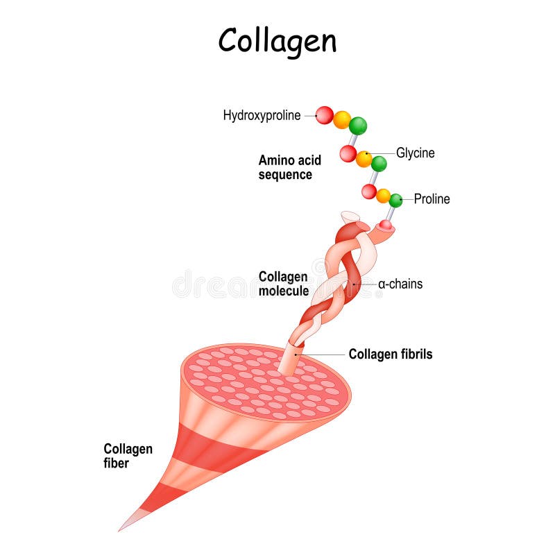
Free with trial Collagen anatomy. Structure of collagen fibers from fibrils and molecule to chains and Amino acid sequence Hydroxyproline, Proline, Glycine. Extracellular matrix. Medical scheme. Vector illustration. Protein chain vectors Collagen Structure. Collagen anatomy. Structure of collagen fibers from fibrils and molecule to chains and Amino acid sequence Hydroxyproline, Proline, Glycine. Extracellular matrix. Medical scheme. Vector illustration
Free with trial Computer generated illustration of hemoglobin molecule. Protein chain illustrations Hemoglobin molecule
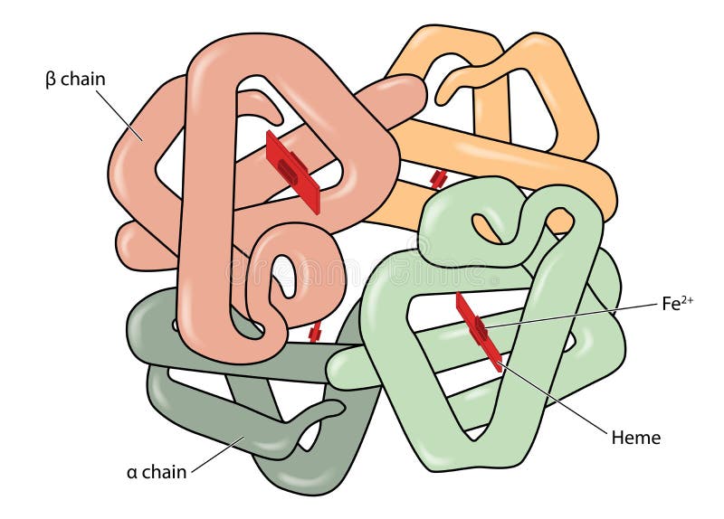
Free with trial Structure of the haemoglobin (hemoglobin) molecule showing alpha and beta chains, heme groups and iron atoms. Created in Adobe Illustrator. EPS 10. Protein chain vectors Haemoglobin molecule. Structure of the haemoglobin (hemoglobin) molecule showing alpha and beta chains, heme groups and iron atoms. Created in Adobe Illustrator. EPS 10
Free with trial Set of nine DNA molecule icons suitable for logo design. Protein chain vectors Set of DNA molecule Icons. Set of nine DNA molecule icons suitable for logo design.
Free with trial Computer generated 3D illustration of deoxyribonucleic acid (DNA). Theme of medicine, genetic engineering, DNA nanotechnology, chemistry, biochemistry, biotechnology, biology. Protein chain illustrations DNA - deoxyribonucleic acid. Computer generated 3D illustration of deoxyribonucleic acid (DNA). Theme of medicine, genetic engineering, DNA nanotechnology, chemistry, biochemistry, biotechnology, biology ...
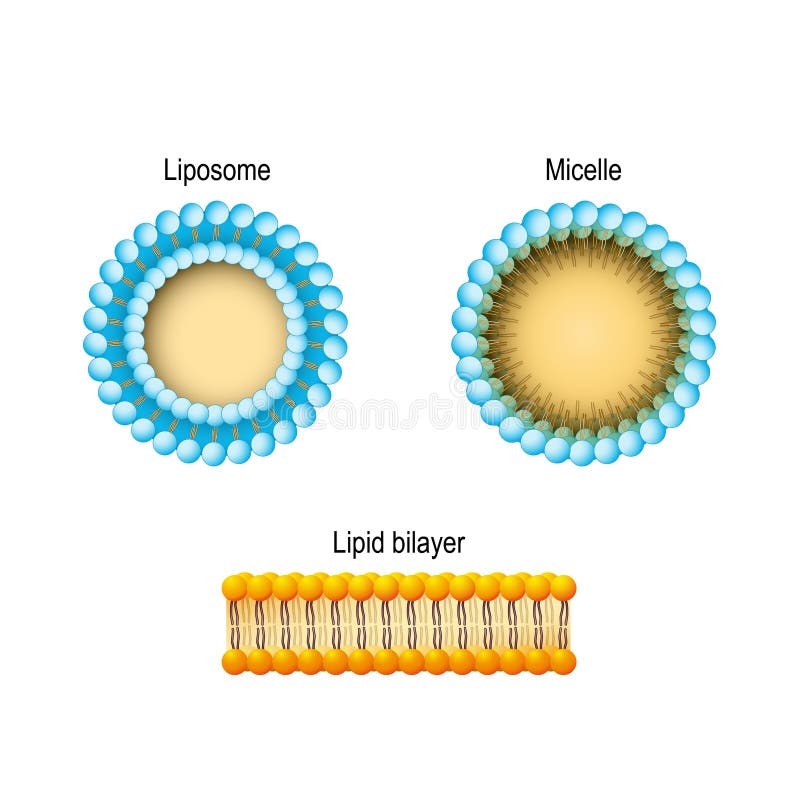
Free with trial Cell membrane Lipid bilayer, Micelle, Liposome. Phospholipids. Protein chain vectors Cell membrane Lipid bilayer, Micelle, Liposome. Phospholipids
Free with trial A nucleosome is a basic unit of DNA packaging in eukaryotes, consisting of a segment of DNA wound in sequence around eight[1] histone protein cores. [2] This structure is often compared to thread wrapped around a spool. Nucleosomes form the fundamental repeating units of eukaryotic chromatin. Protein chain illustrations Nucleosome
Free with trial Computer generated illustration of hemoglobin molecule with heme atoms. Protein chain illustrations Hemoglobin molecule with heme
Free with trial Set of six Infinity Symbol Icons and DNA Molecule Icons. Vector Illustration for Logo Designing. Protein chain vectors Set of Infinity Symbol Icons / DNA Molecule Icons. Set of six Infinity Symbol Icons and DNA Molecule Icons. Vector Illustration for Logo Designing.
Free with trial Chimeric antigen receptor T cell and Antibody molecule. IgE and CAR. Artificial T cell receptors are proteins that have been engineered for cancer therapy. genetically engineered. Vector diagram for medical, educational and science use. Protein chain vectors Chimeric antigen receptor T cell and Antibody molecule. IgE and CAR
Free with trial IgM type antibody molecule pentamer vector illustration. Protein chain vectors IgM antibody pentamer molecule. IgM type antibody molecule pentamer vector illustration
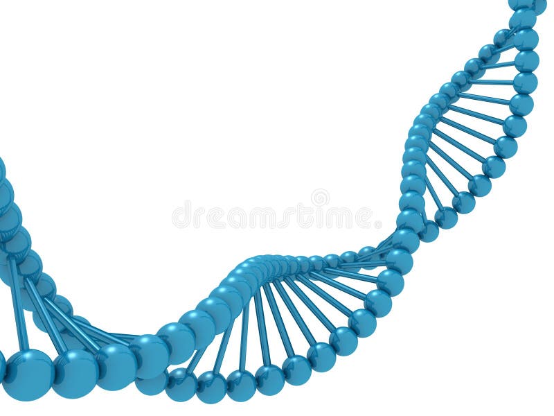
Free with trial Blue model molecule dna helix on white background 3d. Protein chain illustrations Blue model molecule dna helix on white background
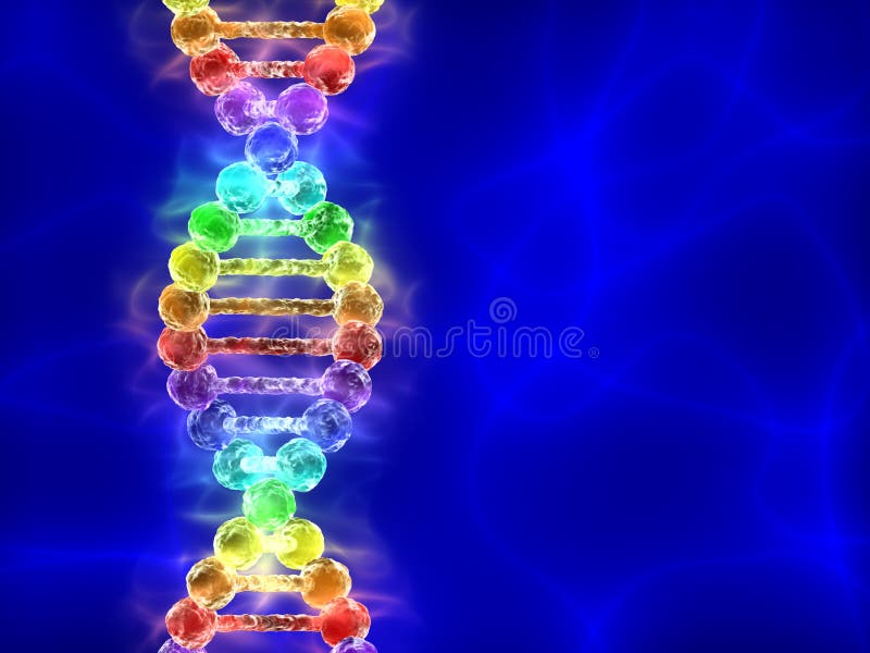
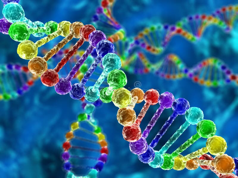
Free with trial Illustration of rainbow DNA (deoxyribonucleic acid) with blue background. Protein chain illustrations Rainbow DNA (deoxyribonucleic acid) on blue background. Illustration of rainbow DNA (deoxyribonucleic acid) with blue background
Free with trial Krebs cycle vector illustration. Cellular respiration labeled outline scheme. Educational diagram with cell, cellular respiration and ATP. Human power molecular metabolism. Protein chain vectors Krebs cycle vector illustration. Cellular respiration labeled outline scheme
Free with trial Amino acids vector illustration. List with food and its essential acids. Healthy and well balanced nutrition meal to get necessary chemical elements like histidine, lysing, valine, leucine and others. Protein chain vectors Amino acids vector illustration. List with food and essential acids. Amino acids vector illustration. List with food and its essential acids. Healthy and well balanced nutrition meal to get necessary chemical elements like histidine, lysing, valine, leucine and others.
Free with trial DNA polymerase I. An enzyme that participates in the DNA replication. Shown with the part of DNA molecule. Protein chain illustrations DNA polymerase I
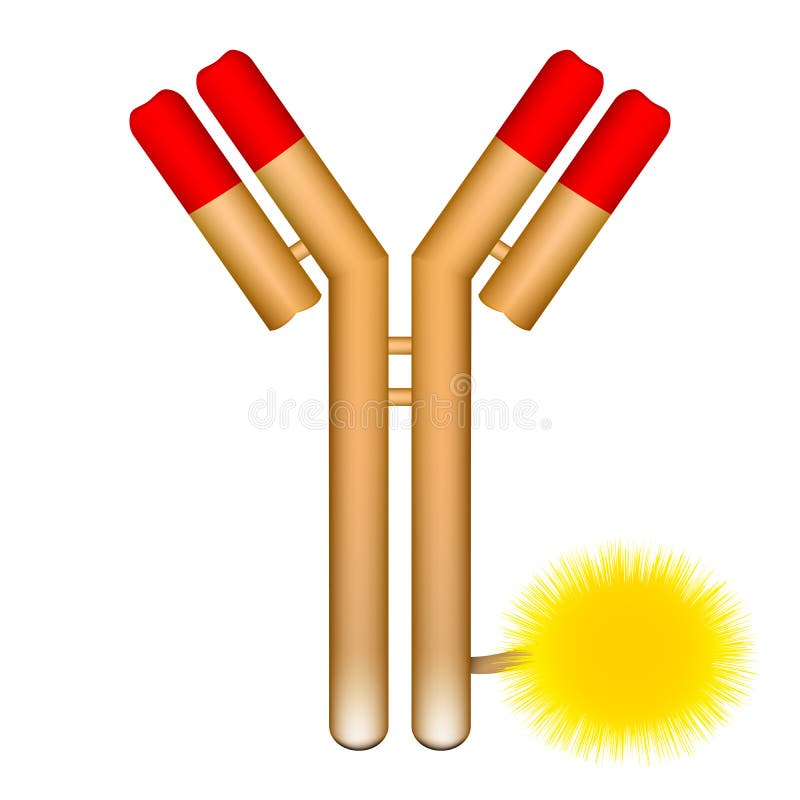
Free with trial Vector illustration of antibody molecule with conjugated fluorophore. Protein chain illustrations Antibody molecule conjugated with fluorophore. Vector illustration of antibody molecule with conjugated fluorophore
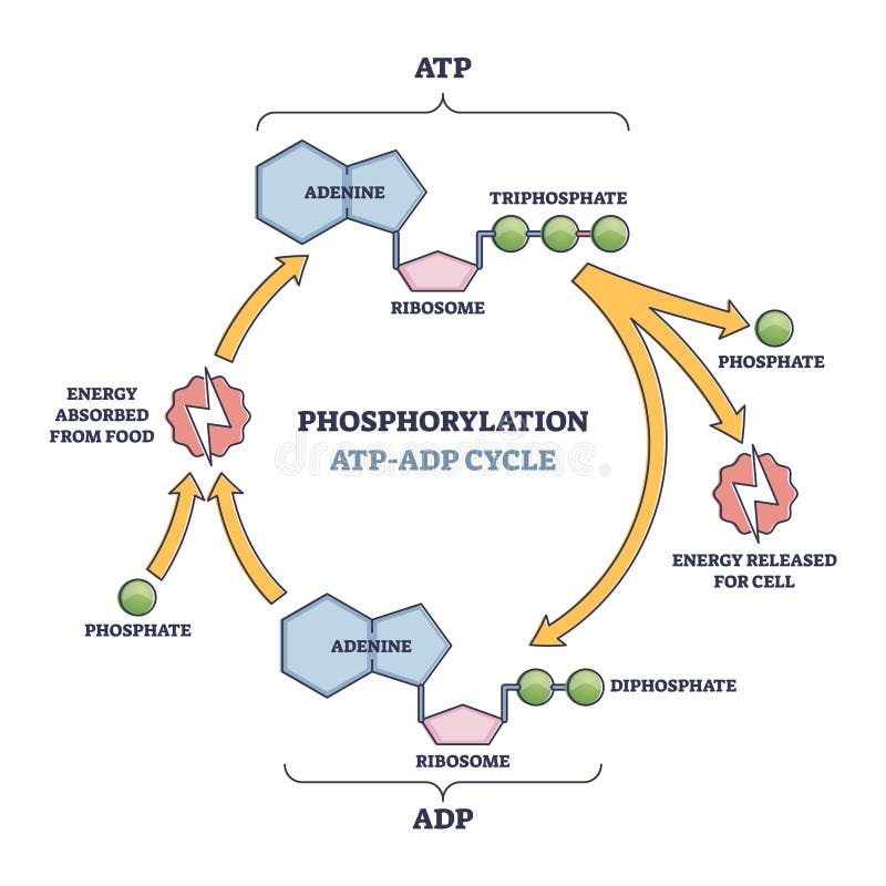
Free with trial Phosphorylation ATP, ADP cycle with detailed process stages outline diagram. Labeled educational energy conversion and absorption from food to make phosphate group bonds formation vector illustration. Protein chain vectors Phosphorylation ATP, ADP cycle with detailed process stages outline diagram
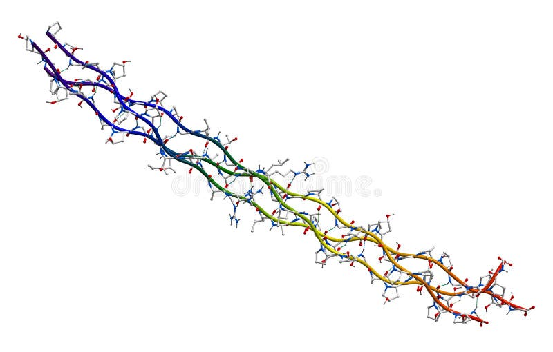
Free with trial Segment of human collagen isolated on a white background. Protein chain illustrations Human collagen molecule (segment). Segment of human collagen isolated on a white background
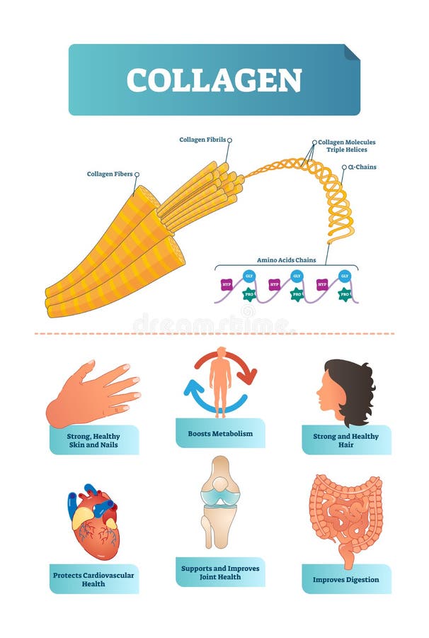
Free with trial Vector illustration about collagen. Metabolism and cardiovascular health diagram. Medical scheme with fibers, fibrils, molecules, helices, alpha and amino acids chains with HYP and GLY visualizations. Protein chain vectors Collagen vector illustration. Scheme with fibers, fibrils, molecules, helices and amino acids chain with HYP and GLY visualisation. Vector illustration about collagen. Metabolism and cardiovascular health diagram. Medical scheme with fibers, fibrils, molecules, helices, alpha and amino acids chains with HYP and GLY visualizations
Free with trial Invertase, an enzyme that catalyzes the hydrolysis (breakdown) of sucrose (table sugar). 3D molecular structure. Protein chain illustrations Enzyme Invertase 3D view. Invertase, an enzyme that catalyzes the hydrolysis (breakdown) of sucrose (table sugar). 3D molecular structure
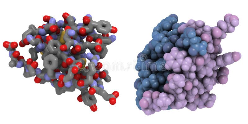
Free with trial Chemical structure of a human insulin molecule. Insuline is used to treat type 1 diabetes. Left: stick representation; right: space-filling representation with coloring by chain. Protein chain illustrations Insulin molecule
Free with trial Alpha-galactosidase (Agalsidase) enzyme. Cause of Fabry's disease. Administered as enzyme replacement therapy. Cartoon & wire representation. Chain gradient coloring. Protein chain illustrations Alpha-galactosidase (Agalsidase) enzyme.
Free with trial Chain of amino acid or biomolecules called protein - 3d illustration. Protein chain illustrations Chain of amino acid or biomolecules called protein
Free with trial Chain of amino acid or biomolecules called protein - 3d illustration. Protein chain illustrations Chain of amino acid or biomolecules called protein
Free with trial Chain of amino acid or biomolecules called protein - 3d illustration. Protein chain illustrations Chain of amino acid or biomolecules called protein
Free with trial Protein syntesis schematic illustration. Vector illustration of the DNA, mRNA and polypeptide chain. Protein chain vectors Protein syntesis schematic illustration. Illustration of the DNA, mRNA and polypeptide chain isolated on white. Protein syntesis schematic illustration. Vector illustration of the DNA, mRNA and polypeptide chain
Free with trial Crystal structure of connector protein from bacteriophage phi29. Protein chain illustrations Protein
Free with trial Protein Digestion. Enzymes proteases and peptidases are digestion breaks the protein into smaller peptide chains and into single amino acids, which are absorbed into the blood. Protein chain vectors Protein Digestion. Enzymes
Free with trial Digestion of protein. Enzymes proteases and peptidases are digestion breaks the protein into smaller peptide chains and into single amino acids, which are absorbed into the blood. Protein chain vectors Digestion of protein
Free with trial Molecular model of the protein, rhodopsin, which is involved in vision. A faulty version of this protein can lead to blindness. Protein chain illustrations Membrane protein on white. Molecular model of the protein, rhodopsin, which is involved in vision. A faulty version of this protein can lead to blindness


![A nucleosome is a basic unit of DNA packaging in eukaryotes, consisting of a segment of DNA wound in sequence around eight[1] histone protein cores. [2] This structure is often compared to thread wrapped around a spool. Nucleosomes form the fundamental repeating units of eukaryotic chromatin. Protein chain illustrations](https://thumbs.dreamstime.com/b/nucleosome-basic-unit-dna-packaging-eukaryotes-consisting-segment-dna-wound-sequence-around-eight-histone-49336255.jpg)