Free with trial Typical fungi cell. Fungal Hyphae. Structure fungi. Diagram illustrating the ultrastructure of a septate hypha. Protist cell wall vectors Fungi cell
Free with trial Spirogyra, cell anatomy of an algae, labeling the cell structures with nucleus, pyrenoid, cytoplasmic strand, cytoplasm, cell membrane, cell wall, mucilaginous sheath, and chloroplasts. Protist cell wall vectors Spirogyra Cell Structures of Algae. Spirogyra, cell anatomy of an algae, labeling the cell structures with nucleus, pyrenoid, cytoplasmic strand, cytoplasm, cell membrane, cell wall, mucilaginous sheath, and chloroplasts.
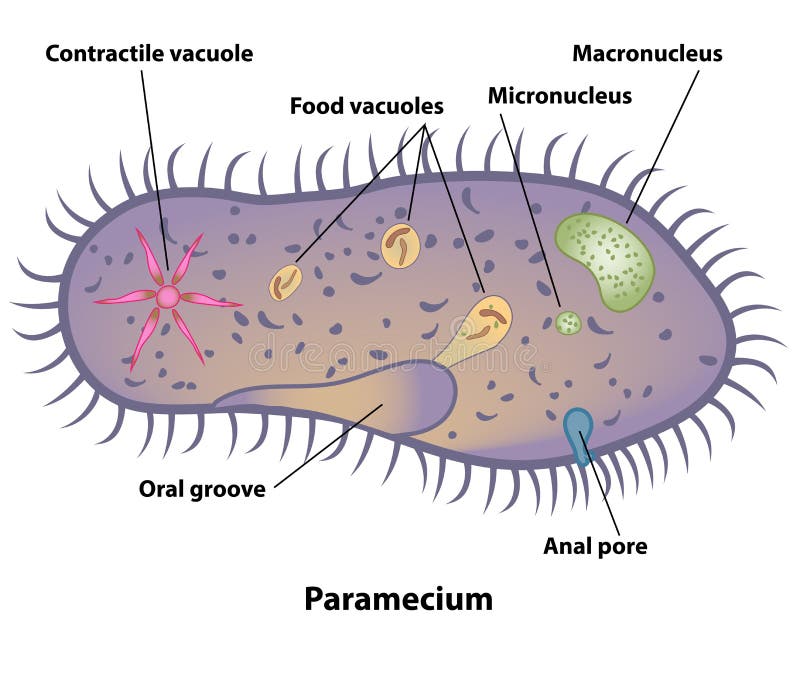
Free with trial Paramecium, cell anatomy of a protozoa, labeling the cell structures with nucleus, oral groove, anal pore, contractile vacuole, and food vacuoles. Protist cell wall vectors Paramecium Cell Structures and Anatomy. Paramecium, cell anatomy of a protozoa, labeling the cell structures with nucleus, oral groove, anal pore, contractile vacuole, and food vacuoles.
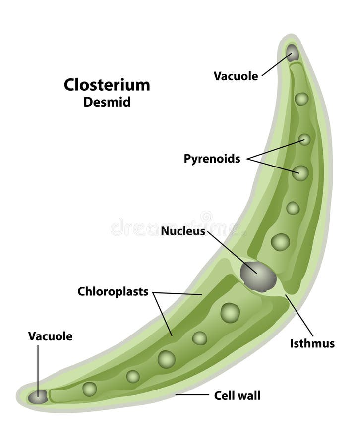
Free with trial Closterium, cell anatomy of a desmid, labeling the cell structures with nucleus, pyrenoid, cell wall, isthmus, vacuole, and chloroplasts. Protist cell wall vectors Closterium Cell Structures and Anatomy. Closterium, cell anatomy of a desmid, labeling the cell structures with nucleus, pyrenoid, cell wall, isthmus, vacuole, and chloroplasts.
Free with trial Euglena, cell anatomy of a protozoa, labeling the cell structures with nucleus, reservoir, photoreceptor, stigma, contractile vacuole, and chloroplasts. Protist cell wall vectors Euglena Protozoan Microscopic Cell Structures. Euglena, cell anatomy of a protozoa, labeling the cell structures with nucleus, reservoir, photoreceptor, stigma, contractile vacuole, and chloroplasts.
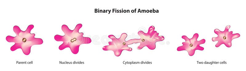
Free with trial Binary fission is a form of asexual reproduction commonly observed in single-celled organisms, such as bacteria and protists. It involves the division of a parent cell into two daughter cells, each of which is genetically identical to the parent cell. Binary fission begins with the replication of the genetic material (DNA) of the parent cell. The DNA molecule, typically a circular chromosome in prokaryotes or a single linear chromosome in some protists, is duplicated to produce two identical copies. Protist cell wall vectors Binary fission
Free with trial A highly magnified image captures a single, intricate diatom with a translucent, silica cell wall known as a frustule, showcasing its complex, symmetrical structure. The central area contains yellowish-brown chloroplasts, viewed against a pale, neutral background under a microscope. Generative AI. Protist cell wall illustrations High Magnification Microscopic View of a Diatom. A highly magnified image captures a single, intricate diatom with a translucent, silica cell wall known as a frustule, showcasing its complex, symmetrical structure. The central area contains yellowish-brown chloroplasts, viewed against a pale, neutral background under a microscope. Generative AI
Free with trial A striking micrograph captures a cluster of amoeba cells, showcasing their complex internal structures. Each amoeba displays a prominent nucleus and visible organelles, some with a distinctive radial arrangement, offering a detailed glimpse into the microscopic world of single-celled organisms. Protist cell wall illustrations Microscopic View of Amoeba Cells with Nucleus and Organelles, Cell Biology. A striking micrograph captures a cluster of amoeba cells, showcasing their complex internal structures. Each amoeba displays a prominent nucleus and visible organelles, some with a distinctive radial arrangement, offering a detailed glimpse into the microscopic world of single-celled organisms.
Free with trial This microscopic image displays two euglena cells, which are single-celled protists. The cells are stained in purple and blue colors, highlighting their internal structures. The nuclei and chloroplasts are clearly visible within each cell. Euglena cells are known for their unique combination of plant-like and animal-like characteristics, as they possess chloroplasts for photosynthesis and can also. Protist cell wall illustrations Microscopic view of two purple and blue stained euglena cells with visible nuclei and chloroplasts. This microscopic image displays two euglena cells, which are. This microscopic image displays two euglena cells, which are single-celled protists. The cells are stained in purple and blue colors, highlighting their internal structures. The nuclei and chloroplasts are clearly visible within each cell. Euglena cells are known for their unique combination of plant-like and animal-like characteristics, as they possess chloroplasts for photosynthesis and can also
Free with trial Explore the intricate world of cells with these captivating abstract illustrations. Perfect for educational posters, these visuals bring complex biological concepts to life, making learning engaging and memorable. From the microscopic details of organelles like mitochondria and ribosomes to the larger structures of cells and tissues, these illustrations offer a unique perspective on cellular. Protist cell wall illustrations Visualizing Cellular Complexity Abstract Biological Illustrations for Educational Posters on Cell Structures and. Explore the intricate world of cells with these captivating abstract illustrations. Perfect for educational posters, these visuals bring complex biological concepts to life, making learning engaging and memorable. From the microscopic details of organelles like mitochondria and ribosomes to the larger structures of cells and tissues, these illustrations offer a unique perspective on cellular
Free with trial A stunning micrograph reveals the intricate beauty of a diatom colony, their golden-brown cells arranged in a delicate arc. This glimpse into the unseen world of plankton highlights the complex structures and patterns of life at a microscopic level, perfect for scientific or educational themes. Protist cell wall illustrations Microscopic View of a Diatom Algae Colony. A stunning micrograph reveals the intricate beauty of a diatom colony, their golden-brown cells arranged in a delicate arc. This glimpse into the unseen world of plankton highlights the complex structures and patterns of life at a microscopic level, perfect for scientific or educational themes
Free with trial A whimsical collection of hand-drawn microbes, viruses, and bacteria, perfect for educational materials or fun designs. The illustrations feature various shapes and expressions. Protist cell wall vectors Collection of Cute Hand-Drawn Microbes, Viruses, and Bacteria Illustrations. A whimsical collection of hand-drawn microbes, viruses, and bacteria, perfect for educational materials or fun designs. The illustrations feature various shapes and expressions.
Free with trial This breathtaking aerial photograph showcases the astonishing detail of a single-celled amoeba. The vibrant colors and dramatic lighting highlight the intricate cellular structures, revealing the amoeba cytoplasm, nucleus, and characteristic pseudopods. Observe the dynamic movement of this microscopic organism as it extends its pseudopods, capturing prey through phagocytosis. Notice the. Protist cell wall illustrations Unveiling the Intricate World of Amoebas Stunning Aerial Photography Reveals Microscopic Details. This breathtaking aerial photograph showcases the astonishing detail of a single-celled amoeba. The vibrant colors and dramatic lighting highlight the intricate cellular structures, revealing the amoeba cytoplasm, nucleus, and characteristic pseudopods. Observe the dynamic movement of this microscopic organism as it extends its pseudopods, capturing prey through phagocytosis. Notice the
Free with trial Delve into the mesmerizing microcosm of life with this captivating microscopic image. Vibrant green and blue hues highlight the intricate structures within organic biological cells, showcasing the beauty and complexity of cellular processes. Observe the detailed textures and dynamic nature of growth and division. This scientific illustration reveals the diversity of cellular life, from the. Protist cell wall illustrations Unveiling the Intricate World of Organic Cells A Microscopic Exploration of Vibrant Green and Blue Biological. Delve into the mesmerizing microcosm of life with this captivating microscopic image. Vibrant green and blue hues highlight the intricate structures within organic biological cells, showcasing the beauty and complexity of cellular processes. Observe the detailed textures and dynamic nature of growth and division. This scientific illustration reveals the diversity of cellular life, from the
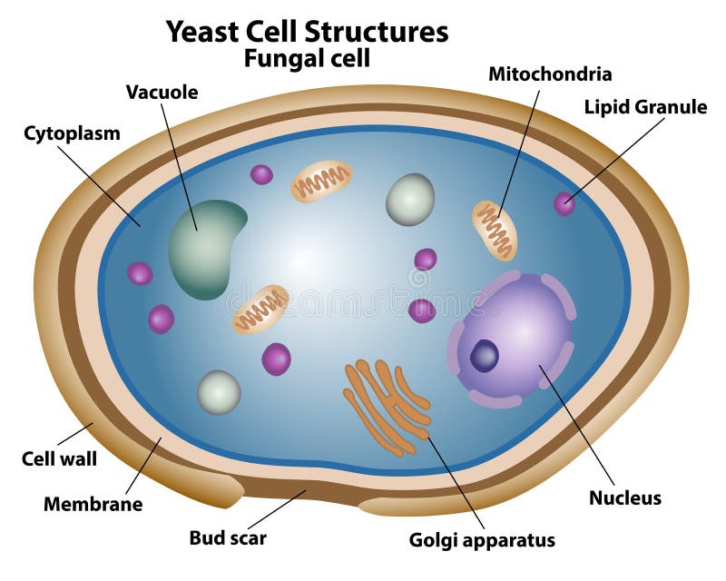
Free with trial Yeast Cell Structures, anatomy of a fungal cell, labeling the nucleus, Golgi apparatus, cell wall, membrane, vacuole, cytoplasm, bud scar, lipid granule, and mitochondria. Protist cell wall vectors Yeast Structures of a Fungal Cell. Yeast Cell Structures, anatomy of a fungal cell, labeling the nucleus, Golgi apparatus, cell wall, membrane, vacuole, cytoplasm, bud scar, lipid granule, and mitochondria.