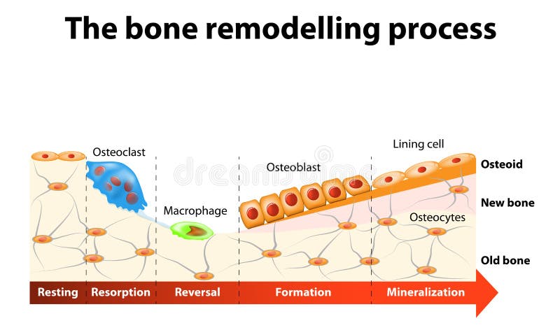
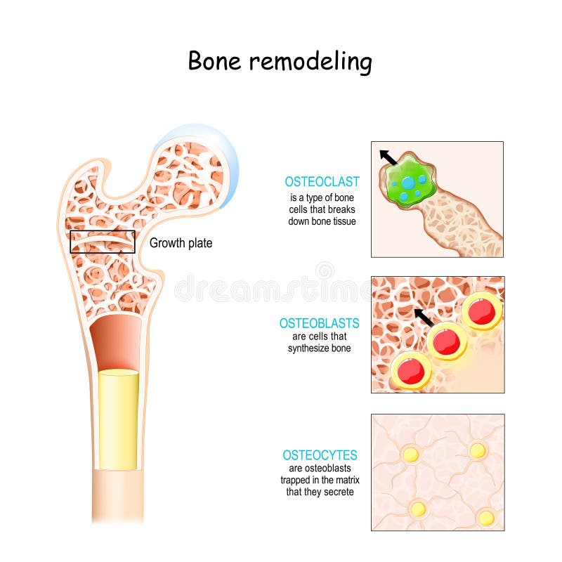
Free with trial The bone remodeling process involves the following steps: resorption, reversal, formation, mineralization and resting. In a healthy body, osteoclasts and osteoblasts work together to maintain the balance between bone loss and bone formation. Section lining vectors Bone remodelling process. The bone remodeling process involves the following steps: resorption, reversal, formation, mineralization and resting. In a healthy. The bone remodeling process involves the following steps: resorption, reversal, formation, mineralization and resting. In a healthy body, osteoclasts and osteoblasts work together to maintain the balance between bone loss and bone formation.
Free with trial Urinary bladder cancer. Stages of urinary bladder cancer, eps8. Section lining vectors Urinary bladder cancer. Stages of urinary bladder cancer, eps8
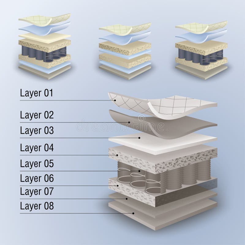
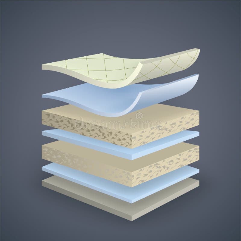
Free with trial Vector mattress section on layers. Section lining vectors Vector mattress section on layers. Vector set mattress section on layers
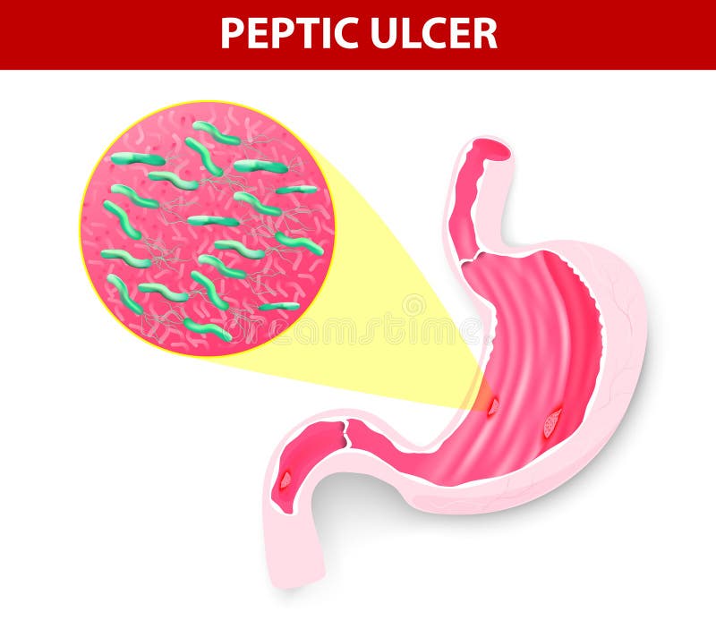
Free with trial Peptic ulcer. A peptic ulcer is a sore on the lining of the stomach or duodenum. Cause of peptic ulcer is bacterial infection. Helicobacter pylori. Vector. Section lining vectors Peptic ulcer. A peptic ulcer is a sore on the lining of the stomach or duodenum. Cause of peptic ulcer is bacterial infection. Helicobacter pylori. Vector
Free with trial Peptic ulcer and helicobacter pylori infographic. With symptoms and causes, stomach cross section diagram, medical illustration. Section lining vectors Peptic ulcer and helicobacter pylori infographic. With symptoms and causes, stomach cross section diagram, medical illustration
Free with trial Small intestine and stomach anatomy. Healthy small intestine and stomach, bright anatomy illustration, white background. Section lining vectors Small intestine and stomach anatomy. Healthy small intestine and stomach, bright anatomy illustration, white background
Free with trial Stomach disorder concept. Fire in human stomach, isolated on a white background. Section lining vectors Stomach disorder concept. Fire in human stomach, isolated on a white background
Free with trial Osteoblast and osteoclast. The bone remodeling process. In a healthy body, osteoclasts and osteoblasts work together to maintain the balance between bone loss and bone formation. Section lining vectors Osteoblast and osteoclast. The bone remodeling process. In a healthy body, osteoclasts and osteoblasts work together to maintain the balance between bone loss
Free with trial Stomach wall. Layers detailed anatomy, beautiful colorful drawing on a white background. Section lining vectors Stomach wall. Layers detailed anatomy, beautiful colorful drawing on a white background
Free with trial Wood slice. Tree rings. Watercolor illustration. Painted texture. Section lining illustrations Wood slice. Tree rings. Watercolor illustration. Painted texture
Free with trial Cross section of two stomach: healthy and Gastritis inflammation of the lining of the stomach. Human anatomy. Vector diagram for medical, science, and educational use. Section lining vectors Gastritis. Cross section of two stomach. Cross section of two stomach: healthy and Gastritis inflammation of the lining of the stomach. Human anatomy. Vector. Cross section of two stomach: healthy and Gastritis inflammation of the lining of the stomach. Human anatomy. Vector diagram for medical, science, and educational use.
Free with trial Stomach erosion and peptic ulcer. Stages infographic: stomach lining and mucosa cross section diagram, medical illustration. Section lining vectors Stomach erosion and peptic ulcer. Stages infographic: stomach lining and mucosa cross section diagram, medical illustration
Free with trial Stomach lining. Stomach cross section anatomy and surrounding organs beautiful colorful drawing. Section lining vectors Stomach lining. Stomach cross section anatomy and surrounding organs beautiful colorful drawing
Free with trial Nasal mucosa anatomy. Nasal mucous membrane lining the respiratory tract. Ciliated, basal and goblet cells. Flat vector illustration. Section lining vectors Nasal mucosa anatomy. Nasal mucous membrane lining the respiratory tract. Ciliated, basal and goblet cells. Flat vector illustration
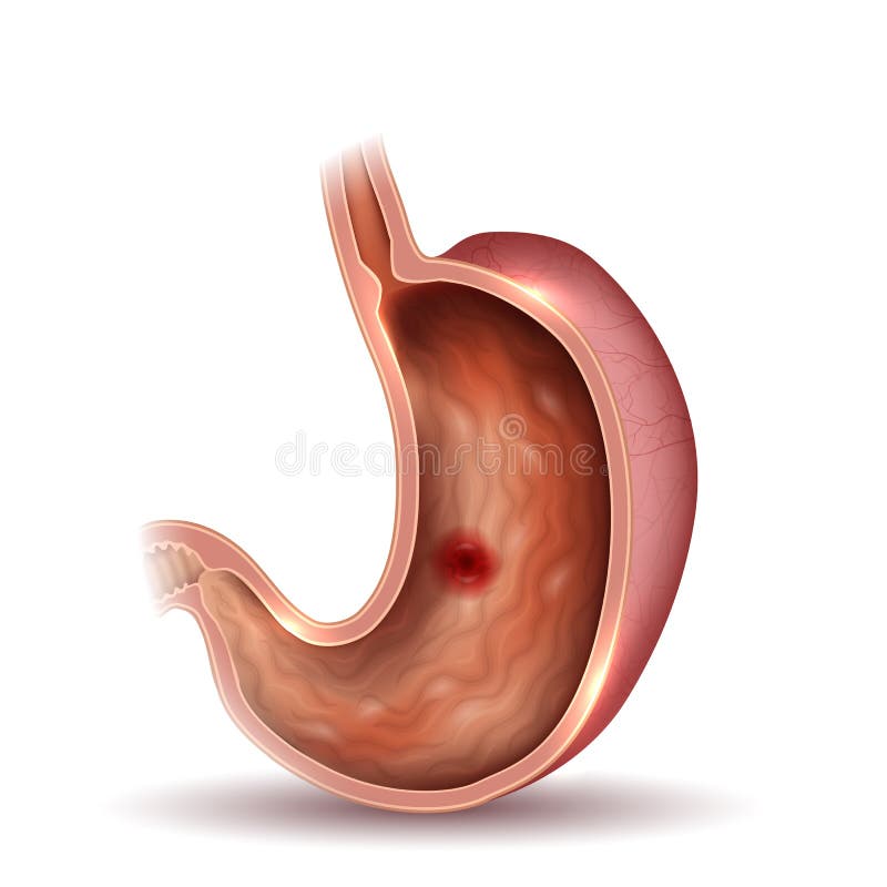
Free with trial Stomach ulcer, interanl organs anatomy colorful drawing. Stomach internal lining and ulcer, colorful drawing. Section lining vectors Stomach ulcer, interanl organs anatomy colorful drawing. Stomach internal lining and ulcer, colorful drawing
Free with trial Peptic ulcer and helicobacter pylori. Helicobacter pylori infecting stomach, causing mucosal damage and peptic ulcer, medical illustration. Section lining vectors Peptic ulcer and helicobacter pylori. Helicobacter pylori infecting stomach, causing mucosal damage and peptic ulcer, medical illustration
Free with trial Ulcer. Stomach ulcer, interanal organs anatomy colorful drawing. Section lining vectors Ulcer. Stomach ulcer, interanal organs anatomy colorful drawing
Free with trial Stomach. 3d rendered anatomy illustration of a human stomach. Section lining illustrations Stomach. 3d rendered anatomy illustration of a human stomach
Free with trial Peptic ulcer. Of the stomach, microscopic colorful anatomy on a white background. Section lining vectors Peptic ulcer. Of the stomach, microscopic colorful anatomy on a white background
Free with trial Intestinal wall anatomy. Intestinal wall medical science vector illustration. Internal human organ: mucosa, muscularis externa, serosa and villi. Human intestinal wall anatomy education illustration. Section lining vectors Intestinal wall anatomy. Intestinal wall medical science vector illustration. Internal human organ: mucosa, muscularis externa, serosa and villi. Human
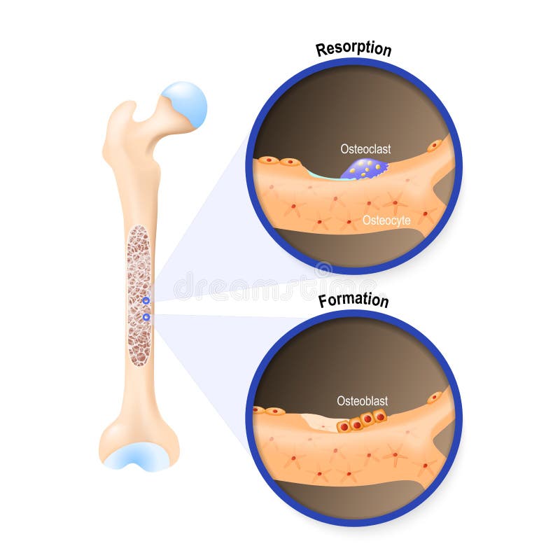
Free with trial Bone remodeling. Close-up of a Osteoclast resorbs bone, and Osteoblasts synthesized bone tissue. Vector illustration. Section lining vectors Bone remodeling. Osteoclast resorbs bone, and Osteoblasts synthesized bone tissue. Bone remodeling. Close-up of a Osteoclast resorbs bone, and Osteoblasts. Bone remodeling. Close-up of a Osteoclast resorbs bone, and Osteoblasts synthesized bone tissue. Vector illustration
Free with trial Bone remodeling process resorption, reversal, formation, and mineralization. Osteoblast, osteoclast, and osteocyte. Vector illustration of human bone cell types. Medical diagram. Section lining vectors Bone remodeling process. Osteoblast, osteoclast, and osteocyte. Bone remodeling process resorption, reversal, formation, and mineralization. Osteoblast. Bone remodeling process resorption, reversal, formation, and mineralization. Osteoblast, osteoclast, and osteocyte. Vector illustration of human bone cell types. Medical diagram
Free with trial Small intestine and stomach anatomy. Healthy small intestine and stomach, bright anatomy illustration, white background. Section lining vectors Small intestine and stomach anatomy. Healthy small intestine and stomach, bright anatomy illustration, white background
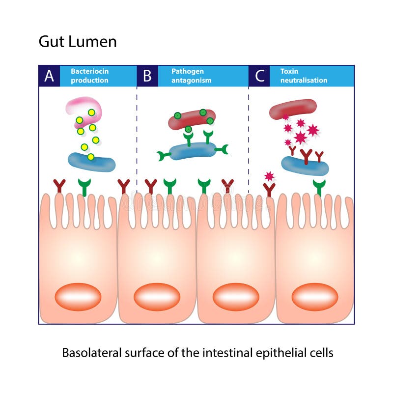
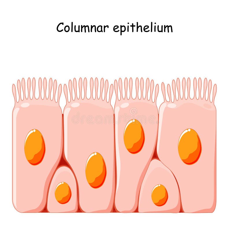
Free with trial Gut Lumen. Columnar epithelial cells. Gut lumen. Enterocytes, or intestinal absorptive cells. Small intestine. Section lining vectors Gut Lumen. Columnar epithelial cells. Gut lumen. Enterocytes, or intestinal absorptive cells. Small intestine. Columnar epithelial cells
Free with trial Intestinal villi in the surface area of intestinal walls. Folds, villus, microvilli, epithelial and goblet cells. Small intestine anatomical poster. Digestive system medical flat vector illustration. Section lining vectors Intestinal villi anatomy. Intestinal villi in the surface area of intestinal walls. Folds, villus, microvilli, epithelial and goblet cells. Small intestine. Intestinal villi in the surface area of intestinal walls. Folds, villus, microvilli, epithelial and goblet cells. Small intestine anatomical poster. Digestive system medical flat vector illustration.
Free with trial Vector mattress section on layers. Vector mattress divided into layers with materials and shadows. Section lining vectors Vector mattress section on layers. Vector mattress divided into layers with materials and shadows
Free with trial Vector mattress section on layers. Vector mattress divided into layers with materials and shadows. Section lining vectors Vector mattress section on layers. Vector mattress divided into layers with materials and shadows
Free with trial Section 3d of the stomach. Peptic ulcer disease, gastric ulcer is a break in the inner lining of the stomach. Anatomy. 3d rendering. Section lining illustrations Section 3d of the stomach. Peptic ulcer disease, gastric ulcer is a break in the inner lining of the stomach. Anatomy. 3d rendering
Free with trial Celiac disease. Cross section of Small intestine lining damage. good and damaged villi. leaky gut. Coeliac sprue disease: an autoimmune disorder against gluten gliadin. cells on gut lining held tightly together. Vector diagram. Section lining vectors Celiac disease. Cross section of Small intestine. Lining damage. good and damaged villi. leaky gut. Coeliac sprue disease: an autoimmune disorder against gluten. Celiac disease. Cross section of Small intestine lining damage. good and damaged villi. leaky gut. Coeliac sprue disease: an autoimmune disorder against gluten gliadin. cells on gut lining held tightly together. Vector diagram
Free with trial Uterine fibroids cross section illustration shows large submucosal leiomyoma protruding into endometrial cavity, thickened lining and heavy menstrual bleeding visualization, medical education. Section lining illustrations Uterine fibroids cross section illustration shows large submucosal leiomyoma protruding into endometrial cavity, thickened lining. And heavy menstrual bleeding. Uterine fibroids cross section illustration shows large submucosal leiomyoma protruding into endometrial cavity, thickened lining and heavy menstrual bleeding visualization, medical education
Free with trial Peptic ulcer medical illustration. Stomach and peptic ulcer: inflamed sore on the stomach mucosa, stomach lining cross section diagram, medical illustration. Section lining vectors Peptic ulcer medical illustration. Stomach and peptic ulcer: inflamed sore on the stomach mucosa, stomach lining cross section diagram, medical illustration
Free with trial Stomach cross section. Anatomy beautiful colorful drawing on a white background. Section lining vectors Stomach cross section. Anatomy beautiful colorful drawing on a white background
Free with trial Stomach lining. Stomach cross section anatomy and surrounding organs on an abstract hexagon background. Section lining vectors Stomach lining. Stomach cross section anatomy and surrounding organs on an abstract hexagon background
Free with trial Small intestine Anatomy. Cross section of a ileum with Internal villi. Close-up of Epithelial cells with Microvilli. Vector illustration. Section lining vectors Small intestine Anatomy. Cross section of a ileum with Internal villi. Close-up of Epithelial cells with Microvilli. Vector illustration
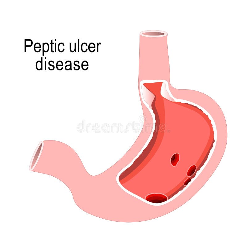
Free with trial Peptic ulcer disease. Cross section of the humans stomach with ulcers inside. flat illustration for medical, science, and educational use. digestive system of the human. Section lining vectors Peptic ulcer disease. Cross section of the humans stomach. With ulcers inside. flat illustration for medical, science, and educational use. digestive system of
Free with trial Stomach lining close up anatomy, beautiful drawing on an abstract blue background. Section lining vectors Stomach lining close up anatomy, beautiful drawing on an abstract blue background
Free with trial Bone remodeling. Cross section of joint. Close-up of Osteoblasts that Synthesize bone tissue, and Osteoclast removes bone tissue. Vector illustration. Medical diagram. Section lining vectors Bone remodeling. Cross section of joint. Close-up of Osteoblasts that Synthesize bone tissue, and Osteoclast removes bone tissue. Vector illustration. Medical
Free with trial Stomach and other internal organs. Stomach cross section anatomy and surrounding organs beautiful colorful drawing. Section lining vectors Stomach and other internal organs. Stomach cross section anatomy and surrounding organs beautiful colorful drawing
Free with trial Ciliated columnar epithelium. epithelial cells forms the lining of the stomach and intestines, duodenum, fallopian tubes, uterus, central canal of the spinal cord, nose, ears and the taste buds. Section lining vectors Ciliated columnar epithelium. Epithelial cells forms the lining of the stomach and intestines, duodenum, fallopian tubes, uterus, central canal of the spinal
Free with trial Small intestine. general view and cross-section. Human anatomy. Section lining vectors Small intestine. Human anatomy. Small intestine. general view and cross-section. Human anatomy. Small intestine. general view and cross-section. Human anatomy.
Free with trial Gastritis. Stomach Inflammation. Gastrointestinal Disorders. Cross section of a human stomach and inflammation of the lining of the stomach under Magnifying glass. Vector illustration. Section lining vectors Gastritis. Stomach Inflammation. Gastrointestinal Disorders. Cross section of a human stomach and inflammation of the lining of the stomach under Magnifying
Free with trial Small intestine with scientific gastrointestinal tract structure outline diagram. Labeled educational anatomy explanation with fold of intestinal lining, microvilli and intestine vector illustration. Section lining vectors Small intestine with scientific gastrointestinal structure outline diagram. Small intestine with scientific gastrointestinal tract structure outline diagram. Small intestine with scientific gastrointestinal tract structure outline diagram. Labeled educational anatomy explanation with fold of intestinal lining, microvilli and intestine vector illustration.
Free with trial An anatomical cross-section illustration of the human nasal cavity, specifically showing the turbinate structures and the lining of the nasal mucosa. Section lining illustrations Cross-section view of human nasal turbinate and nasal mucosa anatomy with turbinate. An anatomical cross-section illustration of the human nasal cavity. An anatomical cross-section illustration of the human nasal cavity, specifically showing the turbinate structures and the lining of the nasal mucosa
Free with trial Scientifically accurate medical illustration of the 28-day menstrual cycle showing ovarian hormone fluctuations, ovulation timing, follicular phase, luteal phase, and endometrial changes throughout the reproductive cycle. Features detailed female reproductive anatomy with uterine cross-section, ovarian development stages, and hormonal regulation. Educational healthcare infographic isolated on a white background, ideal for medical textbooks, gynecology education, fertility awareness materials, healthcare publications, scientific presentations, and pharmaceutical marketing. Section lining illustrations 28-Day Menstrual Cycle Medical Infographic with Ovarian Hormones. Scientifically accurate medical illustration of the 28-day menstrual cycle showing ovarian. Scientifically accurate medical illustration of the 28-day menstrual cycle showing ovarian hormone fluctuations, ovulation timing, follicular phase, luteal phase, and endometrial changes throughout the reproductive cycle. Features detailed female reproductive anatomy with uterine cross-section, ovarian development stages, and hormonal regulation. Educational healthcare infographic isolated on a white background, ideal for medical textbooks, gynecology education, fertility awareness materials, healthcare publications, scientific presentations, and pharmaceutical marketing.
Free with trial Gut flora diagram contrasting healthy vs unhealthy microbiota, central intestinal cross-section, bacteria, and food icons show balance versus dysbiosis. Outline diagram. Section lining vectors Gut flora diagram contrasting healthy vs unhealthy microbiota, central intestinal ... Gut flora diagram contrasting healthy vs unhealthy microbiota, central. Gut flora diagram contrasting healthy vs unhealthy microbiota, central intestinal cross-section, bacteria, and food icons show balance versus dysbiosis. Outline diagram
Free with trial Small intestine anatomical poster. Surface area of intestinal walls. Intestinal villi, cross section, fold, villus and epithelial cells. Digestive system medical flat vector illustration in human body. Section lining vectors Small intestine anatomy. Small intestine anatomical poster. Surface area of intestinal walls. Intestinal villi, cross section, fold, villus and epithelial cells. Small intestine anatomical poster. Surface area of intestinal walls. Intestinal villi, cross section, fold, villus and epithelial cells. Digestive system medical flat vector illustration in human body
Free with trial Intestinal villi diagram. Surface area of intestinal walls. Small intestine cross section, fold, villus, microvilli and epithelial cells. Digestive system medical flat vector illustration for clinic. Section lining vectors Intestinal villi concept. Intestinal villi diagram. Surface area of intestinal walls. Small intestine cross section, fold, villus, microvilli and epithelial. Intestinal villi diagram. Surface area of intestinal walls. Small intestine cross section, fold, villus, microvilli and epithelial cells. Digestive system medical flat vector illustration for clinic
Free with trial Vector set mattress section on layers. Set mattress section on layers. Section lining vectors Vector set mattress section on layers. Set mattress section on layers
Free with trial Vector set mattress section on layers. Set mattress section on layers. Section lining vectors Vector set mattress section on layers. Set mattress section on layers
Free with trial Celiac disease. Damaged and normal Intestinal villi in surface area of intestinal walls. Small intestine cross section, microvilli and epithelial cells. Digestive system medical vector illustration. Section lining vectors Celiac disease inflammation. Celiac disease. Damaged and normal Intestinal villi in surface area of intestinal walls. Small intestine cross section, microvilli. Celiac disease. Damaged and normal Intestinal villi in surface area of intestinal walls. Small intestine cross section, microvilli and epithelial cells. Digestive system medical vector illustration
Free with trial Wood texture Seamless. Generated Wood texture. Lining boards wall. Wooden background pattern. Showing growth rings, Seamless. Section lining illustrations Wood texture Seamless. Generated Wood texture. Lining boards wall. Wooden background pattern. Showing growth rings, Seamless
Free with trial Wood texture Seamless. Generated Wood texture. Lining boards wall. Wooden background pattern. Showing growth rings, Seamless. Section lining illustrations Wood texture Seamless. Generated Wood texture. Lining boards wall. Wooden background pattern. Showing growth rings, Seamless
Free with trial Illustration of the female reproductive system showing the uterine lining. The illustration features a cross-section revealing the layers and blood vessels of the endometrium. The image is in soft pink and light red tones. The graphic style is precise, detailed, and academic. The background is plain white. This medically accurate illustration is suitable for educational purposes. Section lining illustrations Female reproductive system endometrium illustration. Illustration of the female reproductive system showing the uterine lining. The illustration features a cross. Illustration of the female reproductive system showing the uterine lining. The illustration features a cross-section revealing the layers and blood vessels of the endometrium. The image is in soft pink and light red tones. The graphic style is precise, detailed, and academic. The background is plain white. This medically accurate illustration is suitable for educational purposes
Free with trial Medical illustration of stomach lining with bacteria. The image displays an anatomical cross-section of the human stomach, showing internal structures and a cluster of bacteria in a reddish-orange area. The lighting is clinical, highlighting the details of the bacteria and internal organs. The illustration style is detailed and realistic, but not photorealistic. This medical image could be useful. Section lining illustrations Stomach bacteria illustration. Medical illustration of stomach lining with bacteria. The image displays an anatomical cross-section of the human stomach, showing. Medical illustration of stomach lining with bacteria. The image displays an anatomical cross-section of the human stomach, showing internal structures and a cluster of bacteria in a reddish-orange area. The lighting is clinical, highlighting the details of the bacteria and internal organs. The illustration style is detailed and realistic, but not photorealistic. This medical image could be useful
Free with trial A hyperrealistic, high-resolution anatomical cross-section of the human microcirculation system, showcasing the flow of red and white blood cells through plasma-filled capillaries. The highly detailed vessel walls reveal the delicate endothelial structures and subtle textures of the vascular lining, emphasizing the complexity of microcirculatory dynamics. Soft subsurface lighting enhances depth and realism, illuminating the biological textures with scientific accuracy. The color palette consists of rich reds, soft blues, and translucent plasma tones, creating an immersive medical visualization. The perspective provides a microscopic yet anatomically precise representation of the circulatory system, making it perfect for medical education, research, and artistic interpretation of human physiology. Section lining illustrations Anatomical Cross-Section of the Human Microcirculation System. A hyperrealistic, high-resolution anatomical cross-section of the human microcirculation system. A hyperrealistic, high-resolution anatomical cross-section of the human microcirculation system, showcasing the flow of red and white blood cells through plasma-filled capillaries. The highly detailed vessel walls reveal the delicate endothelial structures and subtle textures of the vascular lining, emphasizing the complexity of microcirculatory dynamics. Soft subsurface lighting enhances depth and realism, illuminating the biological textures with scientific accuracy. The color palette consists of rich reds, soft blues, and translucent plasma tones, creating an immersive medical visualization. The perspective provides a microscopic yet anatomically precise representation of the circulatory system, making it perfect for medical education, research, and artistic interpretation of human physiology.
Free with trial Respiratory Epithelium Cell Structure Illustration. Ciliated, Basal, and Goblet Cells Diagram. Mucociliary Epithelium. Cross Section Illustration. Section lining vectors Respiratory Epithelium Cell Structure Illustration. Ciliated, Basal, and Goblet Cells Diagram. Mucociliary Epithelium. Cross Section Illustration
Free with trial Stomach health care. 3d illustration abstract design, human silhouette with internal organs and normal cardiogram at the bottom. Section lining vectors Stomach health care. 3d illustration abstract design, human silhouette with internal organs and normal cardiogram at the bottom
Free with trial Stomach wall isolated on white. Stomach wall layers detailed anatomy, beautiful colorful drawing on a white background. Section lining vectors Stomach wall isolated on white. Stomach wall layers detailed anatomy, beautiful colorful drawing on a white background
Free with trial Intestinal villus. Different between Villi and microvilli. Cross section of a duodenum with submucosa, mucosa and muscularis layers. Vertical section of a villus with Lymph capillary or Lacteal and Blood vessels. Close-up of a Columnar epithelium, and enterocytes. Vector illustration. Section lining vectors Intestinal villus. Different between Villi and microvilli. Cross section of a duodenum with submucosa, mucosa and muscularis layers. Vertical section of a
Free with trial A osteoclast. 3d rendered medically accurate illustration of a osteoclast. Section lining illustrations A osteoclast. 3d rendered medically accurate illustration of a osteoclast
Free with trial Stomach ulcer, internal organs anatomy colorful drawing on an abstract scientific DNA background. Section lining vectors Stomach ulcer. Internal organs anatomy colorful drawing on an abstract scientific DNA background
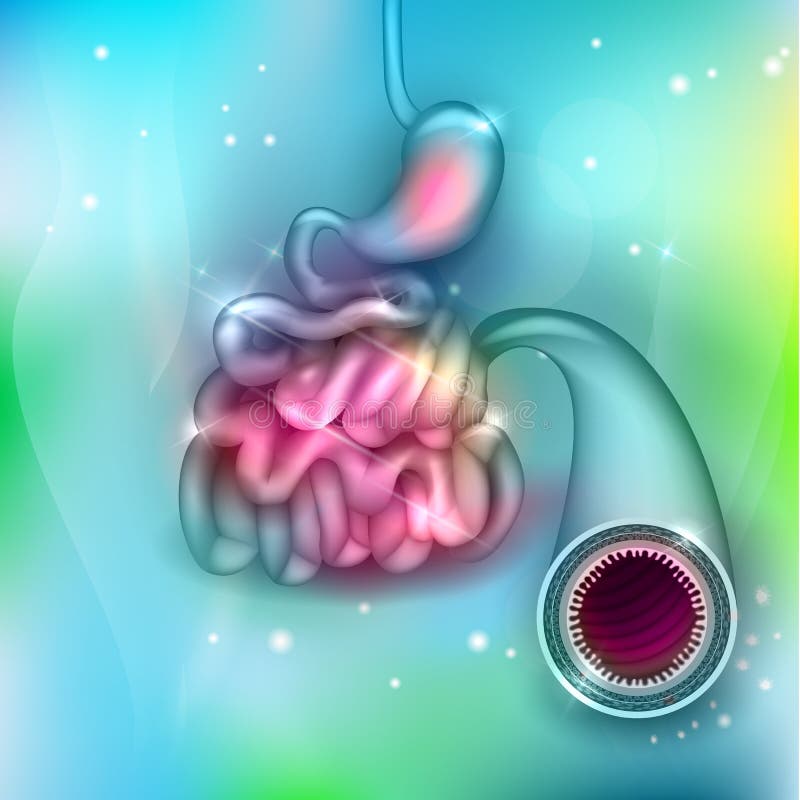
Free with trial Small bowel and stomach. On an abstract light blue mesh background. Section lining vectors Small bowel and stomach. On an abstract light blue mesh background
Free with trial The small intestine is made up of thee sections: the duodenum, the jejunum and the ileum. On its proximal end, the small intestine—beginning with the duodenum—connects to the stomach. On its distal end, the ileum—the last segment of the small intestine—connects to the large intestine. Section lining illustrations Small intestine anatomical structure. Villi and epithelial cell with microvilli illustrations. The small intestine is made up of thee sections: the duodenum. The small intestine is made up of thee sections: the duodenum, the jejunum and the ileum. On its proximal end, the small intestine—beginning with the duodenum—connects to the stomach. On its distal end, the ileum—the last segment of the small intestine—connects to the large intestine
Free with trial Stages of bladder cancer with anatomical oncology model outline diagram. Labeled educational scheme with illness growth and medical condition development vector illustration. Pathology diagnosis. Section lining vectors Stages of bladder cancer with anatomical oncology model outline diagram. Labeled educational scheme with illness growth and medical condition development
Free with trial The frog has three respiratory surfaces on its body that it uses to exchange gas with the surroundings: the skin, in the lungs and on the lining of the mouth. While completely submerged all of the frog`s repiration takes place through the skin. Then the nostrils open allowing air to enter the enlarged mouth. Section lining illustrations Respiratory System of Frog Diagram. Lungs. The frog has three respiratory surfaces on its body that it uses to exchange gas with the surroundings: the skin, in. The frog has three respiratory surfaces on its body that it uses to exchange gas with the surroundings: the skin, in the lungs and on the lining of the mouth. While completely submerged all of the frog`s repiration takes place through the skin. Then the nostrils open allowing air to enter the enlarged mouth.
Free with trial Intestinal villi in the surface area of intestinal walls. Folds, villus, microvilli, epithelial and goblet cells. Small intestine anatomical poster. Digestive system medical flat vector illustration. Section lining vectors Intestinal villi anatomy. Intestinal villi in the surface area of intestinal walls. Folds, villus, microvilli, epithelial and goblet cells. Small intestine. Intestinal villi in the surface area of intestinal walls. Folds, villus, microvilli, epithelial and goblet cells. Small intestine anatomical poster. Digestive system medical flat vector illustration.
Free with trial Celiac disease. Damaged and normal Intestinal villi in surface area of intestinal walls. Small intestine cross section, microvilli and epithelial cells. Digestive system medical vector illustration. Section lining vectors Celiac disease inflammation. Celiac disease. Damaged and normal Intestinal villi in surface area of intestinal walls. Small intestine cross section, microvilli. Celiac disease. Damaged and normal Intestinal villi in surface area of intestinal walls. Small intestine cross section, microvilli and epithelial cells. Digestive system medical vector illustration
Free with trial Wood slice set. Tree rings. Watercolor illustration. Painted texture. Section lining illustrations Wood slice. Tree rings. Watercolor illustration. Wood slice set. Tree rings. Watercolor illustration. Painted texture. Wood slice set. Tree rings. Watercolor illustration. Painted texture.
Free with trial Celiac disease. Damaged and normal Intestinal villi in surface area of intestinal walls. Small intestine cross section, microvilli and epithelial cells. Digestive system medical vector illustration. Section lining vectors Celiac disease inflammation. Celiac disease. Damaged and normal Intestinal villi in surface area of intestinal walls. Small intestine cross section, microvilli. Celiac disease. Damaged and normal Intestinal villi in surface area of intestinal walls. Small intestine cross section, microvilli and epithelial cells. Digestive system medical vector illustration
Free with trial Intestinal villi in the surface area of intestinal walls. Folds, villus, microvilli, epithelial and goblet cells. Small intestine anatomical poster. Digestive system medical flat vector illustration. Section lining vectors Intestinal villi anatomy. Intestinal villi in the surface area of intestinal walls. Folds, villus, microvilli, epithelial and goblet cells. Small intestine. Intestinal villi in the surface area of intestinal walls. Folds, villus, microvilli, epithelial and goblet cells. Small intestine anatomical poster. Digestive system medical flat vector illustration.
Free with trial Celiac disease concept. Damaged and healthy Intestinal villi in human body. Surface area of intestinal walls. Small intestine cross section and microvilli. Digestive system flat vector illustration. Section lining vectors Celiac disease inflammation. Celiac disease concept. Damaged and healthy Intestinal villi in human body. Surface area of intestinal walls. Small intestine cross. Celiac disease concept. Damaged and healthy Intestinal villi in human body. Surface area of intestinal walls. Small intestine cross section and microvilli. Digestive system flat vector illustration
Free with trial Celiac disease. Damaged and normal Intestinal villi in surface area of intestinal walls. Small intestine cross section, microvilli and epithelial cells. Digestive system medical vector illustration. Section lining vectors Celiac disease inflammation. Celiac disease. Damaged and normal Intestinal villi in surface area of intestinal walls. Small intestine cross section, microvilli. Celiac disease. Damaged and normal Intestinal villi in surface area of intestinal walls. Small intestine cross section, microvilli and epithelial cells. Digestive system medical vector illustration
Free with trial Mucous membrane anatomical poster. Stomach wall structure. Soft tissue that lines the canals and organs in the digestive system. Mucosa, submucosa, muscle layer and serosa medical vector illustration. Section lining vectors Mucous membrane structure. Mucous membrane anatomical poster. Stomach wall structure. Soft tissue that lines the canals and organs in the digestive system. Mucous membrane anatomical poster. Stomach wall structure. Soft tissue that lines the canals and organs in the digestive system. Mucosa, submucosa, muscle layer and serosa medical vector illustration.
Free with trial Human Stomach Anatomy Digestion, 3D reander. 3d Illustration of Human Stomach Anatomy Digestion, 3D reander. Section lining vectors Human Stomach Anatomy Digestion, 3D reander. 3d Illustration of Human Stomach Anatomy Digestion, 3D reander
Free with trial Human Stomach Anatomy Digestion, 3D reander. 3d Illustration of Human Stomach Anatomy Digestion, 3D reander. Section lining vectors Human Stomach Anatomy Digestion, 3D reander. 3d Illustration of Human Stomach Anatomy Digestion, 3D reander
Free with trial Intestinal villi in the surface area of intestinal walls. Folds, villus, microvilli, epithelial and goblet cells. Small intestine anatomical poster. Digestive system medical flat vector illustration. Section lining vectors Intestinal villi anatomy. Intestinal villi in the surface area of intestinal walls. Folds, villus, microvilli, epithelial and goblet cells. Small intestine. Intestinal villi in the surface area of intestinal walls. Folds, villus, microvilli, epithelial and goblet cells. Small intestine anatomical poster. Digestive system medical flat vector illustration.
Free with trial Man in rope adventure park moves along cord using a safety harness, flat vector illustration isolated on white background. Climbing extreme entertainment. Section lining vectors Man in rope adventure park moves along cord, flat vector illustration isolated. Man in rope adventure park moves along cord using a safety harness, flat vector. Man in rope adventure park moves along cord using a safety harness, flat vector illustration isolated on white background. Climbing extreme entertainment.
Free with trial Chylothorax disease. Lymphatic fluid between layers of tissue in lungs and chest wall. Severe cough, chest pain, difficulty breathing. Unhealthy internal organs. Respiratory system vector illustration. Section lining vectors Chylothorax anatomical poster. Chylothorax disease. Lymphatic fluid between layers of tissue in lungs and chest wall. Severe cough, chest pain, difficulty. Chylothorax disease. Lymphatic fluid between layers of tissue in lungs and chest wall. Severe cough, chest pain, difficulty breathing. Unhealthy internal organs. Respiratory system vector illustration
Free with trial Pleura anatomy concept. Chest cavity medical poster. Membrane tissue in human body. Respiration system scheme. Pulmonary pleurae diagram. Lungs, trachea, bronchi and diaphragm vector illustration. Section lining vectors Pleura anatomical poster. Pleura anatomy concept. Chest cavity medical poster. Membrane tissue in human body. Respiration system scheme. Pulmonary pleurae. Pleura anatomy concept. Chest cavity medical poster. Membrane tissue in human body. Respiration system scheme. Pulmonary pleurae diagram. Lungs, trachea, bronchi and diaphragm vector illustration.
Free with trial Pleura anatomy concept. Chest cavity medical poster. Membrane tissue in human body. Respiration system scheme. Pulmonary pleurae diagram. Lungs, trachea, and bronchi flat vector illustration. Section lining vectors Pleura anatomical poster. Pleura anatomy concept. Chest cavity medical poster. Membrane tissue in human body. Respiration system scheme. Pulmonary pleurae. Pleura anatomy concept. Chest cavity medical poster. Membrane tissue in human body. Respiration system scheme. Pulmonary pleurae diagram. Lungs, trachea, and bronchi flat vector illustration.
 Sign in