Free with trial The Electrical System of The Heart done in vector. Sinoatrial node vectors The Electrical System of The Heart. The Electrical System of The Heart done in vector.
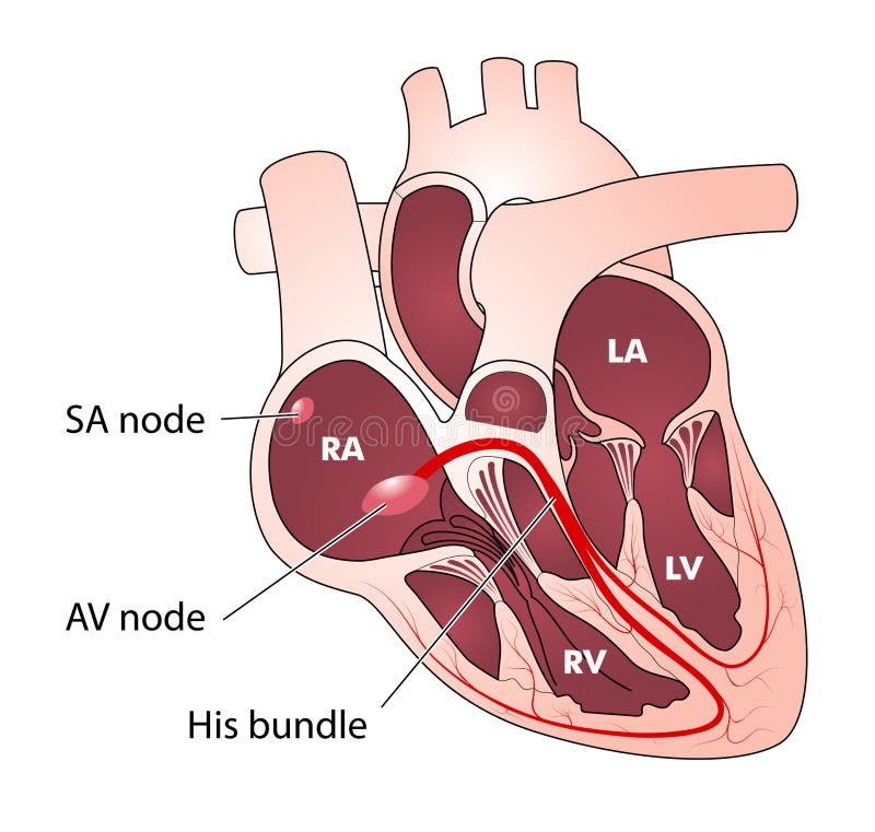
Free with trial Drawing of the heart, showing the SA and AV nodes, the pacemaker electrical conducting system. Sinoatrial node vectors Heart electrical conduction. Drawing of the heart, showing the SA and AV nodes, the pacemaker electrical conducting system
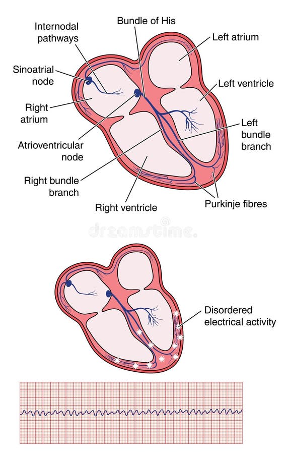
Free with trial Drawing of the heart electrical conduction system showing normal activity and erratic impulses in atrial fibrillation. Sinoatrial node vectors Normal heart electrical conduction and atrial fibrillation. Drawing of the heart electrical conduction system showing normal activity and erratic impulses in atrial fibrillation.
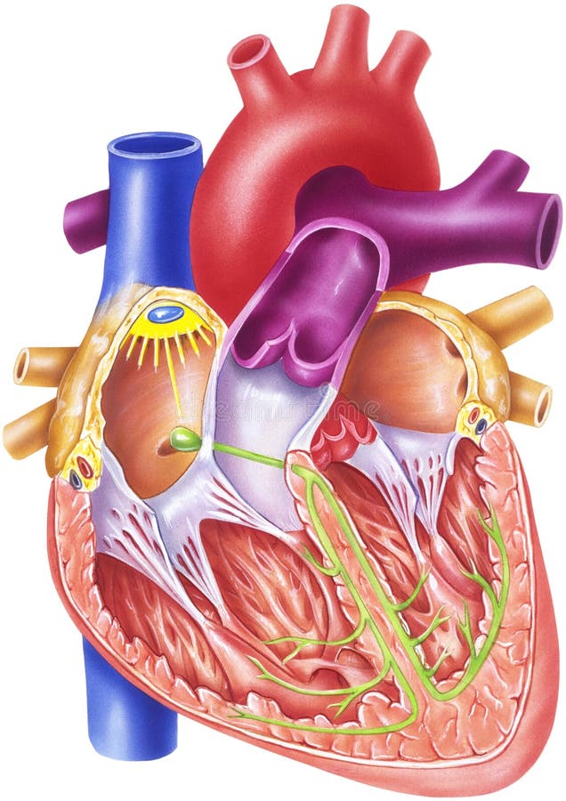
Free with trial Conduction system of heart, showing the SA and AV nodes, bundle branches and bundle of His. Created in Adobe Illustrator. EPS 10. Sinoatrial node vectors Conduction system of heart
Free with trial 1. The cardiac cycle begins at the SA (sinoatrial) node located in the sulcus terminalis between the superior vena cava and the right atrium. 2. From this pacemaker, a wave of negativity (excitation) spreads over both atria and initiates atrial contraction, thereby increasing atrial blood pressure. 3. When atrial pressure exceeds ventricular pressure, both atrioventricular (AV) valves open and bl. Sinoatrial node illustrations Heart - Conduction System. 1. The cardiac cycle begins at the SA (sinoatrial) node located in the sulcus terminalis between the superior vena cava and the right atrium. 2. From this pacemaker, a wave of negativity (excitation) spreads over both atria and initiates atrial contraction, thereby increasing atrial blood pressure. 3. When atrial pressure exceeds ventricular pressure, both atrioventricular (AV) valves open and bl
Free with trial The electrical conducting system of the heart, showing the impulse path from the sino atrial node to the atrioventricular node and the ventricles. Sinoatrial node vectors Heart electrical conducting system. The electrical conducting system of the heart, showing the impulse path from the sino atrial node to the atrioventricular node and the ventricles
Free with trial 1. The cardiac cycle begins at the SA sinoatrial node located in the sulcus terminalis between the superior vena cava and the right atrium. 2. From this pacemaker, a wave of negativity excitation spreads over both atria and initiates atrial contraction, thereby increasing atrial blood pressure. 3. When atrial pressure exceeds ventricular pressure, both atrioventricular AV valves open and blood rushes into both ventricles. Soon the impulse reaches the AV node and is passed along the AV bundle shown in green to the two ventricles, causing them to contract. 4. When ventricular pressure exceeds atrial pressure, the AV valves close, and this can be heard with a stethoscope as the first of the two heart sounds of the heartbeat. 5. Continued ventricular contraction forces the pulmonary and aortic valves to open, and blood rushes simultaneously into the pulmonary artery and the aorta. 6. When the pressure in these vessels exceeds ventricular pressure, blood tends to rush back into the ventricles, but it gets trapped in the sinuses behind the semilunar cusps. This closes both the pulmonary and aortic valves, resulting in the second of the two heart sounds. Sinoatrial node illustrations Heart - Conduction System. 1. The cardiac cycle begins at the SA sinoatrial node located in the sulcus terminalis between the superior vena cava and the right atrium. 2. From this pacemaker, a wave of negativity excitation spreads over both atria and initiates atrial contraction, thereby increasing atrial blood pressure. 3. When atrial pressure exceeds ventricular pressure, both atrioventricular AV valves open and blood rushes into both ventricles. Soon the impulse reaches the AV node and is passed along the AV bundle shown in green to the two ventricles, causing them to contract. 4. When ventricular pressure exceeds atrial pressure, the AV valves close, and this can be heard with a stethoscope as the first of the two heart sounds of the heartbeat. 5. Continued ventricular contraction forces the pulmonary and aortic valves to open, and blood rushes simultaneously into the pulmonary artery and the aorta. 6. When the pressure in these vessels exceeds ventricular pressure, blood tends to rush back into the ventricles, but it gets trapped in the sinuses behind the semilunar cusps. This closes both the pulmonary and aortic valves, resulting in the second of the two heart sounds.
Free with trial Detailed illustrationof heart conduction system and heart chumbers. Sinoatrial node vectors Heart conduction system. Human heart detailed illustration. Detailed illustrationof heart conduction system and heart chumbers
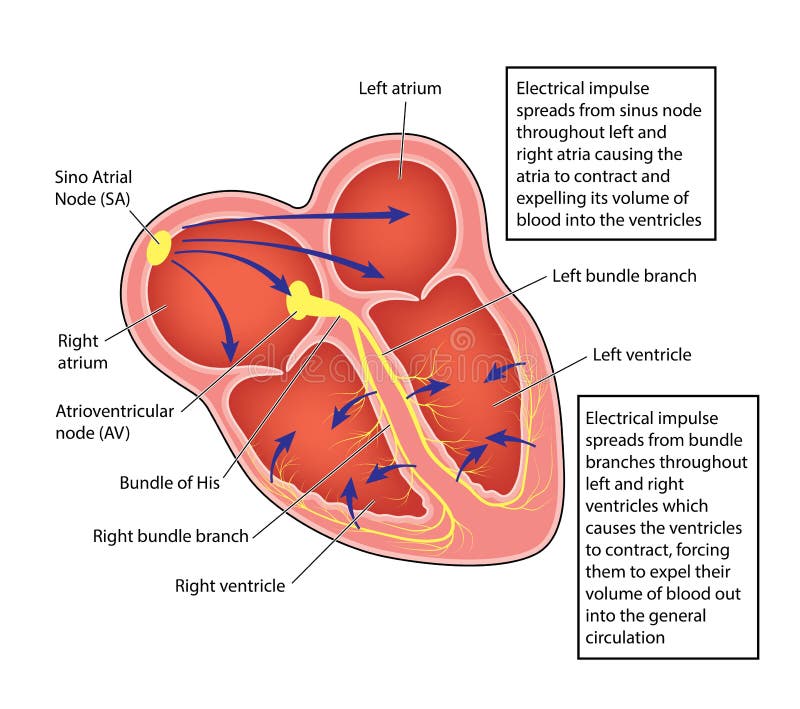
Free with trial The SA sinoatrial node generates an electrical signal that causes the upper heart chambers atria to contract. The signal then passes through the AV atrioventricular node to the lower heart chambers ventricles, causing them to contract, or pump. The SA node is considered the pacemaker of the heart. The heart's natural pacemaker – the SA node – sends out regular electrical impulses from the top chamber (the atrium) causing it to contract and pump blood into the bottom chamber (the ventricle). The electrical impulse is then conducted to the ventricles through a form of 'junction box' called the AV node. Sinoatrial node illustrations Sinoatrial Node in the Heart. The SA sinoatrial node generates an electrical signal that causes the upper heart chambers atria to contract. The signal then passes through the AV atrioventricular node to the lower heart chambers ventricles, causing them to contract, or pump. The SA node is considered the pacemaker of the heart. The heart's natural pacemaker – the SA node – sends out regular electrical impulses from the top chamber (the atrium) causing it to contract and pump blood into the bottom chamber (the ventricle). The electrical impulse is then conducted to the ventricles through a form of 'junction box' called the AV node.
Free with trial The SA sinoatrial node generates an electrical signal that causes the upper heart chambers atria to contract. The signal then passes through the AV atrioventricular node to the lower heart chambers ventricles, causing them to contract, or pump. The SA node is considered the pacemaker of the heart. The heart's natural pacemaker – the SA node – sends out regular electrical impulses from the top chamber (the atrium) causing it to contract and pump blood into the bottom chamber (the ventricle). The electrical impulse is then conducted to the ventricles through a form of 'junction box' called the AV node. Sinoatrial node illustrations Sinoatrial Node in the Heart. The SA sinoatrial node generates an electrical signal that causes the upper heart chambers atria to contract. The signal then passes through the AV atrioventricular node to the lower heart chambers ventricles, causing them to contract, or pump. The SA node is considered the pacemaker of the heart. The heart's natural pacemaker – the SA node – sends out regular electrical impulses from the top chamber (the atrium) causing it to contract and pump blood into the bottom chamber (the ventricle). The electrical impulse is then conducted to the ventricles through a form of 'junction box' called the AV node.
Free with trial The SA & x28;sinoatrial& x29; node generates an electrical signal that causes the upper heart chambers & x28;atria& x29; to contract. The signal then passes through the AV & x28;atrioventricular& x29; node to the lower heart chambers & x28;ventricles& x29;, causing them to contract, or pump. The SA node is considered the pacemaker of the heart. Sinoatrial node illustrations SA Node and AV Node in the Heart. The SA & x28;sinoatrial& x29; node generates an electrical signal that causes the upper heart chambers & x28;atria& x29; to contract. The signal then passes through the AV & x28;atrioventricular& x29; node to the lower heart chambers & x28;ventricles& x29;, causing them to contract, or pump. The SA node is considered the pacemaker of the heart.
Free with trial The SA sinoatrial node generates an electrical signal that causes the upper heart chambers atria to contract. The signal then passes through the AV atrioventricular node to the lower heart chambers ventricles, causing them to contract, or pump. The SA node is considered the pacemaker of the heart. Sinoatrial node illustrations SA Node and AV Node in the Heart. The SA sinoatrial node generates an electrical signal that causes the upper heart chambers atria to contract. The signal then passes through the AV atrioventricular node to the lower heart chambers ventricles, causing them to contract, or pump. The SA node is considered the pacemaker of the heart.
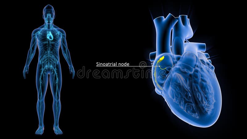
Free with trial The he SA node is the heart's natural pacemaker. The SA node consists of a cluster of cells that are situated in the upper part of the wall of the right atrium (the right upper chamber of the heart). The electrical impulses are generated there. The SA node is also called the sinus node. Sinoatrial node illustrations SA Nodes signal in the Heart or Sinoartial Node signal. The he SA node is the heart's natural pacemaker. The SA node consists of a cluster of cells that are situated in the upper part of the wall of the right atrium (the right upper chamber of the heart). The electrical impulses are generated there. The SA node is also called the sinus node.
Free with trial The AV node is a cluster of cells in the center of the heart between the atria and ventricles, and acts like a gate that slows the electrical signal before it enters the ventricles. This delay gives the atria time to contract before the ventricles do. Sinoatrial node illustrations SA Nodes in the Heart with Human Body. The AV node is a cluster of cells in the center of the heart between the atria and ventricles, and acts like a gate that slows the electrical signal before it enters the ventricles. This delay gives the atria time to contract before the ventricles do.
Free with trial The lower right chamber of the heart that receives deoxygenated blood from the right atrium and pumps it under low pressure into the lungs via the pulmonary artery. ... The pulmonary valve is situated between the right ventricle and the pulmonary artery and performs similarly as a one-way valve. Sinoatrial node illustrations SA Node Signal or Sinoatrial Node Signal of Human Heart. The lower right chamber of the heart that receives deoxygenated blood from the right atrium and pumps it under low pressure into the lungs via the pulmonary artery. ... The pulmonary valve is situated between the right ventricle and the pulmonary artery and performs similarly as a one-way valve.
Free with trial Drawing of heart electrical conducting system, the site of ventricular fibrillation disordered electrical activity and the resulting ecg trace. Created in Adobe Illustrator. EPS 10. Sinoatrial node vectors Ventricular fibrillation of heart. Drawing of heart electrical conducting system, the site of ventricular fibrillation disordered electrical activity and the resulting ecg trace. Created in Adobe Illustrator. EPS 10.
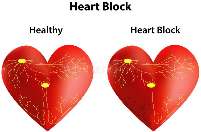
Free with trial A Conducting System of the Heart with Heart Block. Sinoatrial node vectors Heart Block
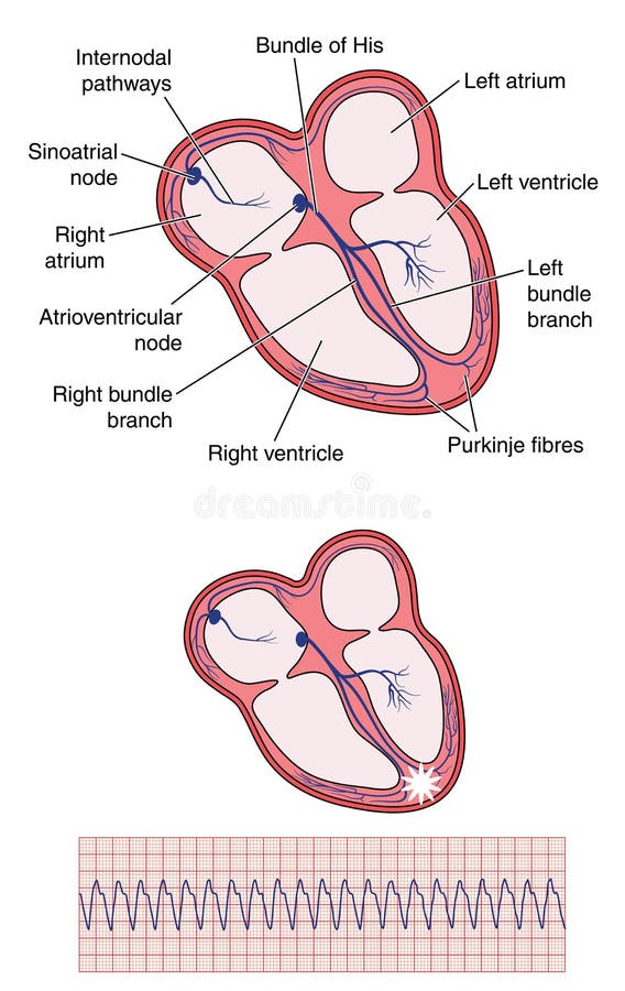
Free with trial Drawing of heart electrical conducting system, the site of ventricular tachycardia and the resulting ecg trace. Created in Adobe Illustrator. EPS 10. Sinoatrial node vectors Ventricular tachycardia of heart. Drawing of heart electrical conducting system, the site of ventricular tachycardia and the resulting ecg trace. Created in Adobe Illustrator. EPS 10.
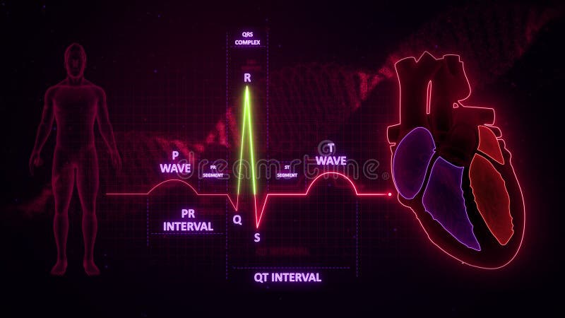
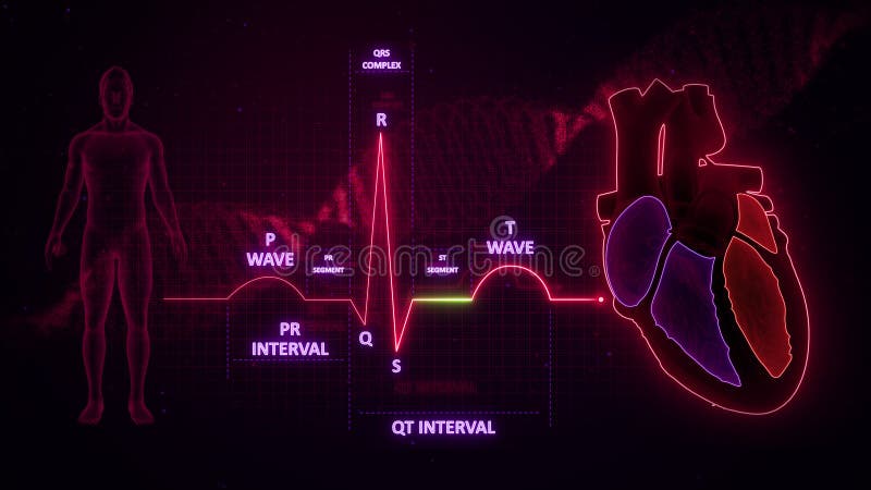
Free with trial The recorded tracing is called an electrocardiogram ECG, or EKG. A `typical` ECG tracing is shown to the right. The different waves that comprise the ECG represent the sequence of depolarization and repolarization of the atria and ventricles. Sinoatrial node illustrations QRS Complex from Electrocardiogram Wave or ECG or EKG. The recorded tracing is called an electrocardiogram ECG, or EKG. A `typical` ECG tracing is shown to the right. The different waves that comprise the ECG represent the sequence of depolarization and repolarization of the atria and ventricles.
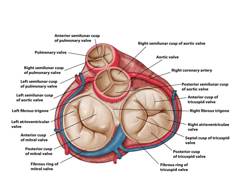
Free with trial Atrioventricular valves: The tricuspid valve and mitral bicuspid valve. They are located between the atria and corresponding ventricle. Semilunar valves: The pulmonary valve and aortic valve. They are located between the ventricles and their corresponding artery, and regulate the flow of blood leaving the heart. Sinoatrial node illustrations Tricuspid and Bicuspid Valve in the Heart. Atrioventricular valves: The tricuspid valve and mitral bicuspid valve. They are located between the atria and corresponding ventricle. Semilunar valves: The pulmonary valve and aortic valve. They are located between the ventricles and their corresponding artery, and regulate the flow of blood leaving the heart.
Free with trial The human heart is a muscular organ roughly the size of a closed fist, located slightly to the left of the center of the chest, behind the sternum. It functions as the central component of the circulatory system, pumping oxygen-rich blood throughout the body and returning oxygen-depleted blood to the lungs for reoxygenation. Sinoatrial node illustrations Chambers of the Heart. The human heart is a muscular organ roughly the size of a closed fist, located slightly to the left of the center of the chest, behind the sternum. It functions as the central component of the circulatory system, pumping oxygen-rich blood throughout the body and returning oxygen-depleted blood to the lungs for reoxygenation.
Free with trial The recorded tracing is called an electrocardiogram ECG, or EKG. A `typical` ECG tracing is shown to the right. The different waves that comprise the ECG represent the sequence of depolarization and repolarization of the atria and ventricles. Sinoatrial node illustrations R Wave from Electrocardiogram Wave or ECG or EKG. The recorded tracing is called an electrocardiogram ECG, or EKG. A `typical` ECG tracing is shown to the right. The different waves that comprise the ECG represent the sequence of depolarization and repolarization of the atria and ventricles.
Free with trial The recorded tracing is called an electrocardiogram ECG, or EKG. A `typical` ECG tracing is shown to the right. The different waves that comprise the ECG represent the sequence of depolarization and repolarization of the atria and ventricles. Sinoatrial node illustrations R Wave from Electrocardiogram Wave or ECG or EKG. The recorded tracing is called an electrocardiogram ECG, or EKG. A `typical` ECG tracing is shown to the right. The different waves that comprise the ECG represent the sequence of depolarization and repolarization of the atria and ventricles.
Free with trial The recorded tracing is called an electrocardiogram ECG, or EKG. A `typical` ECG tracing is shown to the right. The different waves that comprise the ECG represent the sequence of depolarization and repolarization of the atria and ventricles. Sinoatrial node illustrations ST Segment from Electrocardiogram Wave or ECG or EKG. The recorded tracing is called an electrocardiogram ECG, or EKG. A `typical` ECG tracing is shown to the right. The different waves that comprise the ECG represent the sequence of depolarization and repolarization of the atria and ventricles.
Free with trial The recorded tracing is called an electrocardiogram ECG, or EKG. A `typical` ECG tracing is shown to the right. The different waves that comprise the ECG represent the sequence of depolarization and repolarization of the atria and ventricles. Sinoatrial node illustrations PR and QT Intervals of Electrocardiogram Wave or ECG or EKG. The recorded tracing is called an electrocardiogram ECG, or EKG. A `typical` ECG tracing is shown to the right. The different waves that comprise the ECG represent the sequence of depolarization and repolarization of the atria and ventricles.
Free with trial Valves of the Heart and the Origin of the Coronary Vessels (Superior View) medical illustration. Sinoatrial node illustrations Valves of the Heart and the Origin of the Coronary Vessels (Superior View)
Free with trial The human heart is a muscular organ roughly the size of a closed fist, located slightly to the left of the center of the chest, behind the sternum. It functions as the central component of the circulatory system, pumping oxygen-rich blood throughout the body and returning oxygen-depleted blood to the lungs for reoxygenation. Sinoatrial node illustrations Left Dominant Distribution of the Coronary Arteries. The human heart is a muscular organ roughly the size of a closed fist, located slightly to the left of the center of the chest, behind the sternum. It functions as the central component of the circulatory system, pumping oxygen-rich blood throughout the body and returning oxygen-depleted blood to the lungs for reoxygenation.
Free with trial Valves of Tricuspid valve, Mitral valve, Pulmonary valve, Aortic valve, the Heart Anatomy medical illustration. Sinoatrial node illustrations Valves of the Heart Anatomy medical illustration. Valves of Tricuspid valve, Mitral valve, Pulmonary valve, Aortic valve, the Heart Anatomy medical illustration
Free with trial Illustration shows electrical impulse conduction pathway in human heart from sinoatrial node. Heartbeat cardiogram visualization. Anatomy of cardiovascular system, human body, heart. Sinoatrial node illustrations Illustration shows electrical impulse conduction pathway in human heart from sinoatrial node. Heartbeat cardiogram visualization.
Free with trial This vibrant illustration meticulously details the complex electrical conduction system of the human heart. It showcases the key components responsible for coordinating the heart's rhythmic contractions: the sinus node (sinoatrial node), the atrioventricular node (AV node), the bundle of His, and the Purkinje fibers. Each structure is vividly colored and labeled, providing a clear visual. Sinoatrial node illustrations A Colorful Guide to the Human Hearts Electrical Conduction System Understanding the Sinus Node AV Node Bundle of. This vibrant illustration meticulously details the complex electrical conduction system of the human heart. It showcases the key components responsible for coordinating the heart's rhythmic contractions: the sinus node (sinoatrial node), the atrioventricular node (AV node), the bundle of His, and the Purkinje fibers. Each structure is vividly colored and labeled, providing a clear visual
Free with trial The image shows a digital heart rate monitor displaying a sinus complex. The green line represents the heart's electrical activity over time, with peaks indicating heartbeats. The sinus complex is a normal heart rhythm originating from the sinoatrial node. The monitor is likely part of a medical device used to assess heart health. Sinoatrial node illustrations Heart rate monitor displaying sinus complex. The image shows a digital heart rate monitor displaying a sinus complex. The green line represents the heart's electrical activity over time, with peaks indicating heartbeats. The sinus complex is a normal heart rhythm originating from the sinoatrial node. The monitor is likely part of a medical device used to assess heart health
Free with trial This captivating 3D microscopic render showcases the intricate architecture of the human heart's atrioventricular node (AV node). The AV node, a crucial component of the heart's electrical conduction system, acts as a vital gatekeeper, regulating the rhythmic flow of electrical impulses that orchestrate the heartbeat. This detailed visualization provides a unique perspective on the microscopic. Sinoatrial node illustrations Detailed 3D Visualization of the Human Hearts Atrioventricular Node Unveiling the Microscopic Architecture of. This captivating 3D microscopic render showcases the intricate architecture of the human heart's atrioventricular node (AV node). The AV node, a crucial component of the heart's electrical conduction system, acts as a vital gatekeeper, regulating the rhythmic flow of electrical impulses that orchestrate the heartbeat. This detailed visualization provides a unique perspective on the microscopic
Free with trial Detailed illustration showcasing the hearts electrical conduction system, highlighting the sinoatrial SA and atrioventricular AV nodes, and Purkinje fibers for medical education and awareness. Sinoatrial node illustrations Hearts Electrical System - A Detailed Anatomical Illustration. Detailed illustration showcasing the hearts electrical conduction system, highlighting the sinoatrial SA and atrioventricular AV nodes, and Purkinje fibers for medical education and awareness
Free with trial Electrocardiograms (ECGs) are invaluable tools for evaluating heart health and function. A critical component of the ECG is the PR interval, a measure of the time it takes for the electrical impulse to travel from the sinoatrial (SA) node, the heart's natural pacemaker, through the atria and to the ventricles. Understanding the normal range of the PR interval is essential for detecting potential. Sinoatrial node illustrations Understanding the PR Interval in Electrocardiograms (ECGs): A Comprehensive Guide to Heart Health Assessment. Electrocardiograms (ECGs) are invaluable tools for evaluating heart health and function. A critical component of the ECG is the PR interval, a measure of the time it takes for the electrical impulse to travel from the sinoatrial (SA) node, the heart's natural pacemaker, through the atria and to the ventricles. Understanding the normal range of the PR interval is essential for detecting potential
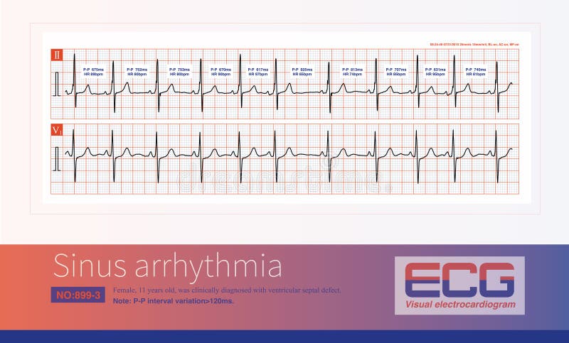
Free with trial When the sinus cycle changes by more than 120ms, the electrocardiogram diagnosis is sinus arrhythmia, which is caused by irregular impulses from the pacemaker cells of the sinoatrial node. Sinoatrial node illustrations Sinus arrhythmia
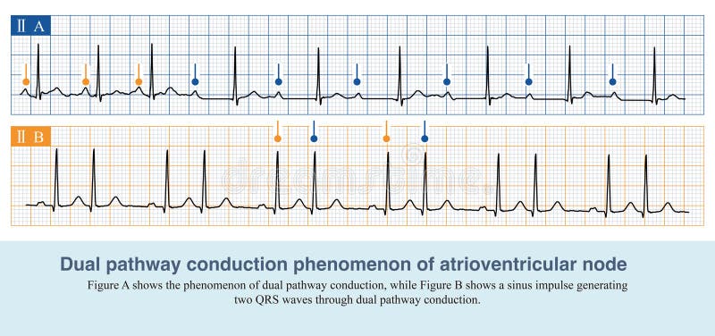
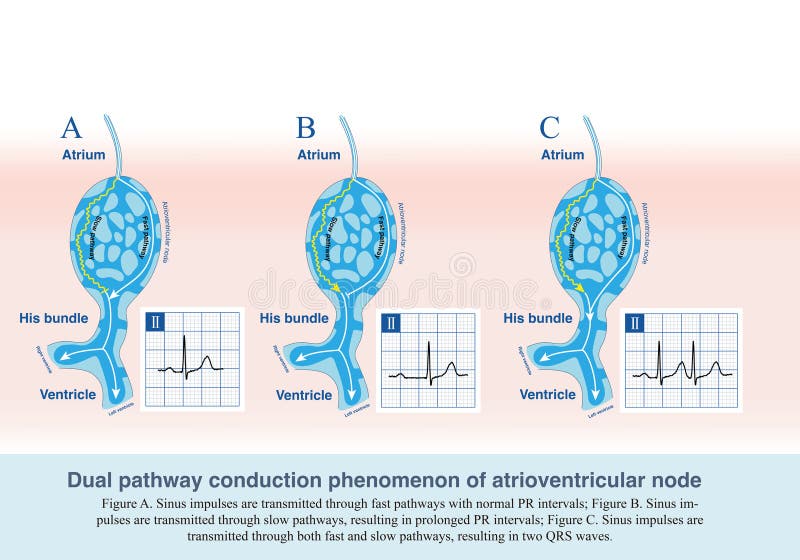
Free with trial On ECG paper, five small squares make up a middle square. Divide 300 by the number of middle squares occupied by adjacent identical ECG waves to get the heart rate. Sinoatrial node illustrations Dual pathway conduction phenomenon of atrioventricular node. On ECG paper, five small squares make up a middle square. Divide 300 by the number of middle squares occupied by adjacent identical ECG waves to get the heart rate.
Free with trial On ECG paper, five small squares make up a middle square. Divide 300 by the number of middle squares occupied by adjacent identical ECG waves to get the heart rate. Sinoatrial node illustrations Dual pathway phenomenon of atrioventricular node. On ECG paper, five small squares make up a middle square. Divide 300 by the number of middle squares occupied by adjacent identical ECG waves to get the heart rate.
Free with trial The human heart is a muscular organ roughly the size of a closed fist, located slightly to the left of the center of the chest, behind the sternum. It functions as the central component of the circulatory system, pumping oxygen-rich blood throughout the body and returning oxygen-depleted blood to the lungs for reoxygenation. Sinoatrial node illustrations Heart Coronary Arteries Medical illustration. The human heart is a muscular organ roughly the size of a closed fist, located slightly to the left of the center of the chest, behind the sternum. It functions as the central component of the circulatory system, pumping oxygen-rich blood throughout the body and returning oxygen-depleted blood to the lungs for reoxygenation.
Free with trial The human heart is a muscular organ roughly the size of a closed fist, located slightly to the left of the center of the chest, behind the sternum. It functions as the central component of the circulatory system, pumping oxygen-rich blood throughout the body and returning oxygen-depleted blood to the lungs for reoxygenation. Sinoatrial node illustrations Anterior view Heart Anatomy medical illustration. The human heart is a muscular organ roughly the size of a closed fist, located slightly to the left of the center of the chest, behind the sternum. It functions as the central component of the circulatory system, pumping oxygen-rich blood throughout the body and returning oxygen-depleted blood to the lungs for reoxygenation.
Free with trial The human heart is a muscular organ roughly the size of a closed fist, located slightly to the left of the center of the chest, behind the sternum. It functions as the central component of the circulatory system, pumping oxygen-rich blood throughout the body and returning oxygen-depleted blood to the lungs for reoxygenation. Sinoatrial node illustrations Heart, Blood Supply (Anterior and Superior Surfaces). The human heart is a muscular organ roughly the size of a closed fist, located slightly to the left of the center of the chest, behind the sternum. It functions as the central component of the circulatory system, pumping oxygen-rich blood throughout the body and returning oxygen-depleted blood to the lungs for reoxygenation.
Free with trial The human heart is a muscular organ roughly the size of a closed fist, located slightly to the left of the center of the chest, behind the sternum. It functions as the central component of the circulatory system, pumping oxygen-rich blood throughout the body and returning oxygen-depleted blood to the lungs for reoxygenation. Sinoatrial node illustrations Balanced Distribution of the Left and Right Coronary Arteries. The human heart is a muscular organ roughly the size of a closed fist, located slightly to the left of the center of the chest, behind the sternum. It functions as the central component of the circulatory system, pumping oxygen-rich blood throughout the body and returning oxygen-depleted blood to the lungs for reoxygenation.
Free with trial Heart anatomy. Part of the human heart. vector illustration. Sinoatrial node vectors Heart anatomy
Free with trial Atrial fibrillation diagram shows heart outline, electrical pathways, and rhythm differences, highlighting irregular heartbeat. Outline diagram. Sinoatrial node vectors Atrial fibrillation diagram shows heart outline, electrical pathways, and rhythm ... Atrial fibrillation diagram shows heart outline, electrical pathways, and rhythm differences, highlighting irregular heartbeat. Outline diagram
Free with trial The human heart is a muscular organ roughly the size of a closed fist, located slightly to the left of the center of the chest, behind the sternum. It functions as the central component of the circulatory system, pumping oxygen-rich blood throughout the body and returning oxygen-depleted blood to the lungs for Reoxygenation. Sinoatrial node illustrations Posterior view Heart Anatomy medical illustration. The human heart is a muscular organ roughly the size of a closed fist, located slightly to the left of the center of the chest, behind the sternum. It functions as the central component of the circulatory system, pumping oxygen-rich blood throughout the body and returning oxygen-depleted blood to the lungs for Reoxygenation.
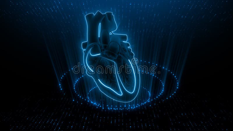
Free with trial Heart's Rhythm Anatomical 3D Render with Energy Waves , This photo was created using generative AI. Sinoatrial node illustrations Heart\'s Rhythm Anatomical 3D Render with Energy Waves. Heart's Rhythm Anatomical 3D Render with Energy Waves , This photo was created using generative AI.
Free with trial Usually, one of the working parameters of an electrocardiogram machine is set to a calibration voltage of 1mV 10mm, which means that when a 1mV voltage is input to the electrocardiogram machine, the amplitude of the electrocardiogram wave deviates by 10mm. Sinoatrial node illustrations Calibration voltage of electrocardiograph. Usually, one of the working parameters of an electrocardiogram machine is set to a calibration voltage of 1mV 10mm, which means that when a 1mV voltage is input to the electrocardiogram machine, the amplitude of the electrocardiogram wave deviates by 10mm.
Free with trial Some patients with severe sinus bradycardia have triggers that can disappear after treatment, while others are permanent and require treatment with ventricular pacemakers. Sinoatrial node illustrations Clinical evaluation of severe sinus bradycardia. Some patients with severe sinus bradycardia have triggers that can disappear after treatment, while others are permanent and require treatment with ventricular pacemakers.
Free with trial This image shows an electrocardiogram (ECG or EKG) strip labeled as 'Normal Sinus'. The ECG depicts a regular heart rhythm with consistent P waves, QRS complexes, and T waves, indicative of a normal sinus rhythm. The strip includes two leads and a clear timeline at the bottom, marked in seconds, to measure the intervals between heartbeats. The regularity and uniformity of the waveform suggest a. Sinoatrial node illustrations Electrocardiogram displaying normal sinus rhythm. This image shows an electrocardiogram (ECG or EKG) strip labeled as 'Normal Sinus'. The ECG depicts a regular. This image shows an electrocardiogram (ECG or EKG) strip labeled as 'Normal Sinus'. The ECG depicts a regular heart rhythm with consistent P waves, QRS complexes, and T waves, indicative of a normal sinus rhythm. The strip includes two leads and a clear timeline at the bottom, marked in seconds, to measure the intervals between heartbeats. The regularity and uniformity of the waveform suggest a
Free with trial This detailed illustration showcases the anatomy of the heart, highlighting its intricate structure and functions. The image features the four chambers of the heart, the valves, and the major blood vessels that supply oxygenated and deoxygenated blood throughout the body. The vibrant colors and precise details make this illustration a valuable resource for medical professionals and students alike. The heart is a symbol of love, passion, and life, and this illustration captures its beauty and complexity. This image would be perfect for educational materials, medical publications, or any project that requires a detailed understanding of the heart's anatomy. Sinoatrial node illustrations Anatomy of the Heart: A Detailed Illustration. This detailed illustration showcases the anatomy of the heart, highlighting its intricate structure and functions. The image features the four chambers of the heart, the valves, and the major blood vessels that supply oxygenated and deoxygenated blood throughout the body. The vibrant colors and precise details make this illustration a valuable resource for medical professionals and students alike. The heart is a symbol of love, passion, and life, and this illustration captures its beauty and complexity. This image would be perfect for educational materials, medical publications, or any project that requires a detailed understanding of the heart's anatomy.
Free with trial Right Dominant Distribution of the Coronary Arteries (Left atrium, Pulmonary veins, Right atrium, Superior vena cava, Inferior vena cava, Posterior interventricular sulcus, Coronary sinus, Pulmonary arteries). Sinoatrial node illustrations Right Dominant Distribution of the Coronary Arteries
Free with trial The human heart is a muscular organ roughly the size of a closed fist, located slightly to the left of the center of the chest, behind the sternum. It functions as the central component of the circulatory system, pumping oxygen-rich blood throughout the body and returning oxygen-depleted blood to the lungs for reoxygenation. Sinoatrial node illustrations Layers of the Heart Wall Anatomy medical illustration. The human heart is a muscular organ roughly the size of a closed fist, located slightly to the left of the center of the chest, behind the sternum. It functions as the central component of the circulatory system, pumping oxygen-rich blood throughout the body and returning oxygen-depleted blood to the lungs for reoxygenation.
Free with trial This detailed 3D illustration provides a comprehensive view of the human heart, showcasing its complex internal structure. The intricate network of blood vessels, the four chambers (left atrium, left ventricle, right atrium, right ventricle), and the valves responsible for blood flow are meticulously depicted. The illustration highlights the layers of the heart wall, including the endocardium,. Sinoatrial node illustrations A Comprehensive 3D Cutaway Illustration of the Human Heart Unveiling the Intricate Anatomy and Function of the. This detailed 3D illustration provides a comprehensive view of the human heart, showcasing its complex internal structure. The intricate network of blood vessels, the four chambers (left atrium, left ventricle, right atrium, right ventricle), and the valves responsible for blood flow are meticulously depicted. The illustration highlights the layers of the heart wall, including the endocardium,
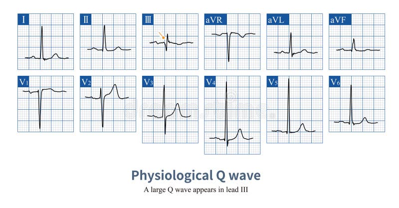
Free with trial Male, 23 years old, healthy. When the initial excitation potential of the ventricle deviates from a certain lead axis, a Q wave will be projected onto that lead, which is a physiological Q wave. Sinoatrial node illustrations Physiological Q wave
Free with trial In the spatial anatomy of the heart, the axis from the base of the heart to the apex of the heart is called the long axis, that is, the upper right side faces the lower left side. Sinoatrial node illustrations The long axis of the heart. In the spatial anatomy of the heart, the axis from the base of the heart to the apex of the heart is called the long axis, that is, the upper right side faces the lower left side.
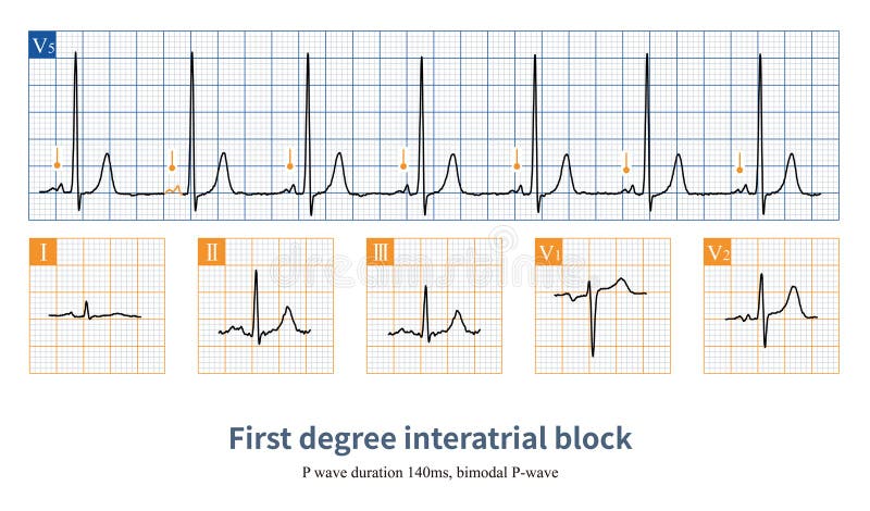
Free with trial When the first degree interatrial block occurs, the conduction time from the right atrium to the left atrium is prolonged, the P wave widens, and bimodal P wave ECG changes appear. Sinoatrial node illustrations First degree interatrial block
Free with trial Detailed 3D rendering of a human heart, showcasing its internal chambers, valves, and major blood vessels. The image highlights the intricate network of pathways within the heart, providing a clear visual representation of its complex structure and function. Sinoatrial node illustrations Human Heart Anatomy: Internal Structure. Detailed 3D rendering of a human heart, showcasing its internal chambers, valves, and major blood vessels. The image highlights the intricate network of pathways within the heart, providing a clear visual representation of its complex structure and function.
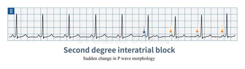
Free with trial When intermittent conduction dysfunction occurs in the Bachmann bundle, intermittent left atrial abnormality may be seen on the ECG, which can be differentiated from anatomical left atrial enlargement. Sinoatrial node illustrations Second degree interatrial block. When intermittent conduction dysfunction occurs in the Bachmann bundle, intermittent left atrial abnormality may be seen on the ECG, which can be differentiated from anatomical left atrial enlargement.
Free with trial A Detailed Anatomical Rendering Of The Human Heart. AI Generative Image. Sinoatrial node illustrations A Detailed Anatomical Rendering Of The Human Heart
Free with trial Atrial fibrillation is an arrhythmia with an absolutely irregular ventricular rhythm, and the ventricular rate can only be estimated based on the average cardiac cycle or the number of QRS waves. Sinoatrial node illustrations Estimate the average ventricular rate of atrial fibrillation. Atrial fibrillation is an arrhythmia with an absolutely irregular ventricular rhythm, and the ventricular rate can only be estimated based on the average cardiac cycle or the number of QRS waves.
Free with trial This cutting-edge electrocardiogram (ECG) machine showcases the remarkable precision of modern cardiac monitoring. The clear display of a steady heartbeat rhythm highlights the machine's ability to capture and interpret the complex electrical signals of the heart, providing crucial data for diagnosing and managing various cardiac conditions. Surrounding the sophisticated ECG device are essential. Sinoatrial node illustrations Precise Heartbeat Monitoring Advanced Electrocardiogram Technology for Accurate Cardiac Activity Assessment in a Modern. This cutting-edge electrocardiogram (ECG) machine showcases the remarkable precision of modern cardiac monitoring. The clear display of a steady heartbeat rhythm highlights the machine's ability to capture and interpret the complex electrical signals of the heart, providing crucial data for diagnosing and managing various cardiac conditions. Surrounding the sophisticated ECG device are essential
Free with trial Chalk drawing of a human heart on black chalkboard and inscription Sinoatrial node. Sinoatrial node illustrations Chalk sketch of human heart on black desc and inscription Sinoatrial node. Chalk drawing of a human heart on black chalkboard and inscription Sinoatrial node
Free with trial The AV node is a cluster of cells in the center of the heart between the atria and ventricles, and acts like a gate that slows the electrical signal before it enters the ventricles. This delay gives the atria time to contract before the ventricles do. Sinoatrial node illustrations SA and AV Node signal in the Heart. The AV node is a cluster of cells in the center of the heart between the atria and ventricles, and acts like a gate that slows the electrical signal before it enters the ventricles. This delay gives the atria time to contract before the ventricles do.
Free with trial The AV node is a cluster of cells in the center of the heart between the atria and ventricles, and acts like a gate that slows the electrical signal before it enters the ventricles. This delay gives the atria time to contract before the ventricles do. Sinoatrial node illustrations SA and AV Node signal in the Heart. The AV node is a cluster of cells in the center of the heart between the atria and ventricles, and acts like a gate that slows the electrical signal before it enters the ventricles. This delay gives the atria time to contract before the ventricles do.
Free with trial The AV node is a cluster of cells in the center of the heart between the atria and ventricles, and acts like a gate that slows the electrical signal before it enters the ventricles. This delay gives the atria time to contract before the ventricles do. Sinoatrial node illustrations SA and AV Node signal in the Heart. The AV node is a cluster of cells in the center of the heart between the atria and ventricles, and acts like a gate that slows the electrical signal before it enters the ventricles. This delay gives the atria time to contract before the ventricles do.
Free with trial The AV node is a cluster of cells in the center of the heart between the atria and ventricles, and acts like a gate that slows the electrical signal before it enters the ventricles. This delay gives the atria time to contract before the ventricles do. Sinoatrial node illustrations SA Nodes in the Heart or Sinoartial Node. The AV node is a cluster of cells in the center of the heart between the atria and ventricles, and acts like a gate that slows the electrical signal before it enters the ventricles. This delay gives the atria time to contract before the ventricles do.
Free with trial The AV node is a cluster of cells in the center of the heart between the atria and ventricles, and acts like a gate that slows the electrical signal before it enters the ventricles. This delay gives the atria time to contract before the ventricles do. Sinoatrial node illustrations SA Node signal in the Heart with Human Body. The AV node is a cluster of cells in the center of the heart between the atria and ventricles, and acts like a gate that slows the electrical signal before it enters the ventricles. This delay gives the atria time to contract before the ventricles do.
Free with trial The atrioventricular node AVN is a complex structure that performs a variety of functions in the heart. The AVN is primarily an electrical gatekeeper between the atria and ventricles and introduces a delay between atrial and ventricular excitation, allowing for efficient ventricular filling. Sinoatrial node illustrations AV Node Signal or Atrioventricular Node Signal of Human Heart. The atrioventricular node AVN is a complex structure that performs a variety of functions in the heart. The AVN is primarily an electrical gatekeeper between the atria and ventricles and introduces a delay between atrial and ventricular excitation, allowing for efficient ventricular filling.
Free with trial The atrioventricular node AVN is a complex structure that performs a variety of functions in the heart. The AVN is primarily an electrical gatekeeper between the atria and ventricles and introduces a delay between atrial and ventricular excitation, allowing for efficient ventricular filling. Sinoatrial node illustrations AV Node Signal or Atrioventricular Node Signal of Human Heart. The atrioventricular node AVN is a complex structure that performs a variety of functions in the heart. The AVN is primarily an electrical gatekeeper between the atria and ventricles and introduces a delay between atrial and ventricular excitation, allowing for efficient ventricular filling.
Free with trial The atrioventricular node AVN is a complex structure that performs a variety of functions in the heart. The AVN is primarily an electrical gatekeeper between the atria and ventricles and introduces a delay between atrial and ventricular excitation, allowing for efficient ventricular filling. Sinoatrial node illustrations AV Node Signal or Atrioventricular Node Signal of Human Heart. The atrioventricular node AVN is a complex structure that performs a variety of functions in the heart. The AVN is primarily an electrical gatekeeper between the atria and ventricles and introduces a delay between atrial and ventricular excitation, allowing for efficient ventricular filling.
Free with trial The AV node is a cluster of cells in the center of the heart between the atria and ventricles, and acts like a gate that slows the electrical signal before it enters the ventricles. This delay gives the atria time to contract before the ventricles do. Sinoatrial node illustrations SA and AV Node signal in the Heart with Human Body. The AV node is a cluster of cells in the center of the heart between the atria and ventricles, and acts like a gate that slows the electrical signal before it enters the ventricles. This delay gives the atria time to contract before the ventricles do.
Free with trial The atrioventricular node AVN is a complex structure that performs a variety of functions in the heart. The AVN is primarily an electrical gatekeeper between the atria and ventricles and introduces a delay between atrial and ventricular excitation, allowing for efficient ventricular filling. Sinoatrial node illustrations AV Node Signal or Atrioventricular Node Signal of Human Heart. The atrioventricular node AVN is a complex structure that performs a variety of functions in the heart. The AVN is primarily an electrical gatekeeper between the atria and ventricles and introduces a delay between atrial and ventricular excitation, allowing for efficient ventricular filling.
Free with trial The atrioventricular node AVN is a complex structure that performs a variety of functions in the heart. The AVN is primarily an electrical gatekeeper between the atria and ventricles and introduces a delay between atrial and ventricular excitation, allowing for efficient ventricular filling. Sinoatrial node illustrations AV Node Signal or Atrioventricular Node Signal of Human Heart. The atrioventricular node AVN is a complex structure that performs a variety of functions in the heart. The AVN is primarily an electrical gatekeeper between the atria and ventricles and introduces a delay between atrial and ventricular excitation, allowing for efficient ventricular filling.
Free with trial The atrioventricular node AVN is a complex structure that performs a variety of functions in the heart. The AVN is primarily an electrical gatekeeper between the atria and ventricles and introduces a delay between atrial and ventricular excitation, allowing for efficient ventricular filling. Sinoatrial node illustrations AV Node Signal or Atrioventricular Node Signal of Human Heart. The atrioventricular node AVN is a complex structure that performs a variety of functions in the heart. The AVN is primarily an electrical gatekeeper between the atria and ventricles and introduces a delay between atrial and ventricular excitation, allowing for efficient ventricular filling.
Free with trial The atrioventricular node AVN is a complex structure that performs a variety of functions in the heart. The AVN is primarily an electrical gatekeeper between the atria and ventricles and introduces a delay between atrial and ventricular excitation, allowing for efficient ventricular filling. Sinoatrial node illustrations AV Node Signal or Atrioventricular Node Signal of Human Heart. The atrioventricular node AVN is a complex structure that performs a variety of functions in the heart. The AVN is primarily an electrical gatekeeper between the atria and ventricles and introduces a delay between atrial and ventricular excitation, allowing for efficient ventricular filling.
Free with trial The atrioventricular node AVN is a complex structure that performs a variety of functions in the heart. The AVN is primarily an electrical gatekeeper between the atria and ventricles and introduces a delay between atrial and ventricular excitation, allowing for efficient ventricular filling. Sinoatrial node illustrations AV and SA Node Signals or Atrioventricular and Sinoartial Node Signal of Human Heart. The atrioventricular node AVN is a complex structure that performs a variety of functions in the heart. The AVN is primarily an electrical gatekeeper between the atria and ventricles and introduces a delay between atrial and ventricular excitation, allowing for efficient ventricular filling.
Free with trial The atrioventricular node AVN is a complex structure that performs a variety of functions in the heart. The AVN is primarily an electrical gatekeeper between the atria and ventricles and introduces a delay between atrial and ventricular excitation, allowing for efficient ventricular filling. Sinoatrial node illustrations AV and SA Node Signals or Atrioventricular and Sinoartial Node Signal of Human Heart. The atrioventricular node AVN is a complex structure that performs a variety of functions in the heart. The AVN is primarily an electrical gatekeeper between the atria and ventricles and introduces a delay between atrial and ventricular excitation, allowing for efficient ventricular filling.
Free with trial The atrioventricular node AVN is a complex structure that performs a variety of functions in the heart. The AVN is primarily an electrical gatekeeper between the atria and ventricles and introduces a delay between atrial and ventricular excitation, allowing for efficient ventricular filling. Sinoatrial node illustrations AV and SA Node Signals or Atrioventricular and Sinoartial Node Signal of Human Heart. The atrioventricular node AVN is a complex structure that performs a variety of functions in the heart. The AVN is primarily an electrical gatekeeper between the atria and ventricles and introduces a delay between atrial and ventricular excitation, allowing for efficient ventricular filling.
Free with trial The atrioventricular node AVN is a complex structure that performs a variety of functions in the heart. The AVN is primarily an electrical gatekeeper between the atria and ventricles and introduces a delay between atrial and ventricular excitation, allowing for efficient ventricular filling. Sinoatrial node illustrations AV and SA Node Signals or Atrioventricular and Sinoartial Node Signal of Human Heart. The atrioventricular node AVN is a complex structure that performs a variety of functions in the heart. The AVN is primarily an electrical gatekeeper between the atria and ventricles and introduces a delay between atrial and ventricular excitation, allowing for efficient ventricular filling.
Free with trial The atrioventricular node AVN is a complex structure that performs a variety of functions in the heart. The AVN is primarily an electrical gatekeeper between the atria and ventricles and introduces a delay between atrial and ventricular excitation, allowing for efficient ventricular filling. Sinoatrial node illustrations AV and SA Node Signals or Atrioventricular and Sinoartial Node Signal of Human Heart. The atrioventricular node AVN is a complex structure that performs a variety of functions in the heart. The AVN is primarily an electrical gatekeeper between the atria and ventricles and introduces a delay between atrial and ventricular excitation, allowing for efficient ventricular filling.
Free with trial The atrioventricular node AVN is a complex structure that performs a variety of functions in the heart. The AVN is primarily an electrical gatekeeper between the atria and ventricles and introduces a delay between atrial and ventricular excitation, allowing for efficient ventricular filling. Sinoatrial node illustrations AV and SA Node Signals or Atrioventricular and Sinoartial Node Signal of Human Heart. The atrioventricular node AVN is a complex structure that performs a variety of functions in the heart. The AVN is primarily an electrical gatekeeper between the atria and ventricles and introduces a delay between atrial and ventricular excitation, allowing for efficient ventricular filling.