Free with trial Diagram of the alkaline mucous layer in the stomach. Diagram of the histological cross-section of the stomach Layers of the Stomach. Stomach mucus layer vectors Mucous layer in the stomach
Free with trial 3D Illustration showing helicobacter pylori bacteria damaging mucus layer, gastritis. Stomach mucus layer illustrations Gastritis, helicobacter pylori bacteria damaging mucus layer, medically accurate 3D illustration. 3D Illustration showing helicobacter pylori bacteria damaging mucus layer, gastritis
Free with trial The tongue also serves as a natural means of cleaning the teeth. It is of importance in the digestive system and is the primary organ of taste in the gustatory system. Education info graphic. Vector design. Stomach mucus layer vectors Tongue. Education info graphic. Vector design. The tongue also serves as a natural means of cleaning the teeth.It is of importance in the digestive system and is the primary organ of taste in the gustatory system. Education info graphic. Vector design.
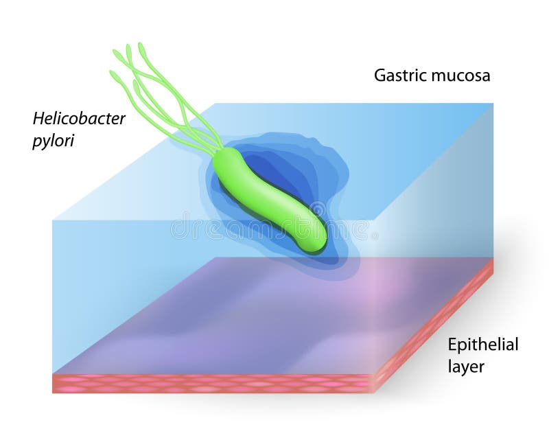
Free with trial Helicobacter pylori - Ulcer-causing bacterium. Stomach mucus layer vectors Helicobacter pylori
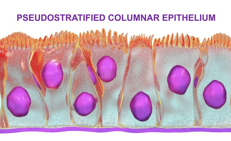
Free with trial Pseudostratified columnar epithelium, 3D illustration. Epithelium found in trachea and upper part of digestive tract. Stomach mucus layer illustrations Pseudostratified columnar epithelium
Free with trial GERD Acid Reflux Helicobacter pylori medical visualization features corkscrew shaped bacteria penetrating stomach mucus layer, scientific pathology education image. Stomach mucus layer illustrations GERD Acid Reflux Helicobacter pylori medical visualization features corkscrew shaped bacteria penetrating stomach mucus layer,. Scientific pathology education
Free with trial 3D Illustration showing helicobacter pylori bacteria damaging mucus layer, gastritis. Stomach mucus layer illustrations Gastritis, helicobacter pylori bacteria damaging mucus layer, medically accurate 3D illustration. 3D Illustration showing helicobacter pylori bacteria damaging mucus layer, gastritis
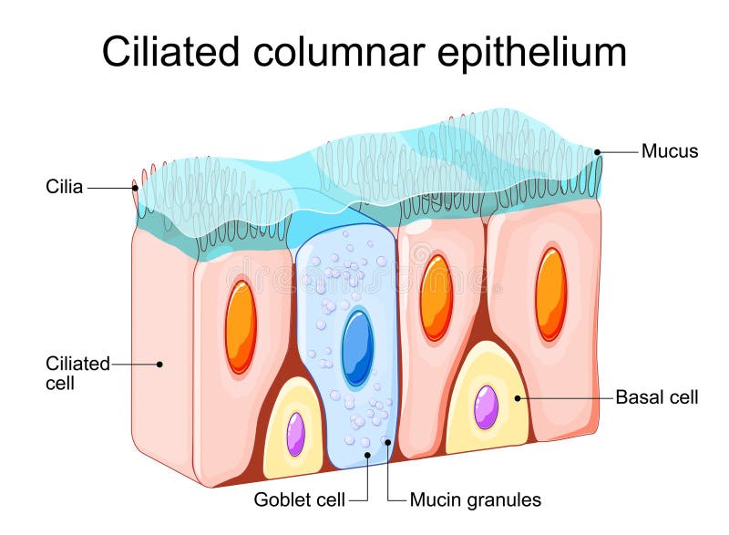
Free with trial Ciliated columnar epithelium. epithelial cells forms the lining of the stomach and intestines, duodenum, fallopian tubes, uterus, central canal of the spinal cord, nose, ears and the taste buds. Stomach mucus layer vectors Ciliated columnar epithelium
Free with trial Ciliated columnar epithelium. Epithelial cells forms the lining of the stomach and intestines, duodenum, fallopian tubes, uterus, central canal of the spinal cord, nose, ears and the taste buds. Nasal epithelium. Ciliated cells. Respiratory defense mechanisms. Vector illustration. Medical poster. Schematic diagram. Stomach mucus layer vectors Ciliated columnar epithelium. Nasal epithelium. Ciliated columnar epithelium. Epithelial cells forms the lining of the stomach and intestines, duodenum, fallopian tubes, uterus, central canal of the spinal cord, nose, ears and the taste buds. Nasal epithelium. Ciliated cells. Respiratory defense mechanisms. Vector illustration. Medical poster. Schematic diagram
Free with trial Nasal epithelium. Ciliated columnar epithelium. epithelial cells forms the lining of the stomach and intestines, duodenum, fallopian tubes, uterus, central canal of the spinal cord, nose, ears and the taste buds. Ciliated cells. Respiratory defense mechanisms. Vector poster. Isometric Flat illustration. Stomach mucus layer vectors Nasal epithelium. Ciliated columnar epithelium
Free with trial The small intestine extends from the pyloric sphincter to the ileocecal valve, where it empties into the large intestine. The small intestine finishes the process of digestion, absorbs the nutrients, and passes the residue on to the large intestine. The liver, gallbladder, and pancreas are accessory organs of the digestive system that are closely associated with the small intestine. Stomach mucus layer illustrations Difference Between Small Intestine and Large Intestine. The small intestine extends from the pyloric sphincter to the ileocecal valve, where it empties into the large intestine. The small intestine finishes the process of digestion, absorbs the nutrients, and passes the residue on to the large intestine. The liver, gallbladder, and pancreas are accessory organs of the digestive system that are closely associated with the small intestine.
Free with trial Pseudostratified columnar epithelium, 3D illustration. Epithelium found in trachea and upper part of digestive tract. Stomach mucus layer illustrations Pseudostratified columnar epithelium
Free with trial This microscopic image showcases the intricate layered structure of the stomach wall after prolonged exposure. The alkaline mucus layer, a crucial protective barrier against stomach acid, is clearly visible. The image reveals the distinct layers of the gastric wall �' mucosa, submucosa, muscularis externa, and serosa �' providing a detailed view of the cellular components. Goblet cells,. Stomach mucus layer illustrations Prolonged Exposure Microscopy Reveals Stomachs Multilayered Structure and Alkaline Mucus Barrier. This microscopic image showcases the intricate layered structure of the stomach wall after prolonged exposure. The alkaline mucus layer, a crucial protective barrier against stomach acid, is clearly visible. The image reveals the distinct layers of the gastric wall �' mucosa, submucosa, muscularis externa, and serosa �' providing a detailed view of the cellular components. Goblet cells,
Free with trial A macro scientific scene showing acid and bicarbonate interaction inside the gastric mucus layer, ideal for illustrating stomach protection, mucosal defense, or antacid mechanisms. This illustration was created using AI Technology. Stomach mucus layer illustrations Gastric Mucosal pH Gradient with Bicarbonate Secretion and SLC26A9. A macro scientific scene showing acid and bicarbonate interaction inside the gastric mucus layer, ideal for illustrating stomach protection, mucosal defense, or antacid mechanisms. This illustration was created using AI Technology
Free with trial H. pylori is a spiral-shaped bacteria that infects the stomach's mucus layer, causing gastritis. It can lead to ulcers and increases the risk of stomach cancer. Stomach mucus layer vectors Helicobacter pylori (H. pylori) Infection. H. pylori is a spiral-shaped bacteria that infects the stomach's mucus layer, causing gastritis. It can lead to ulcers and increases the risk of stomach cancer.
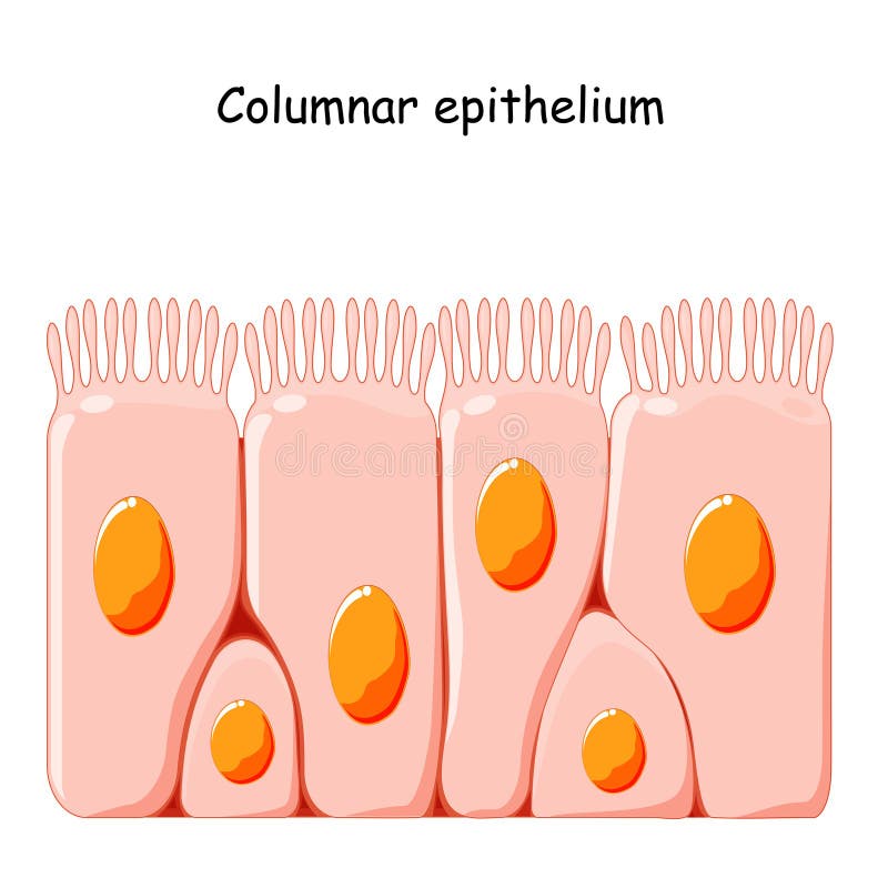
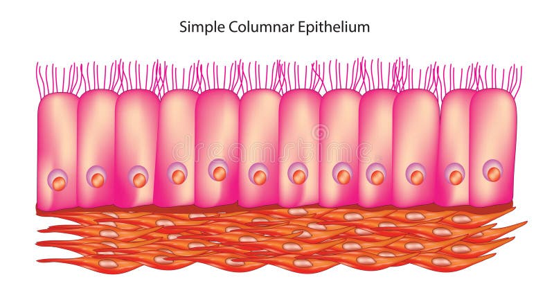
Free with trial Simple columnar epithelium is a type of epithelial tissue characterized by a single layer of tall, column-shaped cells. These cells are closely packed together and have elongated nuclei located near the basal surface. Simple columnar epithelium lines various structures in the body and performs specialized functions. Stomach mucus layer vectors Simple columnar epithelium
Free with trial Cream-white mucosal shield repels acidic sparks on a warm-red stomach, symbolizing barrier protection and mucosal protectants for pharmaceutical and clinical marketing. This illustration was created using AI Technology. Stomach mucus layer illustrations The Fortified Mucosal Shield - Gastric Protection Concept. Cream-white mucosal shield repels acidic sparks on a warm-red stomach, symbolizing barrier protection and mucosal protectants for pharmaceutical and clinical marketing. This illustration was created using AI Technology
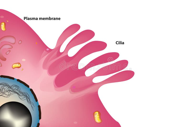
Free with trial Cilia structure with plasma membrane. Stomach mucus layer vectors Cilia of cell. Cilia structure with plasma membrane
Free with trial Medically 3D Illustration showing woman with painful stomach. Stomach mucus layer illustrations Painful stomach, gastritis, helicobacter pylori bacteria damaging mucus layer, medically accurate 3D illustration. Medically 3D Illustration showing woman with painful stomach